Neurochemical Mechanisms in Disease P3 pdf
Bạn đang xem bản rút gọn của tài liệu. Xem và tải ngay bản đầy đủ của tài liệu tại đây (112.06 KB, 10 trang )
Mechanisms Versus Diagnoses 5
and Thomas had to make a living. He did so by practicing the profession whose
literature he couldn’t really read. Fortunately, he was a genius. Science at that time
was heavily a matter of classification, including classification of the new species
of plants and animals being discovered in the Americas and other continents pre-
viously unexplored by Europeans. Sydenham classified illnesses analogously to the
way Linnaeus was classifying plants.
Sydenham used two sets of criteria f or his classifications: clinical signs and
symptoms and response to specific therapy. His model was what we now recog-
nize to be malaria. This disease typically causes high fevers every third or fourth
day, reflecting the life cycle of the parasites which we now recognize to cause it.
Sydenham characterized this clinical entity as “tertian fever” or “quartan fever.” He
recognized the value of treating it with extracts of cinchona bark, whose active prin-
ciple we now recognize to be quinine. This eminently practical approach—clinical
signs and symptoms indicating the need for a particular treatment—proved so useful
that it came to dominate medicine, not only in Britain but also in other countries.
Because Sydenham’s concept included response to a particular treatment (often a
medicine containing one or more critical molecules) it was to some extent a chem-
ical classification. However, it is worth noting that Sydenham himself felt that his
friend Dalton’s ideas about atoms had no significance for clinical medicine. Given
the medical ignorance of the time, his conclusion was correct when he made it.
1.2.4 Chemical and Biological Refinements of Sydenham’s Concepts
Over the succeeding centuries, developments in chemistry and biology led the con-
cept of what constituted a “disease” to depend less on purely clinical observations
and instead on more putatively “scientific” characteristics. The growth of chemistry,
especially the development of the chemistry of dyes during the nineteenth century,
led to the discovery of chemicals that stained human tissues obtained at autopsy.
The increased information that then came from pathology led to the definition of
diseases as “clinical-pathological” entities, that is, conditions in which a clincal pat-
tern was associated with a more or less specific anatomic pathology. This approach
still dominates neurology textbooks. Confusing clinical entities such as Alzheimer
disease are considered to be based on hard scientific definitions, inasmuch as they
are associated with characteristic neuropatholgical changes revealed by microscopic
examination after staining with appropriate dyes. Psychiatry has been considered a
“soft” specialty in part because of the lack of recognized anatomic pathology in the
brains of people with such major disorders as schizophrenia and depression. Now
that modern imaging techniques are increasingly identifying “objective” abnormal-
ities in the major mental illnesses, psychiatry has been described as becoming more
scientific.
Bacteriology and subsequently virology also led to important modifications of
Sydenham’s concept of diseases. Instead of such general classifications as “pthisis”
for inflammation of the lungs, physicians came to recognize more specific entities
such as “tuberculosis” or “diplococcus pneumoniae pneumonia.” The development
of convenient modern techniques for culturing pathogenic infectious agents and
6 J.P. Blass
determining their sensitivities to specific antibiotics has allowed this biological
knowledge to become part of the daily practice of medicine. We recognize an infec-
tion with “multiple antibiotic-resistant staphylococcus aureus” (MRSA) as an entity
independent of the organ infected and the resulting anatomic pathology. However,
it is worth noting that the sensitivities to particular antibiotics of different strains of
the same species of bacteria now vary so much that rational treatment of infec-
tions still involves direct laboratory studies of the patient being treated, namely
culture of the responsible micro-organisms from the specific patient and tests of
its sensitivity to specific antibiotics. You and I may both have pneumonia, and both
your and my pneumonia may be due to infection with diplococcus pneumoniae,
but your pneumonia may respond to and be appropriately treated with penicillin
whereas mine needs to be treated with another antibiotic. Knowing that clini-
cally important difference early in the course of our treatment requires laboratory
tests.
1.2.5 Molecular Studies and Clinical Specificity
Modern biochemistry and particularly modern nucleic acid chemistry (molecular
genetics) are forcing practitioners to re-evaluate their concepts of what constitutes
specific diseases. Nowhere is this more evident than in diseases of the nervous sys-
tem. (Hereditary ataxias are an example discussed above; psychoses are an example
discussed below.)
It would have been convenient if abnormalities in specific genes were to have
led reliably to specific clinical symptoms and signs, that is, to s pecific “diseases.”
Unfortunately, they do not. The general pattern has been that when an abnormal
gene is associated with a clinically defined entity, investigators at first assume that
it is more or less specifically associated with the “disease” in which it was discov-
ered. Subsequent studies of larger populations with a larger variety of “diseases”
typically show that abnormalities of the gene in question turn out to occur in a
variety of different diseases and usually even in people who have no clinically sig-
nificant disability. “Diseases” defined clinically or even by a combination of clinical
and laboratory findings generally turn out, on extensive study, to be genetically
heterogeneous in two senses: different genes can lead to the same clinical pat-
tern, and abnormalities of a single gene can lead to different clinical syndromes.
Stated technically, epidemiologically based studies typically reveal that diseases
defined clinically are genetically heterogeneous, and the consequences of mutations
in a single gene most often turn out to be clinically heterogeneous. The following
paragraphs give examples of these complexities.
1.3 Example 1: Tay–Sachs Disease
A classical “homogeneous” inborn error of metabolism, namely Tay–Sachs disease
(GM
2
gangliosidosis), provides a clear example.
6
Mechanisms Versus Diagnoses 7
1.3.1 Clinical Patterns
This condition was recognized clinically in the nineteenth century in infants of
Ashkenazi Jewish heritage, who suffered from a form of severe psychomotor
retardation in infancy and early death. These children were hard to distinguish
clinically from other infants who had other forms of devastating, early psychomo-
tor retardation with blindness, that is, other forms of “familial amaurotic idiocy.”
Differential clinical diagnosis depended on such clinical signs as an “exaggerated
startle response,” that is, an infant with Tay–Sachs disease was supposed to cry even
more than usual if startled by something like a loud clap of the physician’s hands.
1.3.2 Neuropathology, Neurochemistry, and Molecular Biology
Neuropathological observations subsequently allowed a more biological definition
of the subgroup of children with “true” Tay–Sachs disease. Light and then elec-
tron microscopy revealed characteristic “whorls” of material stored in their brain.
Subsequent neurochemical studies identified that material as GM
2
ganglioside.
Enzymatic studies showed that Tay–Sachs disease was due to a lack of a functional
form of an enzyme that catalyzes the breakdown of GM
2
ganglioside, namely hex-
osaminidase A. Molecular genetic studies demonstrated that this lack was due to
mutations in the HEXA gene that encodes this enzyme. Definitive clinical diagnosis
of Tay–Sachs disease now requires molecular genetic confirmation. The clinical
overlap among patients with “lipid storage diseases” is so great that a specific
diagnosis based on history and physical examination is no more than an informed
guess.
Thus neurochemistry and molecular biology appeared to have identified a bio-
logically homogeneous population who suffered from a particular constellation
of clinical signs and symptoms due to homozygous recessive inheritance of a
mutation-specific gene in a particular ethnic group, that is, from a specific “molecu-
lar disease.” Clinical applications of these neurochemical discoveries have allowed
Ashkenazi Jewish couples to be tested for carrier status even before the woman
becomes pregnant. Prenatal testing of cells obtained at amniocentesis from fetuses
at risk for this disease has allowed termination of the affected pregnancies in this
ethnic group for whom therapeutic abortion is religiously acceptable. This triumph
of modern medicine appeared to hold up as long as the chemical analyses were so
expensive and tedious that they were done largely in children who fit or were at
risk for the expected clinical characteristics. However, as cheaper and more auto-
mated procedures were developed that allowed testing of larger populations, the
associations between gene and ethnicity and gene and clinical syndrome both broke
down.
1.3.3 Genetic Variability
HEXA deficiency has turned out to be neither genetically nor ethnically homo-
geneous. A variety of different alterations—mutations—in the HEXA gene have
been associated with classic, infantile Tay–Sachs disease. The existence of different
8 J.P. Blass
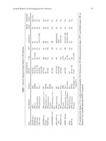
Table 1 Clinical presentations of HEXA deficiency
Psychomotor retardation Gravel et al. (1995)
Early infantile form
Late infantile form
Sandhoff disease Hendriksz et al. (2004)
Infantile form
Juvenile form
Juvenile progressive dystonia Meek et al. (1984)
Spinocerebellar disease (several clinical
syndromes)
Argov and Navon (1984), Oonk et al. (1979), and
Rapin et al. (1976)
Motor neuron disease Argov and Navon (1984) and Drory et al. (2003)
Focal muscular atrophy Iype et al. (2006)
Dementia Hammer (1998) and O’Neill et al. (1978)
Depression Hammer (1998) and Renshaw et al. (1992)
Schizophrenia Hammer (1998) and Rosebush et al. (1995)
Postpartum psychosis Lichtenberg et al. (1988)
Asymptomatic Navon et al. (1973)
This list is not exhaustive, nor is the citation of references. New syndromes associated with HEXA
deficiency are still appearing in the medical literature.
mutations in a single gene among different individuals and among different popula-
tions is, of course, the rule rather than the exception in studies of inherited diseases.
Clinically typical, HEXA-deficient Tay–Sachs disease occurs in a number of ethnic
groups. In some, the mutations tend to differ from those most frequently found in
Ashkenazi Jews. For instance, there is a “French Canadian” mutation as well as an
“Ashkenazi Jewish” mutation. (See Gravel et al., Table 1, for discussion and ref-
erences.) However, the overlaps in mutations among ethnic groups are wide. They
confirm the principle, well known to human geneticists, that genome studies tell
many of us things that we did not know, or want to know, or want our spouses
to know. (Genetic counselors are obligated to warn a family for whom molecular
genetic studies are recommended that for perhaps 15% of children, the supposed
father is not the biological father.)
1.3.4 Variations in Clinical Phenotype
More important for this discussion, abnormalities of the responsible HEXA gene
have now been associated with a dozen or more clinically distinct patterns (Table 1),
including “schizophrenia” and including people with no clinically significant dis-
ability. Put technically, “adult onset Tay–Sachs disease” is clinically pleomorphic.
A steady stream of reports continues to appear describing variant neurological
abnormalities in people with “adult onset Tay–Sachs disease.”
Systematic large studies of the incidence of HEXA abnormalities among patients
in common diagnostic categories such as “schizophrenia” are in short supply.
What studies have been done encourage further work (Goodman, 1994). Studies
of another inborn error of metabolism associated with “schizophrenia syndromes,”
metachromatic leukodystrophy, have shown a high incidence of abnormalities in
Mechanisms Versus Diagnoses 9
sulfatide metabolism. The abnormalities have been attributed to “pseudosulfatase
deficiency” (Alvarez et al., 1995; Galbraith et al., 1989; Herska et al., 1987).
Molecular genetic investigations have not been reported. There appears not to
have been a systematic study at the molecular genetic level of the incidence of
abnormalities in genes responsible for storage disorders such as Tay–Sachs dis-
ease and metachromatic leukodystrophy, what have previously been called “Type II
schizophrenics.” These unfortunate patients suffer from relentless, generally drug-
unresponsive, progressive psychoses which sooner or later turn into dementia, and
are associated with brain atrophy with enlarged ventricles. Type II schizophrenics
appear to have a progressive brain disease. One may speculate that some of them
have as yet unelucidated variants of disorders that in other, more severe forms lead
to progressive psychomotor failure in infancy or early childhood.
1.4 Example 2: Psychoses
The neurochemical and molecular genetic study of psychoses including schizophre-
nia is beset by problems of clinical definition.
1.4.1 Recognizing Psychosis
The first of these problems is deciding who is psychotic. The difficulty in defining
precise criteria for whether someone is crazy is summarized in an old Quaker saying:
“All the world is mad but me and Thee, and sometimes I doubt Thee.” Whole nations
can go mad, for instance, the paranoia of the highly educated German-speaking
world during the time of the Nazis. (The review of the movie Saving Private Ryan
in the Süddeutsche Zeitung pointed out that using the Nazis as a horrible exam-
ple is less controversial than using more up-to-date examples of insane cruelty
masking itself as politics.) Sets of diagnostic criteria for psychosis and for specific
psychoses continue to be promulgated, not least in the volumes of the Diagnostic
and Statistical Manual of the American Psychiatric Association (most recently DSM
IV-TR). Applying these criteria well requires skill and training.
Although the lines between mad, odd, and sane are hard to draw precisely,
common sense often allows easy classification. As usual, a clinical anecdote is
illustrative.
A woman suffering from a severe (masked) depression lost her appetite to the point where
her body weight fell to a dangerously low level (body mass index of 14.3). The neurologist
who saw her immediately arranged admission to a psychiatric hospital. The admitting resi-
dent there was concerned about whether the patient met DSM IV-TR criteria for depression,
and if so of what type. The neurologist responded, somewhat rudely: “Look, this lady has
nearly succeeded in starving herself to death. Please admit her and feed her, if necessary
through a tube, and treat her for depression. There will be plenty of time to worry about
how to classify her mental disease once she is no longer at imminent risk of death from an
infection or other complication of starvation.”
10 J.P. Blass
1.4.2 Classification of Psychoses
Clinical observers have classified forms of madness in different ways over the cen-
turies. The Hippocratic physicians used the single category “delirium” for madness
whether an external cause could be recognized or not. In modern terms, they did not
distinguish between endogenous and exogenous psychoses, and between endoge-
nous and exogenous neurointoxicants. Instead they described what was happening
in individual patients or groups of patients. As knowledge of the molecular bases
of psychotic behavior increases, including endogenous chemical imbalances in the
brain, we may yet go back to a modernized version of their formulation.
The modern classification of madness goes back about a century, to the
German psychiatrist Bleuler. Among the mental disorders he classified that had
no neuropathological stigmata at that time were schizophrenia (thought disor-
der) and depression and manic-depressive disease (mood disorders). This dis-
tinction has persisted in psychiatry. It continues to be widely although not uni-
versally accepted. However, modern molecular studies are bringing the biology
of the distinction between “thought disorder” and “mood disorder” into serious
question.
1.4.3 The DISC1 Locus
DISC1 is an example of a gene that predisposes to both thought disorder and mood
disorder (Craddock et al., 2006; Porteus et al., 2006). The association of this gene
with schizophrenia was discovered in a family in whom the insanity was associ-
ated with a balanced translocation. A number of studies then demonstrated and
confirmed that abnormalities in this gene were associated with “typical” schizophre-
nia. Further studies showed that abnormalities in DISC1 were also associated with
manic-depressive (bipolar) psychosis. This was not too surprising, because the clini-
cal differential diagnosis between schizophrenia and bipolar disease can be difficult,
particularly while the sufferers are very crazy. Further studies then showed that
abnormalities in DISC1 were also associated with recurrent unipolar depression,
which is relatively easy to tell from schizophrenia and, with careful examination,
even from bipolar disorder. Porteus et al. (2006) concluded that: “DISC1 is a gen-
eralizable genetic risk factor for psychiatric illness that also influences cognition in
healthy subjects.”
1.4.4 Other Loci
Other loci also contribute to the risk of schizophrenia as well as other dis-
eases. Mutations in the neuroregulin 1 gene (NRG-1) are also associated with
both thought disorders and mood disorders (Green et al., 2005). Abnormalities in
the 15q13-q14 region of chromosome 15 predispose replicably to the existence
of schizophrenia, but also to bipolar disorder, schizoaffective disorder, Prader–
Willi syndrome (a developmental disorder associated with psychosis), and some
forms of juvenile epilepsy (Leonard and Freedman, 2006). Other loci associated
Mechanisms Versus Diagnoses 11
with both “schizophrenia” and “bipolar disease” have been described on chromo-
some 13 and chromosome 22 and in relation to genes encoding components of
myelin.
1.4.5 Modifier Loci
Presumably, the variable clinical presentations of a single genetic abnormality
reflect the influences of other genes as well as of varying environmental events.
The effect of other parts of an individual’s genome have been referred to as
actions of “modifier genes” or “genetic background.” The effects of specific envi-
ronmental influences on the clinical expression of a variation in DNA are also
being delineated. A striking example is the combination of the interaction of the
Val
158
allele of catechol-O-methyl transferase (COMT) and cannabis use in causing
“schizophrenia.” Whether the effects of this allele by itself are clinically signifi-
cant is controversial. However, it is clear that those who carry this allele and also
use cannabis during their adolescence have a tenfold increased risk of becom-
ing psychotic, compared to the general population (Caspi et al., 2005). Is their
psychosis “schizophrenia” or “cannabis toxicity?” Does it matter? As the late
Houston Merritt said about a patient presented to him as a diagnostic problem,
“We all know what is wrong with this person; we are just debating what to call
it.” These semantic problems can be an entertaining exercise in medical erudition,
but semantic distinctions should not alter the quality of the care we give to individual
patients.
1.4.6 Implications for Research on Mental Illness
The recognition that the same fundamental biological changes can lead to both
thought disorders (schizophrenias) and mood disorders (bipolar disease and some-
times unipolar depression) does not contradict clinical experience as much as
it brings into question interpretations of neurochemical research on these disor-
ders. Bleuler, one of the psychiatrists most responsible for the distinction between
thought disorders and mood disorders, himself recognized that the clinical distinc-
tion (“differential diagnosis”) between these conditions can be extremely difficult.
The standard emergency room pharmacological treatment for a patient with an acute
psychosis involves treatment suitable for both conditions. However, researchers
have in the past used patients with the diagnosis of “bipolar disorder” as “dis-
ease controls” for studies of “schizophrenia,” and vice versa. Several metabolites
discovered decades ago in the urine of mentally ill people were dismissed as “non-
specific findings” because their excretion was associated with both conditions. In
the light of modern molecular genetic discoveries, that interpretation may have been
wrong. The patients in the two categories may have had different clinical manifes-
tations of the same biological, neurochemical abnormality. Skolnick (2006), who
has extensive experience and expertise in the development of new pharmaceuticals,
has proposed that the best way now available for developing innovative treatments
12 J.P. Blass
for complex illnesses such as psychoses is to define genetic risk factors and then
develop innovative new drugs based on the genetic information.
1.5 Example 3: Multiple Sclerosis and Demyelination
1.5.1 Clinical Patterns
The medical school definition of “multiple sclerosis” (MS) is demyelination within
the central nervous system that varies in space and time. The term refers to a dis-
order in which patches of demyelination develop during exacerbations. In the most
common forms of MS, exacerbations are separated by varying periods of time in
which the disease does not appear to progress. Whether progressive demyelination
without clear periods of remission should be considered a form of MS is a matter
of definition, about which clinicians specializing in the care of patients with this
disorder have argued. Conventional medical nomenclature classifies as distinct enti-
ties a number of disorders of myelin that can mimic MS clinically. These include,
for instance, the sometimes devastating demyelination localized to the pons or the
demyelination that can follow infectious diseases and/or vaccinations.
The precise meaning of the term“multiple sclerosis” is “many scars.” The words
themselves do not indicate that the scars are even in the nervous system, let alone in
myelin. The British terminology, “disseminated sclerosis,” is no more descriptive.
Charcot, who first distinguished this condition from other disorders of the nervous
system including syphilis, coined the slightly more precise French term, “sclerose
en plaque.”
An inconvenient but more descriptive name for this MS is “intermittent, patchy
demyelination.” This clumsy term makes clear that “multiple sclerosis” is unlikely
to be a single entity in terms of cause (etiology) or disease mechanisms (patho-
physiology). I n principle, any conditions or combination of conditions that lead to
intermittent, patchy demyelination are forms of “multiple sclerosis.” If a clear cause
can be identified, the condition is by convention not referred to as multiple sclerosis.
The disease is therefore by definition of unclear etiology. The neurology literature
of the last 100 years contains confident declarations that multiple sclerosis has been
proven to be a viral disease, that it has been proven not to be a viral disease, that
it has been proven to be an immune disease, that immune mechanisms in multiple
sclerosis have been shown to be secondary to the disease process, and so on.
1.5.2 Proposed Mechanisms
Current opinions on cause and mechanism include the possibility that MS is often
due to a form of “molecular mimicry,” in which an immune response to an infective
or other exogenous agent leads to the formation of antibodies and/or cells that cross-
react destructively with components of normal myelin. “Molecular mimicry” is well
established in certain other disorders of the nervous system (Candler et al., 2006)
including paraneoplastic syndromes (Posner, 2003).
Mechanisms Versus Diagnoses 13
1.5.3 Chemistry of Demyelination
In contrast to the confusion about what multiple sclerosis is or are, the chemistry
of demyelinating processes is rather well defined. (See Chapters XXX.) Whatever
the causes of “multiple sclerosis” eventually turn out to be, they lead to demyeli-
nating processes which will in all likelihood be very similar to or identical at
the neurochemical level with the demyelinating processes that have already been
elucidated. The information about the mechanisms of demyelination is based on
firm, robust, reproducible experimental observations. This information is likely to
expand, but what is now proven is unlikely to change. This solid science contrasts
with the theoretically rather vague although clinically useful clinical conceptualiza-
tion of “multiple sclerosis” as an entity. Describing the neurochemical correlates
of MS would be an exercise in phenomenology, and unstable phenomenology as
the clinical definition of this syndrome changes. Demyelination can, however, be
meaningfully discussed in terms of mechanistic neurochemistry.
1.6 Implications
The practice of medicine is a service profession, not a science. It has more in
common with a trade than with a branch of scholarship. The physician has the
responsibility of trying to keep in mind all the variables that pertain to the person he
or she is trying to help, and must choose within a relatively short time to do noth-
ing or to do something practical, such as prescribing a medication or giving advice.
The scientist has the responsibility and the luxury of taking the time to think deeply
about one aspect of a problem. There is truth to the cliché that scientists are paid to
think more and more about less and less (at least until they become senior enough
to have “administrative responsibilities” including raising large sums of money).
Science has undoubtedly contributed in major ways to the improvement of human
health, as documented by increasing longevity in developed countries. Not least
have been improvements in nutrition, in the safety of the water and food sup-
plies, and in maternal and child health including vaccination against communicable
plagues of childhood, such as diptheria. The mental health of both patients and
practitioners requires that we also believe in the value of curative medicine (as
do the profits of pharmaceutical companies). Unfortunately, there are observations
which temper that confidence. During a doctors’ strike in Israel some decades
ago, the death rate fell, with no compensatory blip before or after the time dur-
ing which everything but emergency medicine and the refilling of medications was
suspended. These data do not lead to a recommendation that the treatment of ill-
ness be suspended. They do suggest a seemly humility both among those of us who
practice medicine and those in the scientific community who provide the informa-
tion on which those of us who have practiced medicine claim to have based our
recommendations.
Medical practitioners should and will continue to adapt the information made
available by science to improve their treatment of patients. Scientists using the
14 J.P. Blass
experimental method and drawing compelling conclusions from their data will con-
tinue to add to the body of definitive information on which medical practicitoners
can draw. This volume concentrates on the solid scientific studies of neurochemi-
cal mechanisms. It leaves to clinical textbooks the discussion of the classification
(nosology) of diseases and the discussion of biological abnormalities in those often
vaguely defined illnesses, as well as discussions of the interventions now believed
to be appropriate.
References
Alvarez LM, Castillo ST, Perez ZJA, Vargas RI, Sepulveda GR, Zuniga CMA (1995) Activity
of aryl sulfatase a enzyme in patients with schizophrenic disorders. Rev Invest Clin 47:
387–392
Argov Z&Navon R (1984) Ann Neurol 16:14–20
Candler PM, Dale RC, Griffin S, Church AJ, Wait R, C hapman MD, Keir G, Giovannoni G,
Rees JH (2006 Apr) Post-streptococcal opsoclonus-myoclonus syndrome associated with
anti-neuroleukin antibodies. J Neurol Neurosurg Psychiatry 77(4):507–512
Caspi A, Moffit TE, Cannon M, McClay J, Murray R, Harrington H et al (2005) Moderation of the
effect of adolescent-onset cannabis use on adult psychosis by a functional polymorphism in the
catechol-O-methyltransferase gene: longitudinal evidence of a gene X-environment interaction.
Biol Psychiatry 57:1117–1127
Craddock N, O’Donovan MC, Owen MJ (2006) Genes for schizopohrenia and bipolar disorder?
Implications for psychiatric nosology. Schizophr Bull 32:9–16
Drory VE et al (2003) Muscle Nerve 28:109–112
Duenas AM, Gold R, Giunti P (2006) Molecular pathogenesis of spinocerebellar ataxias. Brain
129(Pt 6):1357–1370, Epub 2006 Apr 13
Galbraith DA, Gordon BA, Feleki V, Gordon N, Cooper AJ (1989) Metachromatic leukodystrophy
(MLD) in hospitalized adult schizophrenic patients resistant to drug treatment. Can J Psychiatry
34:299–302
Goodman AB (1994) Medical conditions in Ashkenazi schizophrenic pedigrees. Activity of aryl
sulfatase a enzyme in patients with schizophrenic disorders. Schizophr Bull 20:507–517
Gravel RA et al (1995) In: Scriver CR et al (eds) Metabolic and molecular basis of inherited
disease, 7th edn. McGraw-Hill, New York, NY, pp 2839–2882
Green EK, Raybould R, MacGregor S, Gordon-Smith K, Heron J, Hyde S et al (2005) Operation
of the schizophrenia susceptibility gene, neuregulin 1, across traditional diagnostic bounderies
to increase risk for bipolar disorder. Arch Gen Psychiat 62:642–648
Hammer MB (1998) Psychosomatics 39:446–448
Hendriksz CJ et al (2004) J Inherit Metab Dis 27:241–249
Herska M, Moscovich DG, Kalian M, Gottlieb D, Bach G (1987 Mar) Aryl sulfatase a deficiency
in psychiatric and neurologic patients. Am J Med Genet 26(3):629–635
Iype M et al (2006) Ann Ind Acad Neurol 9:110–112
Kendell RE (1975) The role of diagnosis in psychiatry. Blackwell, London, See also: Kendell
RE (2002) The distinction between personality disorder and mental illness. Br J Psychiat 180:
110–115
Leonard S, Freedman R (2006) Genetics of chromosome 15q13-q14 in schizophrenia. Biol
Psychiatry 60:115–122
Lichtenberg P et al (1988) Br J Psychiat 153:387–389
Lodi R, Tonon C, Calabrese V, Schapira AH (2006) Friedreich’s ataxia: from disease mechanisms
to therapeutic interventions. Antioxid Redox Signal 8:438–443
Meek D et al (1984) Ann Neurol 15:348–352


