Diabeticcpg small 2 (Part 1) pptx
Bạn đang xem bản rút gọn của tài liệu. Xem và tải ngay bản đầy đủ của tài liệu tại đây (711.58 KB, 12 trang )
A Supplement to:
Foot &
Ankle
Surgery
The
Journal
of
An official publication of the
American College of
Foot and Ankle Surgeons
DIABETIC
FOOT
DISORDERS
A CLINICAL
PRACTICE GUIDELINE
SEPTEMBER/OCTOBER 2006
VOLUME 45, NUMBER 5
Development and publication of this Clinical Practice
Guideline was made possible by an Educational Grant
Co-Sponsored by Johnson & Johnson Wound Management,
a division of ETHICON, INC. and KCI USA, Inc.
Robert G. Frykberg, DPM, MPH,
1
Thomas Zgonis, DPM,
2
David G. Armstrong, DPM, PhD,
3
Vickie R. Driver,
DPM, MS
4
John M. Giurini, DPM,
5
Steven R. Kravitz, DPM,
6
Adam S. Landsman, DPM, PhD,
7
Lawrence A.
Lavery, DPM, MPH,
8
J. Christopher Moore, DPM,
9
John M. Schuberth, DPM,
10
Dane K. Wukich, MD,
11
Charles
Andersen, MD,
12
and John V. Vanore, DPM
13
Supplement to:
Foot &
Ankle
Surgery
The
Journal
of
DIABETIC FOOT DISORDERS:
A CLINICAL PRACTICE GUIDELINE (2006 revision)
Address correspondence to: Robert G. Frykberg, DPM, MPH, Chief, Podiatric Surgery, Carl T. Hayden VA
Medical Center, Phoenix, AZ 85012. Email:
1
Chair, Diabetes Panel, Phoenix, AZ;
2
San Antonio, TX;
3
North Chicago, IL;
4
Evanston, IL;
5
Boston, MA;
6
Richboro, PA;
7
Boston, MA;
8
Georgetown, TX;
9
Ashville, NC;
10
San Francisco, CA;
11
Pittsburgh, PA;
12
Seattle, WA;
13
Chair, Clinical Practice Guidelines Core Committee, Gadsden, AL
ABSTRACT: The prevalence of diabetes mellitus is growing at epidemic proportions in the United States and
worldwide. Most alarming is the steady increase in type 2 diabetes, especially among young and obese people. An
estimated 7% of the US population has diabetes, and because of the increased longevity of this population, dia-
betes-associated complications are expected to rise in prevalence.
Foot ulcerations, infections, Charcot neuroarthropathy, and peripheral arterial disease frequently result in gan-
grene and lower limb amputation. Consequently, foot disorders are leading causes of hospitalization for persons
with diabetes and account for billion-dollar expenditures annually in the US. Although not all foot complications
can be prevented, dramatic reductions in frequency have been achieved by taking a multidisciplinary approach to
patient management. Using this concept, the authors present a clinical practice guideline for diabetic foot disor-
ders based on currently available evidence, committee consensus, and current clinical practice. The pathophysiol-
ogy and treatment of diabetic foot ulcers, infections, and the diabetic Charcot foot are reviewed. While these guide-
lines cannot and should not dictate the care of all affected patients, they provide evidence-based guidance for gen-
eral patterns of practice. If these concepts are embraced and incorporated into patient management protocols, a
major reduction in diabetic limb amputations is certainly an attainable goal.
This clinical practice guideline (CPG) is based on the consensus of current clinical practice and review of the clin-
ical literature. This guideline was developed by the Clinical Practice Guideline Diabetes Panel of the American
College of Foot and Ankle Surgeons.
S–2 THE JOURNAL OF FOOT & ANKLE SURGERY
Supplement to:
Foot &
Ankle
Surgery
The
Journal
of
DIABETIC FOOT DISORDERS:
A CLINICAL PRACTICE GUIDELINE (2006 revision)
INTRODUCTION
The prevalence of diabetes mellitus is growing at epidem-
ic proportions in the United States and worldwide (1). Most
alarming is the steady increase in type 2 diabetes, especial-
ly among young and obese persons. An estimated 7% of
Americans are afflicted with diabetes, and with the longevi-
ty of this population increasing, the prevalence of diabetes-
related complications will continue to rise.
Foot disorders are a major source of morbidity and a lead-
ing cause of hospitalization for persons with diabetes.
Ulceration, infection, gangrene, and amputation are signifi-
cant complications of the disease, estimated to cost billions
of dollars each year. Charcot foot, which of itself can lead
to limb-threatening disorders, is another serious complica-
tion of long-standing diabetes. In addition to improving the
management of ulcers—the leading precursor to lower
extremity amputation in diabetic patients (2)—clinicians
must determine how to more effectively
prevent ulceration.
Although not all diabetic foot disorders can be prevented, it
is possible to effect dramatic reductions in their incidence
and morbidity through appropriate evidence-based preven-
tion and management protocols.
Taking a multidisciplinary approach to diabetic foot dis-
orders, many centers from around the world have noted
consistent improvement in limb salvage rates. With this
premise as our central theme, the authors present this clini-
cal practice guideline based on currently available evidence.
Three major pedal complications of diabetes are reviewed:
diabetic foot ulcers, diabetic foot infections, and the diabet-
ic Charcot foot. These guidelines are intended to provide
evidence-based guidance for general patterns of practice
and do not necessarily dictate the care of a particular
patient.
DIABETIC FOOT DISORDERS VOLUME 45, NUMBER 5, SEPTEMBER/OCTOBER 2006 S–3
EPIDEMIOLOGY OF DIABETIC
FOOT DISORDERS
Diabetes is one of the foremost causes of death in many
countries and a leading cause of blindness, renal failure, and
nontraumatic amputation. Global prevalence of diabetes in
2003 was estimated to be 194 million (3). By 2030, this fig-
ure is predicted to rise to 366 million due to longer life
expectancy and changing dietary habits (4).
The estimated incidence of diabetes in the US exceeds 1.5
million new cases annually, with an overall prevalence of
20.8 million people or 7% of the nation’s population (5). An
estimated 14.6 million persons are currently diagnosed with
the disease, while an additional 6.2 million people who
have diabetes remain undiagnosed; this represents a sixfold
increase in the number of persons with diabetes over the
past four decades (6). A higher incidence of diabetes occurs
among non-Hispanic blacks, Hispanic/Latino Americans,
and Native Americans compared with non-Hispanic whites
(7). Diagnosed diabetes is most prevalent in middle-aged
and elderly populations, with the highest rates occurring in
persons aged 65 years and older (8-10). As the sixth leading
cause of death in the US, diabetes contributes to more than
224,000 deaths per year (5).
Four categories of diabetes are recognized (Table 1). Type
1, formerly insulin-dependent diabetes mellitus (IDDM), is
an autoimmune disease affecting the pancreas. Individuals
with type 1 diabetes are prone to ketosis and unable to pro-
duce endogenous insulin. Type 2, formerly non-insulin
dependent diabetes mellitus (NIDDM), accounts for 90% to
95% of cases diagnosed. Type 2 diabetes is characterized by
hyperglycemia in the presence of hyperinsulinemia due to
peripheral insulin resistance. Gestational as well as genetic
defects and endocrinopathies are recognized as other types
of diabetes (11). Diabetes is associated with numerous
complications related to microvascular, macrovascular, and
metabolic etiologies. These include cerebrovascular, cardio-
vascular, and peripheral arterial disease; retinopathy; neu-
ropathy; and nephropathy. Currently, cardiovascular com-
plications are the most common cause of premature death
among patients with diabetes (9, 12). Rates of heart disease
and stroke are 2 to 4 times higher among diabetic adults
compared with nondiabetic adults, accounting for about
65% of deaths in people with diabetes (5). Estimated total
(direct and indirect) annual expenditures for diabetes man-
agement in 2002 was $132 billion, representing 1 of every
10 health care dollars spent in the US (13).
One of the most common complications of diabetes in the
lower extremity is the diabetic foot ulcer. An estimated 15%
of patients with diabetes will develop a lower extremity
ulcer during the course of their disease (14-17). Several
population-based studies indicate a 0.5% to 3% annual
cumulative incidence of diabetic foot ulcers (18-21).
According to one large British study of neuropathic
patients, the 1-year incidence of initial foot ulcer was 7%
(22). The prevalence of foot ulcers reported for a variety of
populations ranges from 2% to 10% (16, 18, 22, 23).
Neuropathy, deformity, high plantar pressure, poor glucose
control, duration of diabetes, and male gender are all con-
tributory factors for foot ulceration (see the following sec-
tion: “Risk for Ulceration”) (24-27). National hospital dis-
charge data indicate that the average hospital length of stay
(LOS) for diabetic patients with ulcer diagnoses was 59%
longer than for diabetic patients without ulcers (16). While
7% to 20% of patients with foot ulcers will subsequently
require an amputation, foot ulceration is the precursor to
approximately 85% of lower extremity of amputations in
persons with diabetes (28-31).
Diabetes continues to be the most common underlying
cause of nontraumatic lower extremity amputations (LEAs)
in the US and Europe (1, 32). More than 60% of LEAs in
the US occur in people with diabetes, averaging 82,000 per
year (5, 10). While the number of diabetes-related hospital
discharges has progressively increased from 33,000 in 1980
to 84,000 in 1997, this number seems to have leveled off
during the present decade. In 2002, there were 82,000 dia-
betes-related LEA discharges, accounting for 911,000 days
of hospital stay with an average LOS of 11.2 days (10). The
age-adjusted rate of amputation for that year was 5.2 per
1,000 persons with diabetes, a notable decrease from the
highest rate of 8.1 per 1,000 in 1996.
In terms of level of diabetes-related lower limb amputa-
tions, toe amputations comprise the majority of procedures.
The age-adjusted LEA rate in 2002 among persons with dia-
betes was highest for toe LEA (2.6 per 1,000 persons), fol-
lowed by below-knee LEA (1.6 per 1,000 persons). For foot
LEA and above-knee LEA, the age-adjusted rate was 0.8
per 1,000 persons. These trends in amputation level have
essentially remained the same since 1993 (10). Generally,
the LEA rate is 15 to 40 times higher in the diabetic versus
Table 1
Type 1 diabetes - absolute insulin deficiency
Type 2 diabetes - insulin resistant +/- insulin deficiency
Other types - genetic defects of ß-cell function or insulin action
endocrinopathies
drug or chemical
infections
Gestational diabetes
* adapted from: Therapy for Diabetes Mellitus and Related Disorders, 3rd edition,
American Diabetes Association, 1998.
Classification of Diabetes Mellitus *
nondiabetic populations, and the rate is at least 50% higher
in men versus women (8, 10, 12, 33). In 2002, the age-
adjusted LEA rate among men was 7.0 per 1,000 persons
with diabetes compared with to the rate among women
reported at 3.3 per 1000 persons with diabetes (10).
Several ethnic differences occur in the frequency of dia-
betes-related amputations. Mexican (Hispanic) Americans,
Native Americans, and African Americans each have at
least a 1.5- to 2-fold greater risk for diabetes-related ampu-
tation than age-matched diabetic Caucasians (8, 10, 16, 17,
34, 35). When LEA risk is compared between diabetic and
nondiabetic populations worldwide, it is apparent that both
diabetes and ethnicity have profound implications on rates
of lower limb amputation (1, 17).
Survival rates after amputation are generally lower for
diabetic versus nondiabetic patients (16, 17, 29). The 3- and
5-year survival rates are about 50% and 40%, respectively,
with cardiovascular disease being the major cause of death
(8). Although mortality rates following major amputation
are high among both diabetic and nondiabetic patients, a
recent study reported no significant difference between
these two populations. The mean survival was approximate-
ly 6.5 years, with a 68% mortality after 9 years regardless
of diabetes status (36). An earlier study from Sweden
reported a 5-year mortality rate of 68% after lower limb
amputation, with survival rates lower among patients who
underwent higher levels of amputation (29). Similar trends
were found in a review of amputations within the Veterans
Affairs system, but worse survival outcomes were observed
for older patients, those with renal disease, and those with
peripheral arterial disease (37). Researchers have reported a
50% incidence of serious contralateral foot lesion (ie, ulcer)
following an LEA, and a 50% incidence of contralateral
amputation within 2 to 5 years of an LEA (16, 29).
Total (direct and indirect) annual health care costs for per-
sons with diabetes were estimated to be $132 billion in
2002. Direct medical expenditures, including hospitaliza-
tion, medical care, and supplies, accounted for $91.8 billion
(13). The estimated cost for foot ulcer care in the US ranges
from $4,595 per ulcer episode to nearly $28,000 for the 2
years after diagnosis (19, 38). One report estimates 800,000
prevalent ulcer cases in the US, with costs averaging $5,457
per year per patient or total national annual costs of $5 bil-
lion (39). Astudy of Medicare claims data found that expen-
ditures for patients with lower extremity ulcers averaged 3
times higher than expenditures for Medicare beneficiaries
in general. With 24% of their total costs allocated to ulcer-
related expenses, lower extremity ulcer patients cost the
Medicare system $1.5 billion in 1995 (40). According to a
large prospective study of diabetic patients with foot ulcers,
about 7% will subsequently require a lower extremity
amputation (31). While hospital LOSs for diabetes-related
LEA have progressively decreased in the US, the overall
direct costs remain high (10, 16). Direct and indirect costs
of LEA—which range from $20,000 to $40,000 per event—
vary by year, payer, level of amputation, LOS, and attendant
comorbidities (16). If the lower figure is applied to the
82,000 amputations performed in 2002, estimated total
costs of LEA might exceed $1.6 billion annually. When out-
patient costs for ulcer care preceding these amputations is
added, the estimated total costs in the US for diabetic foot
disease can easily approach or exceed $6 billion annually.
Risk for Ulceration
Foot ulceration is the most common single precursor to
lower extremity amputations among persons with diabetes
(28-30). Treatment of infected foot wounds comprises up to
one quarter of all diabetic hospital admissions in the US and
Britain, making this the most common reason for diabetes-
related hospitalization in these countries (41-43). The mul-
tifactorial nature of diabetic foot ulceration has been eluci-
dated by numerous observational studies (16, 22, 24, 26, 27,
44-48). Risk factors identified include peripheral neuropa-
thy, vascular disease, limited joint mobility, foot deformi-
ties, abnormal foot pressures, minor trauma, a history of
ulceration or amputation, and impaired visual acuity (25,
49, 50). These and other putative causative factors are
shown in Figure 1.
Peripheral sensory neuropathy in the face of unperceived
trauma is the primary factor leading to diabetic foot ulcera-
tions (24, 27, 46, 49). Approximately 45% to 60% of all dia-
betic ulcerations are purely neuropathic, while up to 45%
have neuropathic and ischemic components (24, 51).
According to an important prospective multicenter study,
sensory neuropathy was the most frequent component in the
causal sequence to ulceration in diabetic patients (24).
Other forms of neuropathy may also play a role in foot
ulceration. Motor neuropathy resulting in anterior crural
muscle atrophy or intrinsic muscle wasting can lead to foot
deformities such as foot drop, equinus, hammertoe, and
prominent plantar metatarsal heads (25, 26, 52-54). Ankle
equinus with restricted dorsiflexory range of motion is fair-
ly common in patients with diabetic neuropathy and can be
a consequence of anterior crural muscle atrophy (55-60).
The decreased ankle motion, which confers higher-than-
normal plantar pressures at the forefoot, has been implicat-
ed as a contributory cause of ulceration as well as recur-
rence or recalcitrance of existing ulcers (57, 58, 60, 61).
Autonomic neuropathy often results in dry skin with
cracking and fissuring, creating a portal of entry for bacte-
S–4 THE JOURNAL OF FOOT & ANKLE SURGERY
DIABETIC FOOT DISORDERS VOLUME 45, NUMBER 5, SEPTEMBER/OCTOBER 2006 S–5
Figure 1 The risk
factors for ulceration
may be distinguished
by general or systemic
considerations versus
those localized to the
foot and its pathology.
ria (42, 63). Autosympathectomy with attendant sympathet-
ic failure, arteriovenous shunting, and microvascular ther-
moregulatory dysfunction impairs normal tissue perfusion
and microvascular responses to injury. These alterations can
subsequently be implicated in the pathogenesis of ulcera-
tion (63-67).
Foot deformities resulting from neuropathy, abnormal
biomechanics, congenital disorders, or prior surgical inter-
vention may result in high focal foot pressures and
increased risk of ulceration (24, 48, 50, 57, 68-71). The
effects of motor neuropathy occur relatively early and lead
to foot muscle atrophy with consequent development of
hammertoes, fat pad displacement, and associated increases
in plantar forefoot pressures (53, 72-75). Although most
deformities cause high plantar pressures and plantar foot
ulcerations, medial and dorsal ulcerations may develop as a
result of footwear irritation. Common deformities might
include prior partial foot amputations, prominent metatarsal
heads, hammertoes, Charcot arthropathy, or hallux valgus
(69, 76-79). A large prospective population-based study
found that elevated plantar foot pressures are significantly
associated with neuropathic ulceration and amputation (80).
The study also revealed a trend for increased foot pressures
as the number of pedal deformities increased.
Trauma to the foot in the presence of sensory neuropathy
is an important component cause of ulceration (24). While
trauma may include puncture wounds and blunt injury, a
common injury leading to ulceration is moderate repetitive
stress associated with walking or day-to-day activity (69,
76, 81). This is often manifested by callus formation under
the metatarsal heads (48, 82, 83). A recent report suggests
that even with moderate activity, ulceration may be precip-
itated by a higher degree of variability in activity or period-
ic “bursts” of activity (84). Shoe-related trauma has also
been identified as a frequent precursor to foot ulceration
(28, 51, 54, 85, 86).
Peripheral arterial disease (PAD) rarely leads to foot
ulcerations directly. However, once ulceration develops,
arterial insufficiency will result in prolonged healing,
imparting an elevated risk of amputation (28, 87, 88).
Additionally, attempts to resolve any infection will be
impaired due to lack of oxygenation and difficulty in deliv-
ering antibiotics to the infection site. Therefore, early recog-
nition and aggressive treatment of lower extremity ischemia
are vital to lower limb salvage (30, 52, 89-91).
Limited joint mobility has also been described as a poten-
tial risk factor for ulceration (92-94). Glycosylation of col-
lagen as a result of longstanding diabetes may lead to stiff-
ening of capsular structures and ligaments (cheiroarthropa-
thy) (95). The subsequent reduction in ankle, subtalar, and
first metatarsophalangeal (MTP) joint mobility has been
shown to result in high focal plantar pressures with
increased ulceration risk in patients with neuropathy (92,
96, 97). Several reports also attribute glycosylation and
altered arrangement of Achilles tendon collagen to the
propensity for diabetic patients to develop ankle equinus
(98, 99).
Other factors frequently associated with heightened
ulceration risk include nephropathy, poor diabetes control,
duration of diabetes, visual loss, and advanced age (48, 69,
93, 100). Soft tissue changes (other than cheiroarthropathy)
in the feet of diabetic patients might also contribute to ulcer-
ation through the pathway of altered pressure distributions
through the sole of the foot. Such alterations include a
reported increased thickness of the plantar fascia with asso-
ciated limitation of hallux dorsiflexion, decreased thickness
of plantar soft tissue, accentuated hardness/stiffness of the
skin, and a propensity to develop calluses (82, 96, 101-105).
While these changes are presumably caused by glycosyla-
tion of collagen, their sum effect is to enhance plantar pres-
sures in gait. In the presence of neuropathy, the accentuated
plantar pressures can be implicated in the development of
ulceration (70, 80, 92, 106).
Mechanisms of Injury
The multifactorial etiology of diabetic foot ulcers is evi-
denced by the numerous pathophysiologic pathways that
can potentially lead to this disorder (24, 43, 54, 62, 90, 107).
Among these are two common mechanisms by which foot
deformity and neuropathy may induce skin breakdown in
persons with diabetes (69, 108, 109).
The first mechanism of injury refers to prolonged low
pressure over a bony prominence (ie, bunion or hammertoe
deformity). This generally causes wounds over the medial,
lateral, and dorsal aspects of the forefoot and is associated
with tight or ill-fitting shoes. Shoe trauma, in concert with
loss of protective sensation and concomitant foot deformity,
is the leading event precipitating foot ulceration in persons
with diabetes (24, 28, 57, 85).
Figure 2 Diabetes mellitus is responsible for a variety of foot pathologies contributing to the complications
of ulceration and amputation. Multiple pathologies may be implicated, from vascular disease to neuropathy to
mechanical trauma.
S–6 THE JOURNAL OF FOOT & ANKLE SURGERY
Regions of high pedal pressure are frequently associated
with foot deformity (68, 73, 76, 77, 106, 107). When an
abnormal focus of pressure is coupled with lack of protec-
tive sensation, the result can be development of a callus,
blister, and ulcer (110). The other common mechanism
of ulceration involves prolonged repetitive moderate stress
(108). This normally occurs on the sole of the foot and is
related to prominent metatarsal heads, atrophied or anterior-
ly displaced fat pads, structural deformity of the lower
extremity, and prolonged walking. Rigid deformities such
as hallux valgus, hallux rigidus, hammertoe, Charcot
arthropathy, and limited range of motion of the ankle (equi-
nus), subtalar, and MTP joints have been linked to the
development of diabetic foot ulcers (27, 57, 71, 80, 94, 96).
Numerous studies support the significant association
between high plantar pressures and foot ulceration (26, 70,
80, 92, 106, 111, 112). Other biomechanical perturbations,
including partial foot amputations, have the same adverse
effects (57, 68, 80, 113).
Figure 2 summarizes the various pathways and contribut-
ing factors leading to diabetic foot complications.
Risk for Infection
Infections are common in diabetic patients and are often
more severe than infections found in nondiabetic patients.
Persons with diabetes have an increased risk for developing
an infection of any kind and a several-fold risk for develop-
ing osteomyelitis (114). With an incidence of 36.5 per 1,000
persons per year, foot infections are among the most com-
mon lower extremity complications in the diabetic popula-
tion (excluding neuropathy), second only to foot ulcers in
frequency (115).
It is well documented that diabetic foot infections are fre-
quently polymicrobial in nature (30, 116-121).
Hyperglycemia, impaired immunologic responses, neuropa-
thy, and peripheral arterial disease are the major predispos-
ing factors leading to limb-threatening diabetic foot infec-
tions (122-124). Uncontrolled diabetes results in impaired
ability of host leukocytes to fight bacterial pathogens, and
ischemia also affects the ability to fight infections because
delivery of antibiotics to the site of infection is impaired.
Consequently, infection can develop, spread rapidly, and
produce significant and irreversible tissue damage (125).
Even in the presence of adequate arterial perfusion, under-
lying peripheral sensory neuropathy will often allow the
progression of infection through continued walking or delay
in recognition (126, 127).
DIABETIC FOOT DISORDERS VOLUME 45, NUMBER 5, SEPTEMBER/OCTOBER 2006 S–7
Risk for Charcot Joint Disease
It has been estimated that less than 1% of persons with
diabetes will develop Charcot joint disease (128-130). Data
on the true incidence of neuroarthropathy in diabetes are
limited by the paucity of prospective or population-based
studies in the literature. One large population-based
prospective study found an incidence of about 8.5 per 1,000
persons with diabetes per year (115); this equates to 0.85%
per year and is probably the most reliable figure currently
available. Much of the data clinicians rely upon have been
extracted from retrospective studies of small, single-center
cohorts. The incidence of reported Charcot cases is likely to
be underestimated because many cases go undetected, espe-
cially in the early stages (131-134).
Primary risk factors for this potentially limb-threatening
deformity are the presence of dense peripheral sensory neu-
ropathy, normal circulation, and history of preceding trau-
ma (often minor in nature) (50, 135, 136). Trauma is not
limited to injuries such as sprains or contusions. Foot
deformities, prior amputations, joint infections, or surgical
trauma may result in sufficient stress that can lead to
Charcot joint disease (137-140).
Risk for Amputation
The reported risk of lower extremity amputations in dia-
betic patients ranges from 2% to 16%, depending on study
design and the populations studied (19, 21, 32, 115, 141-
144). LEA rates can be 15 to 40 times higher among the
diabetic versus nondiabetic populations (8, 16, 34, 35).
Although one author suggests that amputation may be a
marker not only for disease severity but also for disease
management, it is clear that amputation remains a global
problem for all persons with diabetes (32, 143). The same
risk factors that predispose to ulceration can also generally
be considered contributing causes of amputation, albeit with
several modifications (Fig 3).
While peripheral arterial disease may not always be an
independent risk factor for ulceration when controlling for
neuropathy, it can be a significant risk factor for amputation
(24, 28, 88, 142, 145, 146). PAD affecting the feet and legs
is present in 8% of adult diabetic patients at diagnosis and
in 45 % after 20 years (147, 148). The incidence of ampu-
tation is 4 to 7 times greater for diabetic men and women
than for their nondiabetic counterparts. Impairment of arte-
rial perfusion may be an isolated cause for amputation and
a predisposing factor for gangrene. Early diagnosis, control
of risk factors, and medical management as well as timely
revascularization may aid in avoiding limb loss (30, 52, 77,
88, 149).
While infection is not often implicated in the pathway
leading to ulceration, it is a significant risk factor in the
causal pathway to amputation (24, 28). Lack of wound heal-
ing, systemic sepsis, or unresolved infection can lead to
extensive tissue necrosis and gangrene, requiring amputa-
tion to prevent more proximal limb loss. This includes soft
tissue infection with severe tissue destruction, deep space
abscess, or osteomyelitis. Adequate debridement may
require amputation at some level as a means of removing all
infected material (77, 123, 150, 151).
Another frequently described risk factor for amputation is
chronic hyperglycemia. Results of the Diabetes Control
and Complications Trial (DCCT) and the United Kingdom
Prospective Diabetes Study (UKPDS) support the long-held
theory that chronic poor control of diabetes is associated
with a host of systemic complications (152, 153). The link
between degree of glucose control and incidence or pro-
gression of numerous diabetic complications has been well
established by these and other studies (154, 155). Such
complications include peripheral neuropathy, microan-
giopathy, microcirculatory disturbances, impaired leuko-
cyte phagocytosis, and glycosylation of tissue proteins.
Each has adverse effects on the diabetic foot: They can con-
tribute to the etiology of foot ulceration, delay normal
wound healing, and subsequently lead to amputation (25,
30, 48, 50, 72). Several studies have reported a significant
correlation between elevated glucose and LEA (21, 141,
156-161). Amputation has also been associated with other
diabetes-related comorbidities such as nephropathy,
retinopathy, and cardiovascular disease (21, 48, 144).
Aggressive glucose control, management of associated
comorbidities, and appropriate lower extremity care coordi-
nated in a team environment may indeed lower overall risk
for amputation (30, 90, 162-166).
The best predictor of amputation is a history of previous
amputation. A past history of a lower extremity ulceration
or amputation increases the risk for further ulceration,
infection, and subsequent amputation (29, 142, 157, 167). It
may also be inferred that patients with previous ulceration
possess all the risk factors for developing another ulcera-
tion, having demonstrated that they already have the com-
ponent elements in the causal pathway (24, 27, 28, 57). Up
to 34% of patients develop another ulcer within 1 year after
healing an index wound, and the 5-year rate of developing
a new ulcer is 70% (164, 168). The recurrence rate is high-
er for patients with a previous amputation because of abnor-
mal distribution of plantar pressures and altered osseous
architecture. The cumulative risks of neuropathy, deformity,
high plantar pressure, poor glucose control, and male gen-
der are all additive factors for pedal ulceration in these dia-
betic patients (26, 46, 50, 57, 111). Re-amputation can be
attributed to disease progression, nonhealing wounds, and
additional risk factors for limb loss that develop as a result
of the first amputation. Tragically, the 5-year survival rate
S–8 THE JOURNAL OF FOOT & ANKLE SURGERY
Figure 3 The risk
factors for amputation
are multifactorial and
similar to those for
ulceration.
DIABETIC FOOT DISORDERS VOLUME 45, NUMBER 5, SEPTEMBER/OCTOBER 2006 S–9
PATHWAY #1
after a diabetes-related LEA has been reported to be as low
as 28% to 31% (169, 170). Persons with renal failure or
more proximal levels of amputation have a poor prognosis
and higher mortality rate. Those who undergo a diabetes-
related amputation have a 40% to 50 % chance of undergo-
ing a contralateral amputation within 2 years (36, 171, 172).
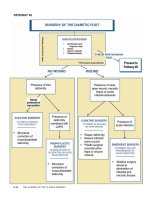
ASSESSMENT OF THE DIABETIC FOOT
(Pathway 1)
The pedal manifestations of diabetes are well document-
ed and potentially limb-threatening when left untreated.
Recognition of risk factors and treatment of diabetic foot
disorders require the skill of a specialized practitioner to
diagnose, manage, treat, and counsel the patient. Integration
of knowledge and experience through a multidisciplinary
team approach promotes more effective treatment, thereby
improving outcomes and limiting the risk of lower extrem-
ity amputation (30, 173).
The evaluation of the diabetic foot involves careful
assimilation of the patient’s history and physical findings
with the results of necessary diagnostic procedures
(Pathway 1). Screening tools may be valuable in evaluating
the patient and determining risk level (Appendix 1). Early
detection of foot pathology, especially in high-risk patients,
can lead to earlier intervention and thereby reduce the
potential for hospitalization and amputation (100). This is
also facilitated by an understanding of the underlying
pathophysiology of diabetic foot disorders and associated
risk factors. Identification of abnormal historical and/or
physical findings can therefore improve the prognosis for a
favorable outcome through appropriate—and early—refer-
ral (91, 174).
History
A thorough medical and foot history must be obtained
from the patient. The history should address several specif-
ic diabetic foot issues (Table 2).
Physical Examination
All patients with diabetes require a pedal inspection
whenever they present to any health care practitioner, and
S–10 THE JOURNAL OF FOOT & ANKLE SURGERY
DIABETIC FOOT DISORDERS VOLUME 45, NUMBER 5, SEPTEMBER/OCTOBER 2006 S–11
they should receive a thorough lower extremity examina-
tion at least once annually (175). Patients with complaints
relating to the diabetic foot require more frequent detailed
evaluations. The examination should be performed system-
atically so that important aspects are not overlooked (62). It
begins with a gross evaluation of the patient and extremi-
ties. Any obvious problem can then receive closer scrutiny.
Key components of the foot examination are presented in
Table 3. Although not specifically mentioned in this sec-
tion, it is assumed that a general medical assessment
(including vital sign measurements) will be obtained.
Diagnostic Procedures
Diagnostic procedures may be indicated in the assess-
ment and care of the diabetic foot. Consideration should be
given to the following tests in concert with those suggested
by members of the consulting team. It should be noted that
many of the following tests lack the ability to impart a
definitive diagnosis, necessitating clinical correlation.
Laboratory Tests
Clinical laboratory tests that may be needed in appropri-
ate clinical situations include fasting or random blood glu-
cose, glycohemoglobin (HbA1c), complete blood count
(CBC) with or without differential, erythrocyte sedimenta-
tion rate (ESR), serum chemistries, C-reactive protein, alka-
line phosphatase, wound and blood cultures, and urinalysis.
Caution must be exercised in the interpretation of laborato-
ry tests in these patients, because several reports have doc-
umented the absence of leukocytosis in the presence of
severe foot infections (117, 122, 151, 176-178). A common
sign of persistent infection is recalcitrant hyperglycemia
despite usual antihyperglycemic regimens (150).
Imaging Studies
The diabetic foot may be predisposed to both common
and unusual infectious or noninfectious processes, partially
because of the complex nature of diabetes and its associat-
ed vascular and neuropathic complications. As a result,
imaging presentations will vary due to lack of specificity in
complex clinical circumstances (179-181). Such variability
creates a challenge in the interpretation of imaging studies.
Therefore, imaging studies should only be ordered to estab-
lish or confirm a suspected diagnosis and/or direct patient
management. Distinguishing osteomyelitis from aseptic
neuropathic arthropathy is not easy, and all imaging studies
(Fig 4) must be interpreted in conjunction with the clinical
findings (123, 151).
Plain radiographs should be the initial imaging study in
diabetic patients with signs and symptoms of a diabetic foot
disorder (180, 182). Radiographs can detect osteomyelitis,
osteolysis, fractures, dislocations seen in neuropathic
arthropathy, medial arterial calcification, soft tissue gas, and
foreign bodies as well as structural foot deformities, pres-
ence of arthritis, and biomechanical alterations (183). Acute
osteomyelitis might not demonstrate osseous changes for up
to 14 days. Serial radiographs should be obtained in the face
of an initial negative radiographic image and a high clinical
suspicion of osseous disease (117, 123).
Technetium-99 methylene diphosphonate (Tc-99 MDP)
bone scans are often used in diabetic foot infection to deter-
mine the presence of osteomyelitis. Although highly sensi-
tive, this modality lacks specificity in the neuropathic foot
(184, 185). Osteomyelitis, fractures, arthritis, and neuro-
pathic arthropathy will all demonstrate increased radiotrac-
er uptake. However, a negative bone scan is strong evidence
against the presence of infection. To improve the specifici-
ty of nuclear imaging, white blood cells can be labeled with
Tc-99 hexamethylpropyleneamineoxime (Tc-99 HMPAO),
indium-111 oxime, or gallium-67 citrate (179, 186-189).
Indium-111 selectively labels polymorphonuclear leuko-
cytes and is more specific for acute infections than Tc-99
MDP scanning. Chronic infections and inflammation are
not well imaged with indium-111, because chronic inflam-
matory cells (ie, lymphocytes) predominate and are not well
labeled with indium. Combining Tc-99 MDP and indium-
111 increases the specificity of diagnosing osteomyelitis
(190). This combined technique is useful, because the Tc-99
MDP scan localizes the anatomic site of inflammation and
the indium-111 labels the infected bone (180, 191). The
indium-111 scan is not typically positive in aseptic neuro-
pathic arthropathy, although false-positive indium scans can
occur (192-194). A 100% sensitivity and 89% specificity
have been reported with the combined technique in evaluat-
ing diabetic infections (190, 191, 195).
In Tc-99 HMPAO scanning, white blood cells are labeled
in a similar manner as in indium scanning. However, with
Tc-99 MHPAO scans, imaging occurs 4 hours following
administration versus 24 hours postadministration with
indium scanning. Tc-99 HMPAO uses a smaller radiation
dose, is less expensive, and offers improved resolution com-
pared with indium scanning. The sensitivity and specificity
of both techniques are comparable (186, 196). Tc-99
HMPAO scans cannot be combined with Tc-99 MDP scans
because of similar labeling characteristics.
Tc-99 sulfur colloid is useful in distinguishing
osteomyelitis from neuropathic arthropathy (183). This
tracer is picked up by the bone marrow and any hemapoet-
ically-active marrow will be positive. Infected bone
replaces normal bone marrow, so it shows up as a relative