several epidemiological characteristics of acute encephalitis syndrome suspected to be caused by banna virus in some provinces of vietnam
Bạn đang xem bản rút gọn của tài liệu. Xem và tải ngay bản đầy đủ của tài liệu tại đây (325.75 KB, 24 trang )
1
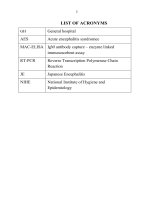
LIST OF ACRONYMS
GH
General hospital
AES Acute encephalitis syndromee
MAC-ELISA
IgM antibody capture – enzyme linked
immunosorbent assay
RT-PCR Reverse Transcription Polymerase Chain
Reaction
JE Japanese Encephalitis
NIHE National Institute of Hygiene and
Epidemiology
2
INTRODUCTION
Acute encephalitis syndromee (AES) suspected to be caused by
viruses has many different causes. There is no specific treatment for
this disease (except for Herpes simplex virus), with high mortality and
severe neurological sequela. Currently, there are about 100 virus types
identified causing AES.
In 1987 and 1992, Banna virus was isolated from AES and
unidentified fever patients’ serum in China. Banna virus was isolated
from Aedes dorsalis mosquitoes in China. According to some studies
in Indonesia, Banna virus was transmitted by two types of
mosquitoes: Anopheles and Culex.
In Vietnam, in 2003 and 2005, viruses of the same group with
Banna virus were isolated from patients in Thanh Hoa and Gia Lai
provinces. Besides, Banna virus was isolated from Culex mosquitoes
from Ha Tay (now part of Ha Noi) and Quang Binh provinces in
2002.
Participating in monitoring, diagnosis, treatment and prevention
of AES suspected to be caused by Banna virus, the study “Some
epidemiological characteristics of acute encephalitis syndrome
suspected to be caused by Banna virus in some provinces of
Vietnam” was conducted with three following specific goals:
1. Describe some epidemiological characteristics, clinical
syndrome of acute encephalitis syndrome suspected to be
caused by Banna virus in some provinces of Vietnam, 2002-
2012.
2. Determine infection ratio of Banna virus in mosquito
population collected in some provinces of Vietnam.
3. Identify some bio-molecular characteristics of Banna virus
isolated in Vietnam.
3
THESIS’ PRACTICAL IMPLICATIONS AND NEW
CONTRIBUTIONS
- New contributions:This is the first study in Vietnam identifying
incidence rate, clinical charateristics of patients with AES caused by
Banna virus, infection rate of Banna virus among mosquito populations
in some provinces and bio-molecular characteristics of Banna virus
identified in Vietnam.
- Practical implications: The research provides completely new
characteristics of AES caused by Banna virus data for scientific
communities of Vietnam and the world. Research results can be applied
in monitoring, diagnosis and prevention of AES caused by Banna
virus, with strong implication in teaching as well as research and
production.
THESIS STRUCTURE
The thesis is 113 pages long (not including references and
appendixes), including 4 chapters, 30 tables, 13 figures, 1 picture.
Introduction is 2 pages long, Chapter 1: Overview (28 pages); Chapter
2: Subject, material and study methodologies (22 pages); Chapter 3:
Study results (33 pages); Chapter 4: Discussions (22 pages);
Conclusion is 3 pages long; Suggestions/proposals is 1 page; List of
published work is 2 page. References: 102 referenced work; 2
appendixes.
4
Chapter I. OVERVIEW
1.1. Characteristics of Banna virus.
Banna virus, belonging to Seadornavirus genus, Reoviridae
family, has genetic material as 12 double-stranded RNA segments.
The first Banna virus strain was isolated from cerebrospinal fluid of
AES patients and blood samples of unidentified fever patients in
Yunnan province, China; and later, isolated in different provinces
from patients and mosquitoes in China, Indonesia and Vietnam.
1.2. Clinical characteristics of AES caused by Banna virus.
Banna virus causing acute infectious disease damaging central
nervous system or unidentified fever was recorded. Typical cases can
be described as following: Onset period: lasts 1-2 days but difficult to
identify when patients do not remember sudden high fever, chills,
headaches, arthralgia, and anorexia symptoms. Full-fledge period: after
3-6 days, patients showing symptoms of high fever, derilium,
autonomic disorder, indifference to surrounding including coma,
dyspnea, photophobia, loss of appetite, nausea. Symptoms of peripheral
nerve injuries include paralysis, chorea Sub-acute progression period:
from days 7-9 of the disease, symptoms reduced such as milder fever,
stable pulse temperature, reduced central and peripheral nervous
syndromee. However, in this period, there are notable complications
from laying for extended period such as pneumonia, sores,
constipation Recovery period: patient only has mild fever, regaining
conciousness, recovered appetite, remaining only sequelae depending
on the severity of the disease such as paralysis, hemorrhage,
myocarditis, pericarditis, reduced memory.
1.3. Epidemiological characteristics of AES caused by Banna virus
Some studies in Vietnam and around the world showed that Banna
virus exist in mosquitoes, there is clear evidence of Banna virus
transmission among animals (pigs) from virus isolation results.
Moreover, Banna virus exist in some migratory birds and the migration
of these birds enables the virus to spread to other areas. Mosquito has
been confirmed as Banna virus transmission vector in a number of
Asian countries from isolation results of Culex tritaeniorhynchus, Culex
5
vishnui, Culex fuscocephalus, Anopheles vagus, Aedes albopictus and
Aedes dorsalis mosquitoes.
Human is the infection target of Banna virus, the study of Liu, et al
(2010) of Banna virus in China from 1987 to 2007 showed that Banna
virus appeared in areas with Japanese Encephalitis (JE) outbreak and
where Culex tritaeniorhynchus mosquito act as the main vector. The
virus is infected through the skin from mosquito bite, once infected, the
virus multiplies in the lymphatic system, virions are passively
transmitted via vascular endothelia or choroid plexus, then to the
central nervous system and remain in the cerebrospinal fluid. Virus
effectiveness peaked in the early days of the onset and decreases
rapidly when neutralizing antibodies apprear. After virus infection, the
body may have immunizing response, neutralizing antibodies increases
from day 12 of the infection, IgM antibodies have higher neutralizing
effect than neutralizing antibodies. IgG antibodies appear from week 3
and lower than IgM antibodies but remain for the whole lifetime.
1.4. Treatment and prevention of AES caused by Banna virus
Treatment: Currently, there is no specific medical treatment for
AES caused by Banna virus, mainly treatment of symptoms and
complications.
Prevention: Banna virus is a mosquito-borne virus, a new virus
discovered in the past few decades mainly in Asian region, studies of
disease burden have not been mentioned, so far, there is no vaccine for
the disease, the most effective prevention method to this day is to
prevent mosquito as transmission vector.
Chapter II.
SUBJECT AND STUDY METHODS
2.1. Regions and time of study
Retrospective study of mosquito samples and specimens were
collected from 01/2002 to 12/2008 and prospectively from 01/2009 to
12/2012.
6
Research locations: Northern region (area previously Ha Tay, Bac
Giang, Thanh Hoa); Central region (Quang Binh); Tay Nguyen region
(Gia Lai, Kon Tum, Dak Lak, Dak Nong); Southern region (Long An,
Can Tho)
2.2. Study subjects:
Patients:
Patients clinically diagnosed with AES suspected to be caused by
viruses according to standards of the World Health Organization:
- Sudden high fever > 38
o
C, accompanied by one of following
symptoms:
- Change of mental state, or
- Nervous symptoms as meningeal signs, movement disorders
Patients diagnosed with AES suspected to be caused by Banna
virus: Cases of AES suspected to be caused by viruses detected
Banna IgM antibodies from cerebrospinal fuild from positive
ELISA technique.
Mosquito species: Study subjects are mosquito samples collected at
study sites in the Northern, Central, Southern and Tay Nguyen regions
from 2001 to 2011.
2.3. Contents of research
Study cases of AES: Collect samples of cerebrospinal fuilds from
patients with AES suspected to be caused by viruses in Infectious
diseases department in provincial hospitals. Test for Banna virus IgM
antibodies. Investigate epidemiological characteristics, clinical
symptoms of AES cases identified (+) with Banna virus antibodies, JE,
ECHO30 positive isolated from retrospective medical record.
Study Culex mosquito vector: Collect mosquito samples in
provinces with high number of patients with AES suspected to be
caused by viruses in Northern, Central, Southern and Tay Nguyen
regions, once per year in the period from March to December.
Mosquitoes are categorized and identified specie compositions, isolated
to identify Banna virus.
7
Banna virus trains isolated from AES patients, from pigs and
mosquitoes are genotype identified based on nucleotide gene sequence
No. 12.
2.4. Study methods
2.4.1. Structure of the study:
The struture of the study is cross-sectional, retrospective and
prosprective epidemiologically combined with laboratory analysis.
2.4.2. Determining investigation of epidemiological characteristics of
AES patients
Study method and sample taking
Sample size: Take cerebrospinal fuild samples of all patients with
AES suspected to be caused by viruses based on diagnostic standards
above when hospitalized. Choose sample based on convenient method,
samples are taken in accordance with regular protocol and surveyed
with pre-designed questionnaire.
Study method for vector mosquito:
Sample size: Sample size is calculated based on regular protocol of
NIHE; 30 household/night x 2 nights x 1 site (district/province) x 1
time/year = 60 household turn/site. Mosquito investigation is conducted
at night according to regular procedure of NIHE (capture mosquitoes
with CDC traps, capture female mosquitoes resting in the house and
barn, from 18h00 to 22h00 in the winter and 19h00 to 23h00 in the
summer.
Laboratory material and testing techniques
Samples including patients' ceresbrospinal fluid samples and
mosquito samples collected at study sites are analyzed in the laboratory.
With cerebrospinal fluid samples, use indirect ELISA technique
detecting specific Banna virus antibodies. With mosquito samples, use
isolation technique detecting mosquito types carrying Banna virus.
8
Isolated Banna virus strains are categorized by RT-PCR technique,
collecting PCR products for product purification techniques and
sequenced by sequencing machine.
Data collected from the study is analyzed by bioinfomatic software
such as: GraphPad, biological software DNA Star (Lasegene), MEGA
4.0
2.4.3. Data processing: Use biostatistic software Epi-info 6.04 and Stata
10 to input and process data.
Chapter III. STUDY RESULTS
3.1. Some epidemiological, clinical characteristics of AES caused
by Banna virus.
3.1.1. Description of ratio of patients with AES caused by Banna virus
Table 3.1. Identification result of Banna virus IgM antibodies in
cerebrospinal fluid sample of patients with AES, 2002 – 2012
Region Province Number of
samples
Number of
positive
Ratio (+)
%
Northern
Bac Giang 216 30 13,63
Ha Tay
(previously)
120 43 35,83
Ha Noi 50 17 34,00
Hai Phong 48 11 22,92
Thai Binh 108 36 33,33
Thanh Hoa 65 21 32,31
Central Hue 18 4 22,22
Tay Nguyen Gia Lai 20 5 25,00
Southern Long An 72 17 23,61
Total 717 184 25,66
There are 1,285 cerebrospinal fluid samples collected from patients
with AES susptected to be caused by viruses in 9 provinces/cities in the
period from 2002 to 2012, excluding causes by JE virus, ECHO30 and
herpes simplex virus type 1 and type 4, there are 717 cerebrospinal
fluid samples with unidentified causes. Using indirect ELISA technique
9
identifying Banna virus IgM antibodies from 717 cerebrospinal fluid
samples, resulting in identification of 184 (+) samples, average (+) ratio
of cerebrospinal fluid samples identified with Banna virus IgM
antibodies is 25.66% (184/717), and 14.32% (184/1285) when
calculated on the total cerebrospinal fluid samples of patients with
AES.
In 9 provinces/cities with sample specimens, identified (+) ratio
ranges from 13.83% to 35.83%. Province/city with highest (+) ratio is
area previously Ha Tay province with 35.83%, then Ha Noi with (+)
ratio of Banna virus antibodies at 34.00%; lowest identified (+) ratio
with Banna virus antibodies is 13.53% in Bac Giang.
According to serological surveillance, AES caused by viruses are
recorded throughout the year, but cases are recorded mainly in May,
June, July and August, with recorded peak of the epidemic is June with
239/717 recoded cases (33.33% of total number of cases)
Table 3.2. Ratio of cases of AES caused by viruses by age group,
2002 – 2012
Age group
<1
n = 61
1 - 4
n = 159
5 - 9
n = 183
10 - 14
n = 141
≥ 15
n = 173
Total
Number of
samples
(+)
11 35 44 42 52 184
Rate of
incidence
(%)
5,98 19,02 23,91 22,83 28,26 100
In 717 cases of AES suspected to be caused by viruses with
unidentified cause having cerebrospinal fluid sample tested with
indirect ELISA technique identifying IgM, resulted in 184 confirmed
cases of Banna virus antigen, cases of confirmed (+) recorded in all age
groups. Among them, incidence rate of AES caused by Banna virus in
age group of <1 year old is lowest at 5.98%, while incidence rate of
AES caused by Banna virus at age group ≥ is highest at 28.26%.
10
In 184 cases of AES identified to be caused by Banna virus, the
ratio of cases of AES caused by Banna virus in men is higher than
women in all age group/
3.1.2. Clinical characteristics of patients with AES caused by Banna
virus
3.1.2.1. Some signs, clinical symptoms at admission
Table 3.3. Some clinical symptoms at admission
Signs,
symptoms
BANNA
virus
n=103
(%)
ECHO30
virus
n=43
(%)
JE virus
(n=5)
(%)
Ratio of
BANNA
and
ECHO30
Ratio of
Banna and
JE
p1 p2
Headache 48,54 88,37 30,51 <0,0001 0,0252
Vomitting 32,04 86,05 28,81 <0,0001 0,6685
Seizures 32,04 2,33 61,02 0,0001 0,0003
Nausea 1,94 30,23 5,08 <0,0001 0,2659
Myalgia 0 0 0 - -
Joint pain 0 2,33 0 - -
Fever
> 37,5
o
C
78,64 74,42 81,36 0,5784 0,6791
Bulging
fontanel
23,30 2,33 0 0,0022 -
Stiff neck 77,45 39,53 50,85 <0,0001 0,0005
Kernig sign 67,96 34,88 38,98 0,0002 0,0003
Mental
disorder
81,55 11,63 88,14 <0,0001 0,2715
Bradykinesia 18,63 4,65 23,73 0,0288 0,4390
Loss of
sensation
0 0 6,78 - -
Analysis of clinical symptoms of AES patients at admission shows
that almost all typical clinical symptoms of AES such as headache,
vomitting, seizures, high fever above 37.5 degrees, bulging fontanel,
stiff neck, Kernig sign, mental disorder and bradykinesia appear in
newly admitted patients with AES caused by Banna virus at high ratio
from 23.3% to 78.64%. However, myalgia, joint pain and loss of
11
sensation are symptoms not observed from patients with AES caused
by Banna virus.
When comparing clinical symptoms in patients with AES caused
by Banna virus with ECHO30 virus and JE virus, there is significant
statistical difference of bulging fontanel (23.3%), stiff neck (77.45%)
and Kernig sign (67,96%) appearing more than patients with AES
caused by ECHO30 virus and JE virus. Especially, bulging fontanel
was noted primarily in patients with AES caused by Banna virus and
rarely in patients with AES caused by ECHO30 virus and particularly
not in patients with AES caused by JE virus.
3.1.2.2. Some signs, clinical symptoms during treatment
Table 3.4. Signs, clinical symptoms after 7 days of treatment of patients
infected with Banna virus comparing to infection with ECHO30 and JE
viruses
Symptoms
BANNA
virus
(n=103)
%
ECHO30
virus
(n=43)
%
JE
virus
(n=59)
%
Ratio due to
Banna and
ECHO30
viruses
Ratio due to
Banna virus
and JEV
p1 p2
Headache 7,77 4,65 30,51 0.4964 0,0001
Vomitting 0,97 2,33 0 0,4882 -
Seizures 0,97 0 0 - -
Nausea 0 0 0 - -
Myalgia 0,97 0 0 - -
Joint paint 0 2,33 0 - -
Fever >37,5
o
8,74 6,98 11,86 0,7242 0,5218
Bulging
fontanel
1,94 0 0 - -
Stiff neck 15,53 6,98 15,25 0,1616 0.9621
Kernig sign 13,59 4,65 8,47 0,1149 0.3297
Mental
disorder
55,34 0 11,86 - <0,0001
Bradykinesia 2,91 0 6,78 - 0,2436
Loss of
sensation
0,97 - - - -
12
After 7 days of treatment, AES symptoms caused by different viral
agents tend to reduce in all patients. However, symptoms such as
headache, fever (>37.5 degrees Celcius), stiff neck, Kernig sign are still
observed in patients infected with Banna virus, ECHO virus and JEV.
Symptoms such as seizures, myalgia, joint paint, bulging fontanel,
loss of sensation only observed in very few patients (1-2 patient) among
the group infected with Banna virus. Among them, seizures, myalgia,
bulging fontanel and loss of sensation only observed from patients
infected with Banna virus. Joint paint only observed from patient
infected with ECHO30 virus.
With Kernig sign, the group infected with Banna virus still records
a high ratio of 13.59%. Conversely, for the group infected with JEV
this rate is 8.47% and for the group infected with ECHO30 the rate is
only 4.65%. With mental disorder symptom, no cases recorded in the
group infected with ECHO30 virus, but the group infected with Banna
virus showed a very high ratio of 55.34% while this ratio for the group
infected with JEV is only 11.86%.
For bradykinesia symptom, the ratio for the group infected with
Banna virus is 2.91%, for the group infected with JEV is 6.78%; nausea
symptom was not observed after 7 days of treatment from all patients in
this study.
3.1.2.3. Result after treatment of AES caused by Banna virus
Table 3.5. Average number of days of treatment of AES caused by
viruses in hospital
Causes of AES Average time (day) Minimum time Maximum time
Banna virus 13,5 1 85
ECHO30 virus 7,4 3 23
JEV 11,3 1 39
F=5,21,
F
P
=0,0062; Bartlett’s test with P<0,0001
13
Study show that average treatment time and maximum treatment
time of AES caused by Banna virus if 13.5 and 85 days, respectively,
these are the longest treatment times compared to AES caused by JEV
(average of 11.3 days) and ECHO30 virus (7.4 days)
Table 3.6. Treatment outcome after Banna virus infection
Causes of AES Number of AES Number of death Ratio (%)
Banna virus 103 15 14,6
ECHO30 virus 43 0 0
JEV 59 1 1,7
Total 205 16 7,8
Retrospective study showed that in 205 cases of AES caused by
Banna virus, ECHO30 virus and JEV, there are 16 cases of death after
treatment. Mortality rate of AES caused by Banna virus is 14.6%
(15/103), followed by mortality rate of AES caused by JEV at 1.7%
(1/59). Conversely, no cases of death recorded in all cases of AES
caused by ECHO30 virus.
3.2. Determining Banna virus prevalence in mosquito polulation
collected in some provinces of Vietnam.
Table 3.7. Rate of Banna virus isolated from mosquitoes
Species
Northern
region
Central
region
Tay Nguyen
region
Southern
region
Species /
samples
Species /
samples
Species /
samples
Species /
samples
An. vagus 2/4 // // //
Ar. subalbalus 2/9 // // //
Cx. pseudovishnui 0/6 // // 13/88
Cx. quinquefaciatus 1/32 0/1 3/16 7/68
Cx. fuscocephalus 1/9 0/1 1/8 //
Cx. gelidus 0/32 0/2 1/18 0/17
Cx.
tritaeniorhynchus 5/551 2/11 2/53 0/27
Cx. vishnui 1/100 2/9 3/28 //
14
Species
Northern
region
Central
region
Tay Nguyen
region
Southern
region
Species /
samples
Species /
samples
Species /
samples
Species /
samples
Total (rate of
positive isolated)
12/744
(1,6%)
4/24
(16,7%)
10/123
(8,1%)
20/200
(10,0%)
In the total of 1,091 mosquito samples collected in provinces/cities
in Northern, Central, Southern and Tay Nguyen regions, there are 46
strains of Banna virus isolated, isolation rate of Banna virus in
mosquitoes in Vietnam is 4.22%.
Isolation rate of Banna virus from mosquitoes is lowest in the
Northern region at 1.61% (12/744), highest in the Central region with
positve isolation at 16.67% (4/24), followed by Tay Nguyen region
with (+) isolation at 10% (20/200) and the Southern region has (+)
isolation rate of 8.13% (10/123).
Table 3.8. Data of Banna virus strains isolated in the Northern region
No. Species Gender
Virus
strain code
Province
1 Cx. vishnui Female 02VN 9 b Ha Tay
2 Cx. tritaeniorhynchus Female
02VN 78 b Ha Tay
3 Cx. quinquefaciatus Female
06 VN 1 Ha Tay
4 Cx. tritaeniorhynchus Female
06 VN 2 Ha Tay
5 An. vagus Female
06VN267 Bac Giang
6 Cx. tritaeniorhynchus Female
06VN268 Bac Giang
7 Cx. tritaeniorhynchus Female
06VN269 Bac Giang
8 Cx. fuscocephalus Female
06VN273 Bac Giang
9 Ar. subalbalus Female
06VN276 Bac Giang
10 An. vagus Female
06VN263 Bac Giang
11 Cx.tritaeniorhynchus Female
08VN117 Bac Giang
12 Ar. subalbatus Female
08VN114 Bac Giang
In 12 Banna virus strains isolated in the Northern region period
2001-2011, there are 4 Banna virus strains isolated in area previously
Ha Tay province, 8 Banna virus strains isolated in Bac Giang province.
15
In this study, no Banna virus strain was isolated in Lang Son, Thai Binh
and Ha Nam province.
Table 3.9. Data on Banna virus strains isolated in the Central region
No Species Gender
Virus strain
code
Province
1 Cx. vishnui
Female 02VN 9
Quang Binh
2 Cx. vishnui
Female
02VN18 b
Quang Binh
3 Cx. tritaeniorhynchus
Female
02VN178 b
Quang Binh
4 Cx. tritaeniorhynchus
Female
02VN180 b
Quang Binh
There are 4 Banna virus strains isolated in Quang Binh province in
the Central region in period 2001-2011, virus strains are isolated mainly
from Culex tritaeniorhynchus and Culex vishnui mosquitoes in 5
species collected in Quang Binh.
Table 3.10. Data on Banna virus strains isolated in Tay Nguyen region
No. Species Gender
Virus strain
code
Province
1 Cx. vishnui Female 06VN 58 Gia Lai
2 Cx. quinquefaciatus Female
06VN 60 Gia Lai
3 Cx. fuscocephalus Female
06VN 63 Gia Lai
4 Cx. tritaeniorhynchus Female
06VN 295 Kon Tum
5 Cx. vishnui Female
06VN 326 Đak Nong
6 Cx. tritaeniorhynchus Female
07VN 287 Đak Nong
7 Cx. quinquefaciatus Female
07VN 300 Đak Lak
8 Cx. quinquefaciatus Female
07VN 307 Kon Tum
9 Cx. vishnui Female
07VN 308 Kon Tum
10 Cx. gelidus Female
07VN 309 Kon Tum
There are 10 Banna virus strains isolated from mosquitoes
collected in 4 provinces of Tay Nguyen region, among them 4/10
Banna virus strains isolated in Kon Tum province from Culex
quinquefaciatus and Culex. vishnui mosquitoes, 3/10 Banna virus
16
strains isolated in Gia Lai province from Culex fuscocephalus, Culex
quinquefaciatus and Culex vishnui mosquitoes. There are 2/10 Banna
virus strains isolated in Dak Nong province from Culex
tritaeniorhynchus and Culex vishnui mosquitoes, only 1/10 strains
isolated in Dak Lak province from Culex quinquefaciatus mosquito.
Table 3.11. Data on Banna virus strains isolated in Southern region
No Species Gender Virus strain code Province
1 Cx. quinquefaciatus
Female
05 VN266
Can Tho
2 Cx. quinquefaciatus
Female
05VN 274
Can Tho
3 Cx. quinquefaciatus
Female
05VN 277
Can Tho
4 Cx. pseudovishnui
Female
05VN 280
Can Tho
5 Cx. quinquefaciatus
Female
05VN 290
Can Tho
6 Cx. quinquefaciatus
Female
05VN 301
Can Tho
7 Cx. quinquefaciatus
Female
05VN 305
Can Tho
8 Cx. pseudovishnui
Female
05VN 486
Can Tho
9 Cx. pseudovishnui
Female
05VN 487
Can Tho
10 Cx. pseudovishnui
Female
05VN 491
Can Tho
11 Cx. pseudovishnui
Female
05VN 492
Can Tho
12 Cx. pseudovishnui
Female
05VN 494
Can Tho
13 Cx. pseudovishnui
Female
05VN 495
Can Tho
14 Cx. pseudovishnui
Female
05VN 496
Can Tho
15 Cx. pseudovishnui
Female
05VN 505
Can Tho
16 Cx. pseudovishnui
Female
05VN 507
Can Tho
17 Cx. pseudovishnui
Female
CT-Mo-P7b
Can Tho
18 Cx. quinquefaciatus
Female
05VN 531
Can Tho
19
Cx. pseudovishnui
Male
05VN 308
Long An
20
Cx. pseudovishnui
Male
05VN 311
Long An
There are 20 Banna virus strains isolated in Can Tho and Long An
provinces, with the majority of Banna virus strains isolated from female
mosquitoes collected in Can Tho province (18/20 strains). In this study,
17
there are 2/20 Banna virus strains isolated from male Culex
pseudovishnui mosquito collected in Long An province. Among 20
Banna virus strains isolated in Can Tho and Long An province in 2005,
there are 7/20 Banna virus strains isolated from Culex quinquefaciatus
mosquito, 13/20 Banna virus strains isolated from Culex pseudovishnui
mosquito.
3.3. Some bio-molecular characteristics of Banna virus isolated in
Vietnam.
3.3.1. Distribution of Banna virus in Vietnam
Identification of Banna virus strains isolated from patients, from
mosquitoes and pigs in some provinces/cities in the Northern, Central,
Southern and Tay Nguyen regions. Banna virus was isolated from
moquitoes in 9 provinces including Bac Giang, area previously Ha Tay
province, Quang Binh, Kon Tum, Gia Lai, Dak Lak, Dak Nong, Can
Tho and Long An. With are previously Ha Tay province, Banna virus is
isolated not only from mosquitoes but also pigs.
Table 3.12. Data on registration number of genetic nucleotide sequence
No. 12 of 5 Banna virus strains in International gene bank
Strain
code
Time
Province Specimen
Registration
number in
gene bank
03VN 99 2003 Thanh Hoa
Human (DNT) AB773281
05VN 225 2005 Gia Lai Human (DNT) AB773282
03VN 45 2003 Ha Tay Pig blood AB773283
05VN 301 2005 Can Tho Culex fatigan AB773284
05VN 305 2005 Can Tho Culex fatigan AB773285
In this study, there are 5 Banna virus strains isolated from
mosquitoes, human, and pigs registered in the gene bank with codes for
lookup and information sharing, among them, 2 Banna virus strains are
isolated from patients, 1 strain isolated from pig and 2 strains isolated
from mosquitoes in provinces in Northern, Southern and Tay Nguyen
region.
18
A
Genealogy tree is built based on entire region genetic sequencing
No. 12 of 5 Banna virus strains isolated from patients with AES, from
pigs and mosquitoes in period 2003-2005 in Vietnam compared with 38
genetic sequencing No. 12 of other Banna virus strains including 5
Banna virus strains isolated in the Northern and Central regions in 2002
and other Banna virus strains in some Asian countries publicized on the
gene bank.
Figure 3.1. Geanology tree describing relationship among Banna virus
strains based on genetic sequence No. 12
Results from comparison of nucleotide genetic sequence No. 12 of
Banna virus strains identified isolated strains belonging to genotype A,
in sub-group genotype A1 and created a separate clade: Vietnam clade.
3.3.2. Result of coding nucleotide genetic sequence No. 12
Nucleotide sequence and amino acid sequence No. 12 were used to
analyze molecular characteristics between two Banna virus strains
isolated from Vietnamese patients and the first Banna virus strain
isolated from Chinese patients in 1987 (Prototype strain).
Comparison of 667 nucleotide genetic sequence No. 12 of Banna
virus strains isolated from patients resulted in 49 confirmed mutations
of genetic sequence No. 12 of Banna virus isolated from Vietnamese
19
patients compared to genetic sequence No. 12 of Banna virus strains
isolated from Chinese patients. Among 49 nucleotide mutations, the
majority is single mutation, only a single double mutation (mutation of
two consecutive nucleotides) observed.
When comparing nucleotide sequence of the entire genetic coding
sequence No. 12 of 2 Banna virus strains isolated from Vietnamese
patients in 2003 and 2005 with Banna virus strain isolated from
Chinese patient show that the Banna virus strains isolated from
Vietnamese patient do not have any close genetic relation to the Banna
virus strain isolated from Chinese patients.
Chapter IV. DISCUSSION
4.1. Describe some epidemiology and clinical symptoms of AES
suspected to be caused by Banna virus in some provinces in
Vietnam, 2002-2012
In this study, there are 717 cerebrospinal fluid samples of patients
with AES suspected to be caused by viruses with unidentified cause
used to identify Banna virus IgM antigen, these are ceresbrospinal fluid
samples of AES patients in 9 provinces/cities in Northern, Central,
Southern and Tay Nguyen regions.
Because the study was designed on the basis of samples collected
and stored when there are cases of patients with AES excluding other
viral-caused AES. On the other hand, due to ecological characteristics
of different regions, cases of AES mainly recorded in the Northern
region of Vietnam. Comparing with serological surveillance of AES
caused by Banna virus in China among cases clinically diagnosed with
JEV, patient serum ratio (+) with Banna virus antigen recombined with
generic region VP9, with ELISA technique identifying IgM about 15%.
So, the result of identifying (+) with Banna virus antigen in cases of
AES suspected to be caused by viruses (excluding cases identified
positively with JEV antigen) in 9 provinces/cities of Vietnam ranges
from 13.83% to 35.83% (averaging 25.66%), showing that in different
geological separations, the ratio of AES infection could be caused by
different agents.
When calculated on the total of 1,285 ceresbrospinal fluid samples
not excluding a number of pathogens, the ratio of confirmed Banna
20
virus antigen is 184/1,285 (14.32%), similar to positive result
confirmed among clinically diagnosed cases of JE in China.
Among confirmed cases of (+), age group <1 year old has the
lowest infection ratio, the highest infection ratio is age group ≥ 15
(28,26 %). This result is consistent with the characteristic of Arbo virus
infection causing AES such as JE, where the infection ratio of age
group <1 year old is very low while Banna virus infection ratio in age
group ≥ 15 years old is higher than other groups at 28.26%, normally,
the infection ratio of AES caused by JE in age group ≥ 15 years old is
about 10% according to previous literature.
Regarding clinical characteristics, symptoms of AES are related to
nervous system such as headaches, vomitting, seizures, nausea, joint
paint appearing with different frequencies in the group of patients with
AES caused by Banna virus, ECHO30 virus or JEV. Mainly, patients
with AES caused by ECHO30 virus showed symptoms of headaches,
vomitting and nausea with much higher frequencies than patients with
AES caused by Banna virus and JEV. Symptom of seizure has high
ratio among the group of patients with AES caused by JEV (61.02%)
while the group of patients with AES caused by Banna virus is only
30.23%, and for the group of patients with AES caused by ECHO30
virus is very low at 2.33%. However, mortality rate among cases with
AES caused by Banna virus recorded in this study is about 15%, higher
than the mortality rate of AES caused by JEV. Cases with AES caused
by Banna virus showed stiff neck syndrome at a high ratio of 77.45%
while this ratio for AES caused by JEV is only 50.85% and for the
group of AES caused by ECHO30 is 39.53%. Mental disorder symptom
ratios appeared in the majority of AES caused by Banna virus and JEV
are 81.55% and 88.14%, respectively, while for the group of patients
with AES caused by ECHO30 this ratio is only 11.63% carrying
specific characteristic of AES caused by Banna virus.
After a week of treatment, the group with AES caused by Banna
virus still showed clinical symptoms especially mental disorder, at a
high rate of 55.34% while this ratio for the group with AES caused by
JEV is only 11.86% and for the group with AES caused by ECHO30
virus is not recorded.
21
4.2. Determining the prevalence of Banna virus in mosquito
population collected in some provinces of Vietnam.
Banna virus is a mosquito-borne virus, highly adaptable in C6/36
cell line. Determination of mosquito species carrying virus/vector must
be based on isolation result and virus identification from mosquitoes.
Mosquitoes collection was conducted from 2002 to 2011 in 4 regions in
Vietnam, resulting in 66,760 specimens of 21 mosquito species divided
into 1091 mosquito samples collected. Among the individual samples
collected, Culex pseudovishnui were predominant at the highest rate of
43.76%. Futhermore, other species of mosquito were also collectes:
Anopheles, Armigeres, Aedes and Mansonia.
Virus isolation result showed that the isolation rate of Banna virus
from mosquito is 4.22% (46/1091). Provinces with Banna virus isolated
from mosquitoes include: Bac Giang, Quang Binh, Kon Tum, Dak Lak,
Dak Nong, Gia Lai, Can Tho, Long An and are previously Ha Tay
province (9/12 provinces).
In this study, Banna virus was isolated from Culex
tritaeniorhynchus mosquito in all regions including Northern, Central
and Tay Nguyen regions. In the Southern region, Banna virus is mainly
isolated from Culex pseudovishnui and Culex quinquefaciatus
mosquitoes. However, the isolation of Banna virus from 8 different
species of mosquitoes showed that the number of mosquito species
being or cound be the vector of Banna virus is bigger that for JEV
vector.
4.3. Determining some bio-molecular characteristics of Banna virus
isolated in Vietnam.
Genetic sequences No. 12 of Banna virus strains were selected for
genotype analysis of Banna virus strains isolated in Vietnam and some
geographical areas in Asia. Result of the analysis identified various
Banna virus strains divided into two different genotype, Chinese and
Vietnamese strains belong to genotype A, while virus strains isolated in
Indonesia belong to genotype B. Genotype A is divided into two
subgroups: A1 and A2; genotype A1 is divided into 4 independent
clades including Banna virus strains isolated in Northern China,
Liaoning and Vietnam; genotype subgroup A2 include Banna virus
strains isolated from the Central region of Vietnam and China.
22
Molecular characteristic of viruses in Reoviridae family is double
stranded ARN including many segments. Depending on different virus
genuses, the number of segments also vary, for example Orthoreovirus
(Reovirus), Orbivirus, Rotavirus are virus genuses with 10 or 11
segments (not including segment No. 12), while Coltivirus and
Seadornavirus have 12 segments. For that reason, in this study, we
select genetic segment No. 12 to conduct molecular epidemiology study
of Banna virus circulation in Vietnam. In this study, there are 10 Banna
virus strains isolated in Vietnam from patients, mosquitoes and pigs
with genetic sequence No. 12 for analysis and comparison with genetic
sequence No. 12 of Banna virus strains isolated in the area. Genetic
sequence No. 12 is derived from human, pigs and mosquitoes with
international gene bank coding, including 5 genetic sequences No. 12
previously registered, 5 genetic sequences registered throughout this
research, including 2 nucleotide genetic sequence No. 12 from Banna
virus isolated from AES patients in Vietnam. This means the study for
genome sequencing of Banna virus isolated from Vietnamese patient
needs to be mentioned in following studies.
CONCLUSION
1. Description of some epidemiological, clinical characteristics of
AES caused by Banna virus in some provinces of Vietnam.
1.1. Ratio of Banna virus infection in Vietnam
Identified rate of (+) with Banna virus antigen ranges from 13.83%
to 35.83% depending on provinces/cities with highest positive rate in
area previously Ha Tay province at 35.83%, lowest in Bac Giang
province at 13.83%. Identified rate of (+) with Banna virus antigen is
recorded throughout the year, concentrating in May, June, July and
August with highest identified rate in June at 34.78%.
Identified rate of (+) with Banna virus antigen differentiates
depending on age group, specifically, the lowest group is <1 year old at
5.98% and the highest group is ≥ 15 years old at 28.26%, the ratios for
age groups 1-4 years old, 5-9 years old and 10-14 years old ranges from
19.02% to 23.91%. The incidence rate among men is higher than
among women, at 60.87% and 39.13%, respectively.
23
1.2. Epidemiological, clinical characteristics of AES caused by Banna
virus
At admission, patients with AES suspected to be caused by viruses
show symptoms of headaches, vomitting, seizures, high fever over
37,5
o
C, bulging fontanel, stiff neck, Kernig sign, mental disorder and
bradykinesia with high ratio of 23.3% - 78.64%, no sign of myalgia,
joint paint and loss of sensation. Especially, symptom of bulging
fontanel mainly observed in patients with AES caused by Banna virus
(23.3%).
After a week of treatment, the majority of patients with AES
caused by Banna virus do not show clinical symptoms observed when
admitted (0.97%-15.53%). However, mental disorder symptom
remained at a high ratio of 55.34% in patients with AES caused by
Banna virus, and rarely in patients with AES of other pathogens.
Average treatment time of patients with AES caused by Banna
virus is 13.5 days and the maximum treatment time is 85 days.
Mortality rate of patients with AES caused by Banna virus is very high
at 14.6% compared to JEV (1.7%) and ECHO30 virus (0%).
2. Determining Banna virus prevalence in mosquito polulation
collected in some provinces of Vietnam.
Average isolation rate of Banna virus from mosquitoes is 4.22% of
collected mosquito samples (46/1,091), among regions, Banna virus
isolation rate ranges from 1.61% to 16.67%.
Among 46 Banna virus strains isolated from mosquitoes, there are
02 strains isolated from male Culex pseudovishnui mosquitoes in Long
An, 44 strains of Banna virus were isolated from female mosquitoes.
3. Determining some bio-molecular characteristics of Banna virus
circulating in Vietnam.
There are 5 Banna virus strains sequenced gene coding No. 12
registered in the gene bank and granted codes AB773281, AB773282,
AB773283, AB773284 and AB773285.
Regarding bio-molecular characteristics, identified Banna virus
strains isolated from patients, mosquitoes and pigs from 2002 to 2005
in the Northern, Central, Southern and Tay Nguyen regions belong to
genotype A, in genotype subgroup A1, forming a separate clade:
Vietnam clade. The 2 Banna virus strains isolated from mosquitoes in
24
the Central region belong to genotype subgroup A2. The similarity with
nucleotide gene sequence No. 12 of Banna virus strains isolated from
Chinese patients (Prototype strain) means that it is only 90.6%
affirmative that Banna virus strains are local.
PROPOSALS
Some proposals based on research results of the thesis:
1. Serological surveillance and retrospective study of clinical
characteristics of AES caused by Banna virus showed that this is a
mosquito-borne disease; incidence rate has been reported in all age
groups, with acute clinical progression, high mortality rate showing
the need for implementation of research of treatment and
prevention methods.
2. High rate of Banna virus carrying in mosquito polulation, the range
of species of Banna virus carrying is broader than JEV, so there
needs to be further study of Banna virus circulation among
mosquito polulation and heightened prevention of mosquito-borne
diseases in general and specifically AES caused by Banna virus
while specific treatment and vaccination are still non-existent.
3.
Analysis of bio-molecular characteristics of of Banna virus strains
isolated in Vietnam identified that they are local strains, not Banna
virus strains invaded from China. They are some of the causes of
recorded AES and unidentified-cause fever, suggesting the need of
further research for diagnosis and treatment biological products to
aid in effective treatment and prevention of the disease.