Báo cáo sinh học: "Complexes between the LKB1 tumor suppressor" pptx
Bạn đang xem bản rút gọn của tài liệu. Xem và tải ngay bản đầy đủ của tài liệu tại đây (286.72 KB, 16 trang )
Research article
Complexes between the LKB1 tumor suppressor, STRAD
␣␣
/

and
MO25
␣␣
/

are upstream kinases in the AMP-activated protein
kinase cascade
Simon A Hawley*
†
, Jérôme Boudeau
‡†
, Jennifer L Reid*
†
, Kirsty J Mustard*,
Lina Udd
§
, Tomi P Mäkelä
§
, Dario R Alessi
‡
and D Grahame Hardie*
Addresses: *Division of Molecular Physiology and
‡
MRC Protein Phosphorylation Unit, Wellcome Trust Biocentre, University of Dundee,
Dundee DD1 5EH, UK.
§
Molecular Cancer Biology Program, Institute of Biomedicine and Helsinki University Central Hospital,
Biomedicum Helsinki, University of Helsinki, Finland.
†
These authors contributed equally to this work.
Correspondence: Dario R Alessi (LKB1). E-mail: D Grahame Hardie (AMPK). E-mail:
Abstract
Background: The AMP-activated protein kinase (AMPK) cascade is a sensor of cellular
energy charge that acts as a ‘metabolic master switch’ and inhibits cell proliferation. Activation
requires phosphorylation of Thr172 of AMPK within the activation loop by upstream kinases
(AMPKKs) that have not been identified. Recently, we identified three related protein kinases
acting upstream of the yeast homolog of AMPK. Although they do not have obvious
mammalian homologs, they are related to LKB1, a tumor suppressor that is mutated in the
human Peutz-Jeghers cancer syndrome. We recently showed that LKB1 exists as a complex
with two accessory subunits, STRAD␣/ and MO25␣/.
Results: We report the following observations. First, two AMPKK activities purified from rat
liver contain LKB1, STRAD␣ and MO25␣, and can be immunoprecipitated using anti-LKB1
antibodies. Second, both endogenous and recombinant complexes of LKB1, STRAD␣/ and
MO25␣/ activate AMPK via phosphorylation of Thr172. Third, catalytically active LKB1,
STRAD␣ or STRAD and MO25␣ or MO25 are required for full activity. Fourth, the
AMPK-activating drugs AICA riboside and phenformin do not activate AMPK in HeLa cells
(which lack LKB1), but activation can be restored by stably expressing wild-type, but not
catalytically inactive, LKB1. Fifth, AICA riboside and phenformin fail to activate AMPK in
immortalized fibroblasts from LKB1-knockout mouse embryos.
Conclusions: These results provide the first description of a physiological substrate for the
LKB1 tumor suppressor and suggest that it functions as an upstream regulator of AMPK. Our
findings indicate that the tumors in Peutz-Jeghers syndrome could result from deficient
activation of AMPK as a consequence of LKB1 inactivation.
BioMed Central
Journal
of Biology
Journal of Biology 2003, 2:28
Open Access
Published: 24 September 2003
Journal of Biology 2003, 2:28
The electronic version of this article is the complete one and can be
found online at />Received: 3 July 2003
Revised: 11 August 2003
Accepted: 9 September 2003
© 2003 Hawley et al., licensee BioMed Central Ltd. This is an Open Access article: verbatim copying and redistribution of this article are permitted in
all media for any purpose, provided this notice is preserved along with the article's original URL.
Introduction
AMP-activated protein kinase kinase (AMPKK) and AMP-
activated protein kinase (AMPK) are the upstream and
downstream components, respectively, of a protein kinase
cascade that acts as a sensor of cellular energy charge [1,2].
AMPK is activated by the elevation in cellular 5-AMP that
accompanies a fall in the ATP:ADP ratio due to the reaction
catalyzed by adenylate kinase (2ADP Ǟǟ ATP + AMP). This
occurs during metabolic stresses such as hypoxia, ischaemia,
glucose deprivation and, in skeletal and cardiac muscle,
during contraction or exercise [1-3]. Once activated by
stress, AMPK switches on the uptake of glucose and fatty
acids and the oxidative metabolism of these fuels to gener-
ate ATP, while switching off biosynthetic pathways that
consume ATP. It achieves this metabolic switching both by
direct phosphorylation of metabolic enzymes and by
longer-term effects on gene expression [1,2].
We have previously partially purified from rat liver an
upstream kinase (AMPKK) that activates AMPK by phospho-
rylation of AMPK residue Thr172 within the activation loop
of the kinase domain [4], but we have been unable to iden-
tify the activity as a defined gene product. As an alternative
approach, we searched for kinases upstream of the Saccha-
romyces cerevisiae homolog of AMPK (the SNF1 complex),
taking advantage of genome-wide approaches available in
that organism. This identified Elm1, Pak1 and Tos3 as alter-
native upstream kinases in yeast that can activate the SNF1
complex in vivo in a partially redundant manner [5]. The
nearest relatives encoded by the human genome are
calmodulin-dependent protein kinase kinase (CaMKK) and
LKB1 (see Additional data file 1 with the online version of
this article). We have previously shown that CaMKK puri-
fied from pig brain could activate AMPK in cell-free assays
(albeit poorly in comparison to the extent to which it acti-
vates its known substrate, calmodulin-dependent protein
kinase I); but the AMPKK previously purified from rat liver
was not calmodulin-dependent [6]. LKB1 is a 50 kDa
serine/threonine kinase that was originally discovered as the
product of the gene mutated in the autosomal dominant
human disorder, Peutz-Jeghers syndrome (PJS) [7,8].
People with PJS develop benign polyps in the gastrointesti-
nal tract but also have a 15-fold increased risk of developing
malignant tumors in other tissues [9,10]. Nearly 100 differ-
ent PJS mutations have been reported, and most are
expected to impair the kinase activity of LKB1 [11]. Several
human tumor cell lines, including HeLa and G361 cells,
lack expression of LKB1. Expression of wild-type LKB1, but
not catalytically inactive LKB1 or PJS mutants, in G361 cells
caused a G1-phase cell-cycle arrest [12] that was associated
with the induction of the cyclin-dependent kinase inhibitor,
p21, and was dependent on p53 [13]. Homozygous LKB1
knockout mice die of multiple defects at mid-gestation [14].
Heterozygotes are viable, but most develop polyps similar
to those found in people with PJS by 45 weeks of age [15-
18], although it is controversial as to whether these are
caused by haploinsufficiency or loss of heterozygosity
(reviewed in [11]). It has also been reported that a signifi-
cant number of LKB1
+/-
mice over 50 weeks of age develop
hepatocellular carcinomas that are associated with loss of
LKB1 expression [19]. These results show that LKB1 acts as a
tumor suppressor and that the catalytic activity of LKB1 is
essential for this function, but the downstream substrate(s)
that LKB1 phosphorylates to mediate the suppression of cell
proliferation remained unknown.
Recently, we reported that LKB1 is associated with two
accessory proteins called Ste20-related adaptor protein-␣
(STRAD␣) [20] and mouse protein 25-␣ (MO25␣) [21], for
each of which there is also a closely related isoform
(STRAD and MO25) encoded in the human genome.
Although STRAD␣ and  are related to the Ste20 protein
kinases, several of the residues expected in active protein
kinases are not conserved, and they appear to be inactive
‘pseudokinases’ [20]. MO25␣ binds to the carboxyl-termi-
nus of STRAD␣ and stabilizes the association between
STRAD␣ and LKB1 [21]. The association of LKB1 with
STRAD␣ and MO25␣ increased the kinase activity of LKB1
against an artificial substrate (myelin basic protein) and
also enhanced its cytoplasmic localization [20,21], which
was previously implicated in the tumor suppressor function
of LKB1 [13]. Here, on the basis of the sequence similarity
between LKB1 and the upstream kinases identified for the
yeast homolog of AMPK [5], we investigate whether
LKB1:STRAD:MO25 complexes could play a role in activat-
ing AMPK in mammalian cells.
Results
Resolution of two AMPKKs from rat liver that both
contain LKB1, STRAD
␣␣
and MO25
␣␣
While experimenting with different conditions to optimize
recovery at the Q-Sepharose step of our previous purifica-
tion protocol for AMPKK [4], we found that we were able to
resolve two peaks of activity (Figure 1a). Because the second
peak corresponds to the AMPKK originally purified [4], we
refer to it as AMPKK1, with the first peak being termed
AMPKK2. On size-exclusion chromatography on Sephacryl
S-200, AMPKK1 and AMPKK2 eluted as proteins of large but
distinct size, with estimated Stokes radii of 5.7 and 5.2 nm
respectively. We probed blots of fractions across the Q-
Sepharose column using antibodies against LKB1, STRAD␣
and MO25␣ (Figure 1b). This revealed that the activity of
AMPKK2 correlated with the presence of the LKB1 polypep-
tide (around 50 kDa), as well as those of STRAD␣ (around
45/48 kDa) and MO25␣ (around 40 kDa). The monoclonal
28.2 Journal of Biology 2003, Volume 2, Issue 4, Article 28 Hawley et al. />Journal of Biology 2003, 2:28
antibody against STRAD␣, which is specific for the ␣
isoform, detected a doublet, as reported previously [20]; the
explanation for this is not known. We did not obtain any
signal of the correct molecular mass in these fractions using
anti-MO25 antibody (not shown), consistent with previ-
ous observations that MO25 is not expressed in mouse
liver [21]. We also obtained a faint signal for LKB1 and
STRAD␣ in fractions containing AMPKK1, but at this
loading MO25␣ was below the limit of detection. However,
the presence of LKB1, STRAD␣ and MO25␣ in these frac-
tions was confirmed by analyzing a higher loading
(Figure 1b, bottom three panels). An interesting finding was
that the LKB1 polypeptide migrated with a significantly
faster mobility in AMPKK1 than in AMPKK2, while LKB1 in
AMPKK2 appeared to run as a doublet (Figure 1b; see also
Figures 1c, 2b and 2d). The results in Figure 1c suggest that
this difference in mobility was not due to a difference in
phosphorylation state of the LKB1 polypeptide. Incubation
Journal of Biology 2003, Volume 2, Issue 4, Article 28 Hawley et al. 28.3
Journal of Biology 2003, 2:28
Figure 1
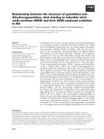
Two AMPKKs can be resolved from rat liver extracts and both contain LKB1, STRAD␣ and MO25␣. (a) Separation of two activities that activate
the GST-AMPK␣1 catalytic domain by Q-Sepharose chromatography. The graph shows AMPKK activity in 4.5 ml fractions (red circles and red line),
absorbance at 280 nm (continuous black line) and conductivity in the eluate (dashed black line) plotted against fraction number. (b) Probing of blots
of column fractions after SDS gel electrophoresis (1 l per lane) using anti-LKB1, anti-STRAD␣ or anti-MO25␣ antibodies. In the three bottom
panels, fractions 26-30 were concentrated from 4.5 ml to 250 l using Amicon Ultra-4 30,000 MWCO centrifugal concentrators, and reanalyzed by
western blotting using 2 l per lane. (c) The effect of protein phosphatase treatment on the mobility of LKB1. The peak fractions of AMPKK1 (0.2
units) or AMPKK2 (0.8 units) were incubated in a final volume of 20 l with or without 5 mM MgCl
2
and 200 M ATP for 15 min at 30
o
C. Protein
phosphatases (PP1␥, 8 mU; or PP2A
1
, 1 mU) or buffer were added and incubation continued for a further 15 min before stopping the reactions in
SDS sample buffer and analyzing by SDS gel electrophoresis and western blotting using anti-LKB1 antibody.
1
2
150
100
50
14 20 30
90
60
30
0
AMPKK2 AMPKK1
LKB1
STRADα
STRADα
MO25α
LKB1
MO25α
1816 22 24 2826 363432
Activity (units per ml) ( )
A280 ( )
Conductivity (mS) ( )
AMPKK1 AMPKK2 AMPK
LKB1
pT172
PP2A1: + + +
MgATP: + ++++++++
PP1γ:+ + +
(a)
(b)
(c)
of the AMPKK1 and AMPKK2 fractions with MgATP, fol-
lowed by treatment with or without the catalytic subunit of
protein phosphatase 1␥ (PP1␥) or the protein phosphatase
2A
1
(PP2A
1
) holoenzyme, did not alter the mobility of any
of the LKB1 polypeptides. The right-hand panel in Figure 1c
shows that these protein phosphatases did dephosphorylate
Thr172 on the ␣ subunit of AMPK when incubated under
identical conditions.
AMPKK activity can be immunoprecipitated from
AMPKK1 and AMPKK2
Using anti-LKB1 antibody but not a pre-immune control
immunoglobulin, we were able to immunoprecipitate
AMPKK activity from fractions containing both AMPKK1
and AMPKK2. Figure 2a shows results of an experiment
where the amount of AMPKK1 or AMPKK2 was limiting and
the antibody was in excess, and shows that we were able to
remove more than 80% of the activity from the peak frac-
tions containing AMPKK1 and AMPKK2 by immunoprecipi-
tating with anti-LKB1 antibody, while no activity was
removed using a pre-immune control immunoglobulin.
We could remove more than 95% of the AMPKK activity of
a recombinant tagged LKB1:STRAD␣:MO25␣ complex (see
below) under the same conditions (Figure 2a). The small
amount of activity remaining in the supernatants of the
AMPKK1 and AMPKK2 immunoprecipitates could be
accounted for by the fact that the immunoprecipitation
was not quantitative, with a small amount of the LKB1
28.4 Journal of Biology 2003, Volume 2, Issue 4, Article 28 Hawley et al. />Journal of Biology 2003, 2:28
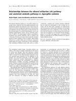
Figure 2
AMPKK activity (that is, the ability to activate AMPK␣1 catalytic domain), and LKB1, STRAD␣ and MO25␣ polypeptides, can be immunoprecipitated
from rat liver AMPKK1 and AMPKK2 using anti-LKB1 antibody. (a) Depletion of AMPKK activity from supernatant. Sheep anti-human LKB1 or pre-
immune control immunoglobulin (IgG) was prebound to Protein G-Sepharose beads and cross-linked with dimethylpimelimidate as described [47],
except that a final wash of the beads with 100 mM glycine, pH 2.5, was performed. Bead-bound antibodies (40 l) were incubated with the peak
fraction of AMPKK1 (0.04 units), AMPKK2 (0.03 units) or recombinant GST-LKB1:STRAD␣:MO25␣ complex (0.06 units) for 120 minutes and the
beads removed in a microcentrifuge (14,000 × g for 2 min). AMPKK activity was determined in the supernatants and is expressed as a percentage of
the value obtained using the control IgG. (b) The pellets from the experiment in (a) were resuspended in the original volume and samples of the
supernatants and pellets analysed by western blotting with anti-LKB1 antibody. The recombinant LKB1 migrates at a higher molecular mass because
of the GST tag. (c) As in (a), except that the amounts of AMPKK1, AMPKK2 and recombinant GST-LKB1:STRAD␣:MO25␣ complex were increased
to 0.44, 0.70 and 1.4 units, respectively, and the activities were determined in the resuspended pellets. In this experiment the amount of antibody
was limiting, so only a fraction of the activity was precipitated. (d) The pellets from the experiment in (c) were resuspended and samples analyzed by
western blotting with anti-LKB1, anti-STRAD␣ and anti-MO25␣ antibodies.
100
50
Activity (%)
Anti-LKB1:
Control IgG:
AMPKK1
AMPKK2
LKB1
+++
+++
Anti-LKB1 SN:
Control IgG SN:
Anti-LKB1 P:
Control IgG P:
+++
+++
+++
+++
LKB1
GST-LKB1
Anti-LKB1:
Control IgG:
AMPKK1
AMPKK2
LKB1
+++
+++
0.01
0.02
Activity (units)
LKB1
STRADα
MO25α
AMPKK1 AMPKK2
(a)
(b)
(c)
(d)
polypeptide remaining in the supernatant. No LKB1
polypeptide was precipitated using the pre-immune control
immunoglobulin (Figure 2b).
Because of the small amount of AMPKK1 and AMPKK2 used
in this experiment, it proved difficult to analyze the pellets for
AMPKK activity and the presence of the other polypeptides.
We therefore repeated the experiment with more AMPKK
(the amount of antibody was now limiting) and analyzed
the pellets only. This showed that we could recover a similar
amount of AMPKK activity in the pellet from the peak frac-
tions containing AMPKK1 and AMPKK2 as we could from
the recombinant LKB1:STRAD␣:MO25␣ complex, with no
activity being recovered in the pellet using the pre-immune
control immunoglobulin (Figure 2c). Western blotting of
the AMPKK1 and AMPKK2 pellets showed that they con-
tained LKB1, STRAD␣ and MO25␣ (Figure 2d).
Recombinant LKB1:STRAD
␣␣
:MO25
␣␣
complexes
activate AMPK
␣␣
1 catalytic domain in cell-free assays
To examine whether LKB1 activated AMPK on its own or
whether the accessory subunits STRAD␣/ and MO25␣/
were required, we expressed LKB1 tagged with glutathione-
S-transferase (GST), FLAG-tagged STRAD␣/ and Myc-
tagged MO25␣/ in various combinations in HEK-293T
cells, and purified the complexes on glutathione-Sepharose.
We also used a GST-tagged kinase-inactive mutant of LKB1
(D194A), and a plasmid expressing GST alone, as controls.
The complexes were purified on glutathione-Sepharose and
incubated with the AMPK␣1 catalytic domain in the pres-
ence of MgATP, and activation of the catalytic domain as
well as phosphorylation of Thr172 (using a phosphospecific
anti-pT172 antibody) was measured. Figure 3a shows that
LKB1 alone did not significantly increase the activity, or
phosphorylation of Thr172, of the AMPK␣1 catalytic
domain above the basal activity observed in the presence of
GST alone (compare lanes 1 and 14). The same result was
obtained with LKB1 that had been co-expressed with
MO25␣ or MO25 (lanes 4 and 5), which was expected as
these proteins do not interact with LKB1 in the absence of
STRAD␣/ [21]. An LKB1:STRAD␣ complex did give a small
but significant activation, and Thr172 phosphorylation, of
the AMPK␣1 catalytic domain above the basal value
(compare lanes 1 and 2). To produce, however, a large acti-
vation and phosphorylation of the AMPK␣1 catalytic
domain, a heterotrimeric complex containing LKB1,
STRAD␣ or STRAD, and MO25␣ or MO25 was required
(lanes 6 to 9). With the heterotrimeric complexes the degree
of activation was in the order LKB1:STRAD␣:MO25␣
> LKB1:STRAD␣:MO25 Ϸ LKB1:STRAD:MO25␣ >
LKB1:STRAD:MO25. The ability of LKB1:STRAD:MO25
complexes to activate AMPK␣1 was dependent on LKB1 cat-
alytic activity, because complexes of a catalytically inactive
mutant of LKB1 (D194A) with the various combinations of
STRAD␣/ and MO25␣/ (lanes 10-13) were unable to acti-
vate or phosphorylate AMPK␣1. The degree of activation
obtained with the various complexes of wild-type LKB1 cor-
related well with the phosphorylation of Thr172, as assessed
by probing blots with a phosphospecific antibody (pT172).
The bottom three panels in Figure 3a, probed with anti-GST,
anti-FLAG or anti-Myc antibodies, confirm that the relevant
STRAD and MO25 subunit co-precipitated with LKB1 when
DNAs encoding these subunits had been co-transfected.
When STRAD␣ was co-expressed with LKB1 in the absence
of a MO25 subunit, the amount of STRAD␣ subunit co-pre-
cipitated with LKB1 was reduced (compare lanes 2 and 3
with lanes 6 and 7).
Figure 3b provides evidence that Thr172 was the only site on
the AMPK␣1 catalytic domain phosphorylated by the
LKB1:STRAD␣:MO25␣ complex. When the two proteins
were incubated together in the presence of [␥-
32
P]ATP, the
wild-type AMPK␣1 catalytic domain became
32
P-labeled, but
a T172A mutant of the AMPK␣1 catalytic domain did not.
AMPKK1, AMPKK2 and recombinant
LKB1:STRAD:MO25 complexes also activate
heterotrimeric AMPK complexes
Although most of the AMPKK assays in this study were
conducted using the AMPK␣1 catalytic domain as sub-
strate, AMPKK1, AMPKK2 and the recombinant GST-
LKB1:STRAD␣:MO25␣ complex also activated heterotrimeric
AMPK complexes. We incubated rat liver AMPK (a mixture
of ␣1 and ␣2 in complexes with 1 and ␥1) with MgATP
with or without each of the three AMPKK preparations. We
then immunoprecipitated with anti-AMPK␣1 or anti-
AMPK␣2 antibodies, and measured the activation of each
isoform in the precipitate. The results (Figure 4a) show that
the AMPK␣1 and AMPK␣2 complexes were activated by all
three AMPKK preparations. Blotting of the three AMPKK
preparations using anti-LKB1, anti-STRAD␣ and anti-
MO25␣ antibodies (Figure 4b) showed that activation of
the heterotrimers was not simply proportional to the
amount of these polypeptides in the preparation. Although
the amounts of each AMPKK preparation used for Figure 4a
had been chosen to yield a comparable degree of AMPK
activation, there was much more LKB1, STRAD␣ and
MO25␣ in the recombinant LKB1:STRAD␣:MO25␣
complex than in either of the native complexes, and more
of all three subunits in AMPKK2 than in AMPKK1. All three
AMPKK preparations also activated recombinant ␣11␥1
and ␣21␥1 complexes prepared [22] by co-expression of
recombinant DNA in CCL13 cells (not shown).
The assays in Figure 4a were conducted in the presence of
200 M AMP. Figure 4c shows that when the AMPK␣11␥1
Journal of Biology 2003, Volume 2, Issue 4, Article 28 Hawley et al. 28.5
Journal of Biology 2003, 2:28
or AMPK␣11␥1 heterotrimers were used as substrate, the
activation of all three AMPKK preparations was stimulated
from 2- to 3.5-fold by AMP. When the AMPK␣1 catalytic
domain was used as substrate, however, the activation was
not affected, or was even slightly inhibited, by AMP. The
activity of the three AMPKK preparations was not signifi-
cantly affected by the direct addition of phenformin to the
assays up to 1 mM concentration, although concentrations
above that started to inhibit AMPKK activity (not shown).
These results indicate that neither AMP nor phenformin
directly stimulates the LKB1:STRAD␣:MO25␣ complex.
Endogenous LKB1 that activates AMPK can be
immunoprecipitated from 293 cells but not from
HeLa cells
Figure 5a shows that AMPKK activity that activated the
AMPK␣1 catalytic domain above the basal activity, and
phosphorylated Thr172, could be immunoprecipitated
28.6 Journal of Biology 2003, Volume 2, Issue 4, Article 28 Hawley et al. />Journal of Biology 2003, 2:28
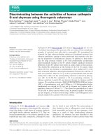
Figure 3
Recombinant LKB1:STRAD:MO25 complexes can efficiently activate the AMPK␣1 catalytic domain via phosphorylation at Thr172. (a) The indicated
combinations of GST-tagged wild-type LKB1 (WT, lanes 1-9), or kinase-dead (D194A; KD, lanes 10-13) LKB1 mutant, or GST-alone (lane 14),
FLAG-tagged STRAD␣ or STRAD, and Myc-tagged MO25␣ or MO25 were coexpressed in HEK-293T cells, purified on glutathione-Sepharose
and tested for their ability to activate GST-AMPK␣1 catalytic domain (top panel). The results are expressed as the increase in the units of AMPK
activity generated per mg full-length GST-AMPK␣1 catalytic domain. Samples from each incubation were also analyzed by western blotting and
probed using the indicated antibodies (from top to bottom): anti-pT172; anti-AMPK␣1 catalytic domain (GST-AMPK␣1); anti-GST to detect GST-
LKB1; anti-FLAG to detect STRAD␣ and STRAD, and anti-Myc to detect MO25␣ and MO25. All proteins migrated with the expected mobility,
taking into account the epitope tags. The bottom three blots were conducted on blank reactions lacking GST-AMPK␣1 catalytic domain, as the latter
appeared to cause some interference with detection. (b) Recombinant GST-LKB1:STRAD␣:MO25␣ complex was used to phosphorylate wild-type
GST-AMPK␣1 catalytic domain (GST-␣1-WT) or a T172A mutant (GST-␣1-T172A) using [␥-
32
P]ATP as described in Materials and methods. The
reaction was analyzed by SDS gel electrophoresis and autoradiography. Arrows show the migration of GST-LKB1 (which autophosphorylates) and
GST-AMPK␣1 catalytic domain.
GST-LKB1
GST-AMPKα1
GST-AMPKα1-WT:
GST-AMPKα1-T172A:
LKB1:STRADα:MO25α:
++
++
+++
200
400
600
Activity (unit per mg)
MO25β:+++++
MO25α:+++++
STRADβ:+++++
STRADα:+++++
LKB1 WT LKB1 KD
GST
pT172
GST-AMPKα1
1 2 3 4 5 6 7 8 9 10111213
14
GST-LKB1
FLAG-STRADα
Myc-MO25β
Myc-MO25α
FLAG-STRADβ
(a)
(b)
Journal of Biology 2003, Volume 2, Issue 4, Article 28 Hawley et al. 28.7
Journal of Biology 2003, 2:28
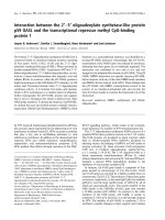
Figure 4
Activation and phosphorylation of heterotrimeric AMPK complexes by AMPKK1, AMPKK2 and recombinant GST-LKB1:STRAD␣:MO25␣
complexes, and the effect of AMP. (a) Activation of ␣1  1␥1 and ␣2  1␥1 complexes separated from purified rat liver AMPK. The AMPK␣1- or
AMPK␣2-containing complexes were purified by immunoprecipitation and activation of the resuspended immunoprecipitates by the three AMPKK
preparations examined. The results are expressed as activation relative to the control without added AMPKK. (b) Quantification by western blotting
of the relative amounts of LKB1, STRAD␣ and MO25␣ polypeptides in the three AMPKK preparations used in (a). A small amount of degradation is
detectable due in part to the heavy loadings of the GST-LKB1 and FLAG-STRAD␣. The identity of the polypeptide labeled ‘?’ in the anti-LKB1 blot is
not known. (c) Effect of AMP on the activation of ␣11␥1 and ␣21␥1 heterotrimers of AMPK, and of GST-AMPK␣1 catalytic domain, by
AMPKK1, AMPKK2 and the recombinant GST-LKB1:STRAD␣:MO25␣ complex. AMPKK activity was measured as in Figure 3 with or without
200 M AMP. The results are expressed as ratios of the activities obtained with and without AMP.
LKB1
GST-LKB1
?
STRADα
FLAG-STRADα
MO25α
++LKB1 complex: LKB1 complex:
++AMPKK2: AMPKK2:
++AMPKK1: AMPKK1:
Myc-MO25α
4
3
2
1
Activation
(relative to control)
+
+
+
LKB1 complex:
AMPKK2:
AMPKK1:
+
+
+
α1β1γ1 α2β1γ1
Activity ratio (+/− AMP)
3
2
1
+
+
α1β1γ1 α1 domainα2β1γ1
+
LKB1 complex: ++
AMPKK2: ++
++AMPKK1:
(a)
(b)
(c)
pT172
α1/α2
from untransfected HEK-293T cell extract using anti-LKB1
antibody (lane 1), but not pre-immune control immunoglob-
ulin (lane 2). As reported previously [20,21], immunopre-
cipitation of endogenous LKB1 resulted in the
co-precipitation of STRAD␣ and MO25␣ (lane 1). As a
further control, we employed normal HeLa cells as an LKB1-
null cell line, as it is known that LKB1 is not expressed in
these cells [12]. Consistent with this, no LKB1, STRAD␣ and
MO25␣ subunits or AMPKK activity were immunoprecipi-
tated from the same amount of HeLa cell-extract protein
using anti-LKB1 antibody (lane 3). AMPKK activity and
Thr172 phosphorylation, as well as detectable STRAD␣ and
MO25␣ subunits, were recovered following immunoprecipi-
tation of LKB1 from a line of HeLa cells that stably express
wild-type LKB1 [23] (lane 5). The LKB1, STRAD␣ and
MO25␣ polypeptides were still recovered in cells expressing
a catalytically inactive mutant of LKB1, but AMPKK activity
was not (lane 7). Although the LKB1 polypeptide was over-
expressed to a large extent in the HeLa cells compared to the
endogenous levels observed in 293 cells (compare lane 1
with lane 5 or 7), it is clear that the availability of STRAD␣
and/or MO25␣ limits the activity in these cells. There was
less AMPKK activity and Thr172 phosphorylation, as well as
less co-precipitated STRAD␣ and MO25␣, in HeLa cells
expressing LKB1 than in 293 cells, even though the LKB1
polypeptide was overexpressed. The AMPKK activity in the
immunoprecipitates from 293 cells and HeLa cells express-
ing wild-type LKB1 correlated with the levels of STRAD␣ and
MO25␣ in the complex, rather than with the levels of LKB1.
Figure 5b shows western blots of total lysates of the same
cells. Although the expression of MO25␣ in HeLa cells was
lower than in 293 cells, it was unaffected by overexpression
of either wild-type or kinase-dead LKB1, as was expression
of the protein kinases ERK1 and ERK2, used as loading con-
trols. Interestingly, however, expression of the STRAD␣
doublet was almost undetectable in the control HeLa cells
but was readily detectable in cells stably expressing wild-
type or kinase-dead LKB1.
Expression of LKB1 restores activation of AMPK in
HeLa cells
The drug 5-aminoimidazole-4-carboxamide (AICA) riboside
activates AMPK in intact cells by being taken up and imme-
diately converted by adenosine kinase to AICA riboside
monophosphate (ZMP), which mimics the effect of AMP on
the AMPK system [24]. The anti-diabetic drug metformin
activates AMPK in intact cells by a mechanism that is not
known, although it does not involve changes in cellular
adenine nucleotide content [25-27]. We have previously
found (unpublished observations) that, although AMPK is
expressed in HeLa cells, it is not activated either by AICA
riboside or by metformin. A potential explanation for this is
that HeLa cells do not express LKB1 ([12]; see also above).
To examine whether expression of recombinant LKB1 might
restore the ability of HeLa cells to respond to these drugs,
we used the HeLa cell line that stably expresses wild-type
LKB1 [23]. In these experiments we used phenformin, a
close relative of metformin that we have found to activate
AMPK more rapidly than metformin in other cell types.
Figure 6a shows that neither AICA riboside nor phenformin
activated AMPK above the basal level in control HeLa cells.
In cells expressing wild-type LKB1 but not the kinase-inac-
tive mutant, however, both AICA riboside and phenformin
caused a robust activation, as well as a small increase in
basal activity. The AMPK activity also correlated with the
phosphorylation of Thr172 on AMPK␣ (shown by probing
with the anti-pT172 antibody, Figure 6b) and with the
phosphorylation of a downstream target of AMPK, acetyl-
CoA carboxylase (ACC; shown by probing with a phosphos-
pecific antibody against Ser-79, the primary AMPK site on
that protein; Figure 6c). Interestingly, stable expression of
wild-type LKB1 in the HeLa cells caused a small but repro-
ducible degree of upregulation of expression of AMPK␣1
and a marked down-regulation of expression of ACC (data
not shown). These effects may be a consequence of the
increase in basal AMPK activity shown in Figure 6a. Because
the expression of these proteins was not uniform in these
cells, in order to accurately quantify their phosphorylation
status we simultaneously probed single blots of lysates
using either anti-pT172 and anti-␣1/␣2 antibodies (to
detect Thr172 phosphorylation and total AMPK␣), or with
anti-pACC and streptavidin (to detect Ser79 phosphoryla-
tion and total ACC). The two probing reagents used in each
of these dual-labeling protocols were labeled with fluores-
cent dyes emitting in different regions of the infra-red spec-
trum, and the results were quantified in two separate
channels using an infra-red laser scanner. In Figure 6b, these
results are expressed as ratios of the signal obtained using
the phosphospecific antibody to the signal obtained for the
total protein, which corrects for different levels of expres-
sion of the proteins. This revealed that there was a good cor-
relation between activation of AMPK and phosphorylation
of Thr172. There was also a correlation with phosphoryla-
tion of ACC, although in this case AICA riboside appeared
to have a larger effect than phenformin.
AMPK activation is defective in immortalized
fibroblasts from LKB
-/-
mouse embryos
We performed further experiments with immortalized mouse
embryo fibroblasts (MEF cells) from embryonic-day (E) 9.5
embryos of LKB1
-/-
knockout mice. In cells from control
LKB1
+/+
embryos, AICA riboside and phenformin caused two-
fold and three-fold activation of endogenous AMPK, but this
was completely absent from the LKB1
-/-
cells (Figure 7). The
basal activity of AMPK was also about 60% lower in the
28.8 Journal of Biology 2003, Volume 2, Issue 4, Article 28 Hawley et al. />Journal of Biology 2003, 2:28
Journal of Biology 2003, Volume 2, Issue 4, Article 28 Hawley et al. 28.9
Journal of Biology 2003, 2:28
Figure 5
Endogenous AMPKK activity (that is, ability to activate AMPK␣1 catalytic domain) can be immunoprecipitated from 293 cells using anti-LKB1
antibody, but activity can only be immunoprecipitated from HeLa cells if they stably express wild-type LKB1, but not a catalytically-inactive mutant.
(a) LKB1 was immunoprecipitated from 0.5 mg cell extract derived from untransfected HEK-293T cells (lanes 1,2), untransfected HeLa cells
(control; lanes 3,4), or HeLa cells stably expressing wild-type LKB1 (WT; lanes 5,6) or a kinase-dead LKB1 mutant (D194A; KD, lanes 7,8).
Immunoprecipitation used anti-LKB1 (lanes 1, 3, 5, 7) or a pre-immune control immunoglobulin (IgG; lanes 2, 4, 6, 8). Samples of each
immunoprecipitate were used to assay activation of GST-AMPK␣1 catalytic domain, to analyze phosphorylation of GST-AMPK␣1 catalytic domain
on Thr172 (middle panel), and to determine by western blotting the recovery of LKB1 and its accessory subunits (bottom panels). In lanes 5 and 7
some immunoglobulin heavy chain (IgG-H) had eluted from the protein G-Sepharose despite the fact that it had been cross-linked: this explains
why LKB1 may not appear to comigrate in lanes 1, 5 and 7. Also shown at left in the top panel is the basal activity obtained when the GST-
AMPK␣1-catalytic domain was incubated with MgATP on its own (no addition). (b) Whole cell lysates from the same cells were analyzed by SDS
gel electrophoresis and blots probed using anti-LKB1, anti-STRAD␣, and anti-MO25␣ antibodies. They were also probed with anti-ERK1/2
antibodies as loading controls.
293 HeLa cells
IP anti-LKB1:
IP control IgG:
++++
++++
Control WT KD
Activity (U.mg
−1
)
18765432
pT172
LKB1
STRADα
MO25α
LKB1
IgG-H
No addition
293 HeLa cells
LKB1
STRADα
MO25α
ERK1
ERK2
Control WT KD
293 HeLa cells
20
40
60
80
(a)
(b)
LKB1
-/-
cells. Figure 7 also confirms, by western blotting of
cell lysates, that the expression of LKB1 was absent from
LKB1
-/-
cells, that the expression of the AMPK␣ subunits was
normal, and that the phosphorylation of Thr172 on the
AMPK␣ subunits correlated with AMPK activity.
Discussion
Our results provide strong evidence that LKB1:STRAD:MO25
complexes represent the major upstream kinases acting on
AMPK, although they do not rule out the possibility that the
complex might contain additional components. The key
evidence may be summarized as follows. First, during previ-
ous extensive efforts to purify from rat liver extracts activi-
ties that activate dephosphorylated AMPK, ([4] and
subsequent unpublished work), we have not detected any
activities other than AMPKK1 and AMPKK2, at least under
the assay conditions used. Second, both AMPKK1 and
AMPKK2 purified from rat liver contained LKB1, STRAD␣
and MO25␣ that were detectable by western blotting and
whose presence correlated with AMPKK activity across the
column fractions (Figure 1). Third, the ability of the
AMPKK1 and AMPKK2 fractions to activate AMPK was
almost completely eliminated by immunoprecipitation with
anti-LKB1 antibody, but not a control immunoglobulin.
Activity was also detected, along with the LKB1, STRAD␣
and MO25␣ polypeptides, in the anti-LKB1 immunoprecip-
itates but not in the control immunoprecipitates (Figure 2).
Fourth, the AMPKK activity in AMPKK1 and AMPKK2 was
not a contaminant that co-precipitated with anti-LKB1 anti-
body, because recombinant complexes of GST-LKB1,
STRAD and MO25 expressed in 293 cells and purified on
glutathione-Sepharose also activated the AMPK␣1 catalytic
domain efficiently, and phosphorylated Thr172 (Figure 3).
Complexes formed from a catalytically inactive mutant
LKB1 failed to activate or phosphorylate AMPK. Phosphory-
lation of the AMPK␣1 catalytic domain by this recombinant
complex occurred exclusively at Thr172, because the wild-
type AMPK␣1 catalytic domain, but not a T172A mutant,
could be phosphorylated using [␥-
32
P]ATP and the GST-
LKB1:STRAD␣:MO25␣ complex. Fifth, although most of the
experiments in this study were conducted using the bacteri-
ally expressed AMPK␣1 catalytic domain as substrate,
AMPKK1, AMPKK2 and recombinant LKB1-STRAD␣-
MO25␣ complexes also efficiently activated heterotrimeric
AMPK complexes, both the ␣11␥1 and ␣21␥1 isoforms
(Figure 4). Sixth, HeLa cells, unlike HEK 293T cells, do not
express LKB1 (Figure 5) and therefore represent a natural
‘knockout’ cell line. The drugs AICA riboside and phen-
formin, which activate AMPK in other cell types via distinct
mechanisms [24,27], did not activate AMPK in HeLa cells.
In cells stably transfected with DNA that expressed wild-
type LKB1 (but not a catalytically inactive mutant),
however, the ability of AICA riboside and phenformin to
activate AMPK, to phosphorylate Thr172 on the AMPK␣
subunit, and to cause phosphorylation of a downstream
target (ACC) was restored (Figure 6). This experiment
proves that (in the presence of STRAD␣ and MO25␣) LKB1
is sufficient for AMPK activation, but does not prove that it
is necessary, because expression of upstream kinases other
than LKB1 might also be defective in HeLa cells. Figure 5
also confirms that STRAD␣ and MO25␣ are necessary to
generate an active complex because, although the LKB1
28.10 Journal of Biology 2003, Volume 2, Issue 4, Article 28 Hawley et al. />Journal of Biology 2003, 2:28
Figure 6
Restoration of the ability of AMPK to be activated, and AMPK and
acetyl-CoA carboxylase to be phosphorylated, by AICA riboside and
phenformin in HeLa cells following expression of LKB1. Control HeLa
cells (lanes 1,2,3), HeLa cells expressing wild-type LKB1 (WT; lanes
4,5,6) or kinase-inactive mutant LKB1 (D194A; KD, lanes 7,8,9) were
incubated for 60 min with no further addition, with 2 mM AICA
riboside or 10 mM phenformin, and lysed. (a) Endogenous AMPK was
immunoprecipitated from the cell extracts and assayed. (b) The cell
lysates was immunoblotted with antibodies recognizing AMPK␣1
phosphorylated at Thr172 or total AMPK␣1; the results were analyzed
using the LI-COR Odyssey™ IR imager as described in the Materials
and methods section, and are expressed as a ratio of the two signals.
(c) The cell lysates were analyzed by western blotting and the
membranes probed with antibodies recognizing ACC phosphorylated at
Ser79, or streptavidin to determine total AMPK␣1. The results were
analyzed using the LI-COR imager as for (b).
0.2
0.1
Activity
(units per mg)
LKB1 induction:
None
WT
KD
AICA riboside:
Phenformin:
+++
+++
187654329
4.0
3.0
pT172/total AMPK
2.0
4.0
pACC/total ACC
(a)
(b)
(c)
polypeptide was greatly overexpressed in the stably trans-
fected HeLa cells compared to the endogenous level in 293
cells, the AMPKK activity, and the amounts of STRAD␣ and
MO25␣, in the anti-LKB1 immunoprecipitate, were actually
less. This suggests that the amount of active LKB1 was
limited by the availability of STRAD␣ and MO25␣. Seventh,
in LKB1
+/+
MEF cells, AMPK became activated in response to
both AICA riboside and phenformin. In LKB1
-/-
MEF cells,
however, the basal activity of AMPK was lower and AICA
riboside and phenformin failed to activate AMPK. These
results show that LKB1 is both necessary and sufficient for
AMPK activation, at least in MEF cells.
All of the assays of the activity of LKB1 and its complexes
described in this article, whether using the AMPK␣1 cat-
alytic domain or AMPK heterotrimers as substrate, utilized
MgATP as the co-substrate. Previous studies of the kinase
activity of LKB1, whether utilizing autophosphorylation
[12], or p53 [28] or myelin basic protein [20] as substrates,
had used MnATP as co-substrate and had reported that there
was no activity with the more physiological MgATP
complex. Thus, unlike previously used substrates, AMPK is a
good substrate for LKB1 complexes even using the physio-
logically relevant divalent metal ion.
We have previously reported that the activation of AMPK by
AMPKK1 (called at that time AMPKK) was stimulated by
AMP, and presented evidence favoring the hypothesis that
AMP acted not only by binding to the downstream kinase
and making it a better substrate, but also by activating the
upstream kinase [6]. The results in Figure 4c support the
first hypothesis but do not support the second. Using
␣11␥1 or ␣21␥1 AMPK complexes as substrate, activa-
tion by AMPKK1, AMPKK2 or LKB1 was stimulated from 2-
to 3.5-fold by AMP. When using the AMPK␣1 catalytic
domain as substrate, however, AMP had no effect, or even
slightly inhibited activation. The AMPK␣1 catalytic domain
is not allosterically activated by AMP [29], and AMP
binding appears to be a function of the ␥ subunit ([30], and
unpublished observations). Taken together with previous
results [6,24,31], these data support the idea that the effects
of AMP on the kinase cascade are all mediated through
binding to the downstream kinase, AMPK. The previous
report that AMP stimulated the upstream kinase was
obtained using a less pure AMPKK preparation [6], and we
have been unable to reproduce this with the more purified
preparations utilized here.
Both AMPKK1 and AMPKK2 appear to contain LKB1,
STRAD␣ and MO25␣, and thus it is not clear at present why
they resolve on Q-Sepharose chromatography. One interest-
ing difference is that the LKB1 polypeptide in AMPKK1
migrated significantly faster on SDS gels than that in
AMPKK2 (Figures 1 and 2). LKB1 is known to be phospho-
rylated at up to eight sites, and is also farnesylated at
Cys433, near the carboxyl-terminus [23,28,32,33], suggest-
ing that the difference in mobility might be due to a differ-
ence in covalent modification. It did not appear to be due to
differential phosphorylation, however, because neither
incubation with MgATP, nor protein phosphatase treat-
ment, produced a shift in mobility of the LKB1 polypeptides
in either AMPKK1 or AMPKK2 (Figure 1c). Another differ-
ence between AMPKK1 and AMPKK2 was their Stokes radii
estimated by size exclusion chromatography (5.7 versus 5.2
nm respectively). By combining estimates of Stokes radius
and sedimentation coefficient, we previously estimated the
molecular mass of AMPKK1 to be 195 kDa [4], and assum-
ing a similar shape our estimate of the Stokes radius of
AMPKK2 would suggest a mass of about 175 kDa. These
values are close to, although slightly larger than, the calcu-
lated mass of 140 kDa for a 1:1:1 LKB1:STRAD␣:MO25␣
complex. Although we cannot rule out the possibility that
AMPKK1 and/or AMPKK2 contain additional associated
protein(s) other than LKB1, STRAD␣/ and MO25␣/, it is
also possible that differences in covalent modification
Journal of Biology 2003, Volume 2, Issue 4, Article 28 Hawley et al. 28.11
Journal of Biology 2003, 2:28
Figure 7
AMPK is activated and phosphorylated in response to AICA riboside
and phenformin in LKB1
+/+
but not in LKB1
-/-
MEF cells. Cells
immortalized from control embryos LKB1
+/+
and LKB1
-/-
knockout
embryos [14], were incubated with AICA riboside (2 mM) or
phenformin (10 mM) for 1 hour. Lysates were prepared and AMPK
activity (expressed as units per mg total lysate protein) determined in
immunoprecipitates made using a mixture of anti-AMPK␣1 and anti-
AMPK␣2 antibodies. Lysates were also analyzed by SDS gel
electrophoresis and blots probed using anti-LKB1, anti-AMPK-␣1/2,
and anti-pT172 antibodies.
Phenformin: + +
AICA riboside: + +
165432
LKB1: −/−
+/+
−/−
+/+
−/−
+/+
0.2
0.1
AMPK activity
(units per mg)
pT172
AMPKα
LKB1
might affect the shape of the complex and hence the Stokes
radius. Whatever the reason for the difference in elec-
trophoretic and chromatographic behavior of AMPKK1 and
AMPKK2, a clear conclusion from Figure 4 is that, for the
same amount of LKB1, STRAD␣ and MO25␣ polypeptides,
the former was more active than the latter. Although further
work is required to explain these differences, they might be
caused by the same covalent modifications that alter the
mobility on SDS gels. Figure 4 also shows that, for the same
amount of LKB1, STRAD␣ and MO25␣ polypeptides, both
AMPKK1 and AMPKK2 were much more active than the
recombinant complex. The low activity of the latter might
be explained by the presence of the purification tag on each
subunit, by imperfect folding or assembly, or by an altered
level of covalent modification, when the complex is overex-
pressed. As mentioned above, our data do not rule out the
possibility that the recombinant LKB1 complex may be
lacking additional subunit(s) present in the endogenous
AMPKK1 and AMPKK2 complexes.
Our present results confirm, using a probable physiological
substrate, previous findings using an artificial substrate
(myelin basic protein) that a STRAD subunit stimulates the
kinase activity of LKB1 [20], and that the MO25 subunit
stimulates the activity further, probably by stabilizing the
LKB1:STRAD complex [21]. No AMPKK activity was
obtained with recombinant LKB1 unless a STRAD subunit
was also expressed, and the activity was increased substan-
tially by the additional presence of a MO25 subunit
(Figure 3). It was also noticeable that the amount of
STRAD␣ and STRAD that co-precipitated with LKB1 was
greatly enhanced by the co-expression of MO25␣ or MO25
(Figure 3), consistent with previous findings [21]. Another
new result in this article is that STRAD␣ protein (unlike
MO25␣) was not detectable in HeLa cells unless either wild-
type or kinase-dead LKB1 was stably expressed (Figure 6b).
These results suggest that STRAD␣ is normally complexed
with LKB1 in the cell, and that STRAD is unstable in the
absence of LKB1. The exact mechanism by which the STRAD
and MO25 subunits activate LKB1 remains unclear, but
these accessory subunits introduce scope for additional reg-
ulation of the kinase. It is already known that LKB1 phos-
phorylates STRAD␣ at two distinct sites [20], and that
STRAD␣ and MO25␣ form a complex that retains LKB1 in
the cytoplasm [21].
People with PJS are heterozygous for mutations in LKB1,
and further work is required to establish whether loss of one
allele of LKB1 could affect AMPK activation in these
patients. An interesting unanswered question is whether
activation of AMPK can explain the ability of LKB1 to act as
a tumor suppressor and to arrest cell growth and prolifera-
tion. This certainly seems plausible, because apart from the
fact that AMPK is a general inhibitor of biosynthesis [1,2],
there is accumulating evidence that it can regulate cell pro-
liferation and apoptosis. For example, activation of AMPK
inhibits proliferation of HepG2 cells by stabilizing p53 [34].
Interestingly, expression of LKB1 in G361 cells that nor-
mally lack expression of the kinase causes an arrest in G1
phase of the cell cycle that is associated with an induction of
p21 and is dependent on p53 [12,13].
Another exciting possibility is that LKB1:STRAD:MO25
complexes might also act as upstream kinases for other
protein kinases, in the same manner that PDK1 phosphory-
lates threonine residues in the activation loop of a number
of kinases of the ‘AGC’ subfamily [35]. A dendrogram
showing the relationships between catalytic domain
sequences of 518 human protein kinases encoded in the
human genome [36] shows that the AMPK␣1 and AMPK␣2
subunits lie on a small sub-branch also containing eight
other protein kinases (NuaK1, NuaK2, BrsK1, BrsK2, SIK,
QIK, QSK and MELK), most of which either have not been
studied previously or have very little known about them. An
alignment of the activation loop sequences of these kinases
is shown in Additional data file 2 with the online version of
this article and show that, as well as conservation of the
threonine residue phosphorylated by LKB1 in AMPK, they
have other conserved motifs that are not present in other
protein kinases known to be activated by other upstream
kinases. It remains to be determined whether the other
kinases in the AMPK subfamily are activated by phosphory-
lation of the conserved threonine residue by
LKB1:STRAD:MO25 complexes, but if this is the case these
other kinases might mediate some of the tumor suppressor
functions of LKB1.
A significant number of inherited forms of PJS found in
certain families do not exhibit mutations in the LKB1 gene
[37,38], indicating that there could be other causative loci
for PJS. On the basis of the results presented here it would
be very interesting to examine whether mutations in the
genes encoding STRAD␣ or , MO25␣ or , or any of the
subunits of AMPK or of the AMPK-like subfamily of kinases,
are found in PJS patients who do not have mutations in the
LKB1 gene.
While this article was under review, two papers were pub-
lished that are relevant to our results. Hong et al. [39]
reported that FLAG-tagged LKB1 expressed in, and purified
from, COS7 cells would activate a recombinant AMPK het-
erotrimer, and phosphorylate the ␣ subunit at Thr172, in
cell-free assays. This is consistent with our results, although
these authors provided no evidence that LKB1 acts on
AMPK in vivo, or that LKB1 required the presence of the
STRAD and MO25 subunits to phosphorylate AMPK. Spicer
28.12 Journal of Biology 2003, Volume 2, Issue 4, Article 28 Hawley et al. />Journal of Biology 2003, 2:28
et al. [40] reported evidence, based on expression of recom-
binant LKB1 in cultured cells, suggesting that it might act
upstream of the PAR1A protein kinase. PAR1A (also known
as MARK-3 [41]) lies with three closely related protein
kinases (MARK-1, MARK-2, MARK-4) on a branch of the
human kinase tree [36] immediately adjacent to AMPK-␣1
and -␣2 and the eight AMPK-like kinases discussed above.
Although Spicer et al. [40] did not provide evidence that
LKB1 directly phosphorylated PAR1A, the results of our
study suggest that this might be the case. The sequence of
the activation segment of PARIA is given in Additional data
file 2 (available with the online version of this article).
Conclusions
Our results provide strong evidence, both in cell-free assays
and in intact cells, that complexes between LKB1, STRAD␣/
and MO25␣/ constitute the long sought-after upstream
kinases that activate AMPK via phosphorylation at Thr172 in
the activation loop. Because it is already known that pharma-
cological activation of AMPK causes a general inhibition of
biosynthesis, as well as a p53-dependent arrest in G1 phase of
the cell cycle, activation of AMPK by LKB1 might explain, at
least in part, the ability of LKB1 to act as a tumor suppressor.
LKB1 may also act as an upstream kinase for other members
of the AMPK-like subfamily of protein kinases.
Materials and methods
Materials, proteins and antibodies
Protein G-Sepharose, glutathione-Sepharose and prepacked
Q-Sepharose columns were from Amersham Pharmacia
Biotech, Little Chalfont, UK. The GST-AMPK␣1 catalytic
domain, and a T172A mutant, were expressed in Escherichia
coli and purified as described previously [29]. Sheep anti-
bodies against the ␣1 and ␣2 subunits of AMPK [22],
human LKB1, MO25␣ and MO25 [21], and phosphospe-
cific antibody against the Thr172 site on the AMPK␣
subunit (anti-pT172) [42] were described previously. Sheep
antibody against AMPK␣1 catalytic domain was raised
against the peptide CDPMKRATpIKDIRE (cysteine +
residues 252-264 of rat AMPK␣1 given in the single-letter
code for amino acids; Tp, phosphothreonine) using
methods described for anti-pT172 [42]. Although designed
as a phosphospecific antibody, it recognizes the GST-
AMPK␣1 catalytic domain expressed in bacteria and recog-
nition is not affected by protein phosphatase treatment. The
monoclonal antibody against STRAD␣ was described previ-
ously [20]. Monoclonal anti-GST and anti-FLAG epitope
antibodies were from Sigma (Poole, UK). Anti-Myc antibod-
ies were prepared by ammonium sulfate precipitation of
medium from Myc1-9E10 hybridoma cells grown in RPMI
1640 medium supplemented with 2 mM glutamine and
15% (v/v) fetal bovine serum. Anti-Erk1/2 antibodies were
from Cell Signaling Technology (New England Biolabs,
Hitchin, UK). The DNA constructs encoding GST-LKB1
(wild-type and kinase-dead) in the pEBG-2T vector [28],
and FLAG-STRAD␣, FLAG-STRAD, Myc-MO25␣ and Myc-
MO25 in the pCMV5 vector [21] have been described pre-
viously. PP1␥ was expressed in E. coli [43], and PP2A
1
was
purified from rabbit skeletal muscle [43]. Sources of other
materials and proteins were as described previously [4].
Enzyme assays
AMPK [4], PP1␥ and PP2A
1
[43] were assayed, and units
defined, as described previously. AMPKK was assayed as
follows (based on [27]). A fusion protein between the
kinase domain of the ␣1 subunit of AMPK and glutathione-
S-transferase (GST-AMPK␣1) was expressed in E. coli [29].
Although some preparations of GST-AMPK␣1 show evidence
of proteolytic degradation, only the full length GST-AMPK␣1
is phosphorylated (J.L.R., unpublished observations). The
amount of full-length GST-AMPK␣1 was quantified by den-
sitometry of Coomassie-Blue-stained gels, using bovine
serum albumin as standard. The E. coli lysate expressing
GST-AMPK␣1 was adsorbed onto glutathione-Sepharose
beads (Amersham-Pharmacia) such that the final concentra-
tion of kinase after maximal activation using MgATP and
AMPKK in the assay below was 1 unit in the standard kinase
assay per 5 l of beads. The slurry was washed with 4 × 1 ml
of IP buffer (50 mM Tris-HCl, pH 7.4 at 4
o
C, 50 mM NaF, 5
mM Na pyrophosphate, 1 mM EDTA, 1 mM EGTA, 1%
Triton X-100, 1 mM dithiothreitol (DTT), 1 mM benzami-
dine, 0.1 mM phenylmethane sulfonyl fluoride, 1 M NaCl)
to remove unbound proteins. It was then washed in 3 × 1
ml of assay buffer (50 mM Na Hepes, pH 7.4, 1 mM DTT,
0.02% Brij-35).
For the kinase kinase assay, the AMPKK preparation was
incubated with 10 l of a 50% slurry of the glutathione-
Sepharose beads with bound GST-AMPK␣1, plus 200 M
AMP, 200 M ATP, 5 mM MgCl
2
in assay buffer in a final
volume of 25 l. After incubation for 20 min at 30
o
C on a
rotary shaker, the beads were washed with 4 × 1 ml of IP
buffer and 3 × 1 ml of assay buffer prior to a standard
AMPK assay. The units of AMPKK are the units of AMPK
generated in the assay, expressed per mg of full length GST-
AMPK␣1 protein used. Rapid lysis of cells for AMPK assays
was as described previously [44]. AMPK and AMPKK assays
were carried out in triplicate and results are expressed as
mean ± standard deviation.
Purification of AMPKK1 and AMPKK2
AMPKK was purified to the Blue-Sepharose stage as
described previously [4]. The flow-through from this
column was adjusted to 160 mM NaCl by dilution in buffer
Journal of Biology 2003, Volume 2, Issue 4, Article 28 Hawley et al. 28.13
Journal of Biology 2003, 2:28
A (50 mM Hepes, 10% (w/v) glycerol, 0.02% (w/v) Brij-35,
1 mM EDTA, 1 mM EGTA, 1 mM DTT, 1 mM benzamidine,
0.1 mM PMSF, 1 g/ml soybean trypsin inhibitor), and
applied to a high performance Q-Sepharose HiLoad 16/10
column in buffer A plus 160 mM NaCl at 3 ml/min. The
column was washed in buffer A plus 160 mM NaCl until the
A
280
was < 0.05, and AMPKK activity was then eluted with a
linear gradient (120 ml) from 160-400 mM NaCl in buffer A.
Expression of recombinant LKB1 complexes in HEK-
293T cells
Various combinations of GST-tagged LKB1, FLAG-tagged
STRAD␣ or STRAD, and Myc-tagged MO25␣ or MO25
were expressed in HEK293 cells and the complexes purified
on glutathione-Sepharose as described previously [21].
Phosphorylation of GST-
␣␣
1 catalytic domain using
[
␥␥
-
32
P]ATP
GST-AMPK␣1 catalytic domain (50 g/ml), either wild-type
or a T172A mutant [29] was incubated for 30 min at 30°C
with 5 mM MgCl
2
and [␥-
32
P]ATP (200 M; approximately
750 cpm/pmole) in the presence or absence of the GST-
LKB1:STRAD␣:MO25␣ complex (20 units/ml). The reaction
was terminated by the addition of SDS sample buffer (Invitro-
gen, Paisley, UK), the polypeptides resolved by SDS gel elec-
trophoresis and the dried gel subjected to autoradiography.
Preparation of and activation of AMPK
heterotrimers
AMPK was purified from rat liver as described previously
[4]. Dephosphorylation,with PP2A, addition of okadaic
acid to inhibit the phosphatase, and incubation with
AMPKK was as described previously [4]. The reaction was
stopped by adding 5 l of 0.5 M EDTA to 20 l of the
dephosphorylated AMPK, and 20 l of the mixture was
then incubated for 2 h at 4°C with 75 l of a 15% suspen-
sion of anti-AMPK␣1 or anti-AMPK␣2 antibodies bound to
protein G-Sepharose [44] plus 200 l of IP buffer. The
beads were recovered by centrifugation (14,000 × g for 2
min) and washed twice with IP buffer and twice with 50
mM Na Hepes buffer, pH 7.4. AMPK assays were then
carried out on the resuspended immunoprecipitates [44].
To obtain recombinant AMPK, plasmids encoding Myc-
AMPK␣1 or Myc-AMPK␣2, AMPK1 and AMPK␥1 were co-
expressed in CCL13 cells [22], and cells harvested by the
rapid lysis method [44]. Lysates were immunoprecipitated
with anti-Myc antibody and resuspended in 50 mM Na
HEPES, 1 mM dithiothreitol, 0.02% (w/v) Brij-35, pH 7.5,
and assayed as above.
Immunoprecipitation of endogenous LKB1
Immunoprecipitation of endogenous LKB1 using anti-
human antibody and protein G-Sepharose has been
described previously [21]. The kinase kinase assays were
conducted using GST-AMPK␣1 catalytic domain as substrate
in shaking incubators as described previously for immuno-
precipitate assays of AMPK [44].
HeLa cells expressing LKB1
The generation and culture conditions of HeLa cells stably
expressing inducible (Tet-ON) wild-type or kinase-inactive
mutant LKB1, and conditions for their culture, has been
described previously [23].
Production of immortalized mouse embryo
fibroblasts
Wild-type and LKB1
-/-
E9.5 embryos [14] were minced into
small fragments and placed in culture in Dulbecco’s modified
Eagle’s Medium supplemented with penicillin, streptomycin,
glutamine, 10% fetal bovine serum (AutogenBioclear,
Santa Cruz, USA), and 10% conditioned medium collected
from day-3 cultures of wild-type fibroblasts. The cultures
were subsequently allowed to expand for 5 days, after which
they were passaged according to a modified 3T3 protocol
[45]. High-passage cultures that expanded were considered
immortalized.
Protein analysis and electrophoresis
Protein concentration was determined using the dye-
binding method of Bradford [46]. SDS gel electrophoresis
used precast Bis-Tris 4-12% gradient polyacrylamide gels, in
the MOPS buffer system (Invitrogen), except for analysis of
acetyl-CoA carboxylase, where pre-cast 3-8% Tris-acetate
gels (Invitrogen) were used. Proteins were transferred to
nitrocellulose membranes (BioRad, Hemel Hempstead, UK)
using the Xcell II Blot Module (Invitrogen).
Detection of western blots by infra-red imaging
To analyze phosphorylation of ACC, membranes were incu-
bated in LI-COR Odyssey
TM
Blocking buffer for 1 h. Anti-
pACC antibody (1.46 g/ml in blocking buffer containing
Tween-20 0.2% v/v) was then added and left shaking for
1 h. The membranes were washed 6 × 5 min with TBS
(10 mM Tris-HCl, pH 7.4, 0.5 M NaCl) plus Tween-20
(0.2% v/v). The membranes were immersed in blocking
buffer containing Tween-20 (0.2% v/v) and 1 g/ml anti-
sheep IgG conjugated to IR dye 680 (Molecular Probes,
Leiden, The Netherlands) and 1 g/ml streptavidin conju-
gated to IR Dye 800 (Rockland Inc., from Lorne Laborato-
ries, Reading, UK) and left shaking for 1 h, protected from
light. The membranes were then washed 6 × 5 min using
TBS-Tween (0.2%) and 1 × 5 min in PBS. The membranes
were scanned in two different channels using the Odyssey
IR imager, the results quantified using Odyssey software and
expressed as a ratio of the signal obtained with the pACC
antibody to that obtained with streptavidin. Analysis of
28.14 Journal of Biology 2003, Volume 2, Issue 4, Article 28 Hawley et al. />Journal of Biology 2003, 2:28
phosphorylation of GST-␣1 catalytic domain was similar
except that the 4-12% Bis-Tris gels were used, and the mem-
branes were simultaneously probed for 1 h with the sheep
anti-pT172 and anti-AMPK␣1 catalytic domain antibodies,
directly labeled with the IR dye 680 and IR dye 800 respec-
tively, according to manufacturers’ instructions.
Acknowledgements
This study was supported by the Wellcome Trust (D.G.H.) the UK
Medical Research Council (D.G.H. and D.R.A.), the Association for
International Cancer Research (D.R.A), Diabetes UK (D.R.A.), and by
the Finnish Cancer Organization, Sigrid Juselius Foundation, and the
Academy of Finland (T.P.M.). J.L.R. was supported by a studentship from
the MRC and L.U is a student of the Helsinki Biomedical Graduate
School. We thank Philip Cohen for helpful discussions, Moustapha
Aoubala for preparation of antibodies, Agnieszka Kieloch for assistance
with tissue culture, Debbie Mander for preparation of
LKB1:STRAD:MO25 complexes, Annette Baas and Hans Clevers for
providing us with the STRAD␣ cDNA and STRAD␣ antibodies, Greg
Stewart for the preparation of the T172A mutant of the AMPK␣1 cat-
alytic domain, and Antti Ylikorkala and Derrick Rossi for helping to gen-
erate the MEF cultures.
Additional data files
The following additional materials are available with the
online version of this article: Additional data file 1, showing
an alignment of the kinase domains of Tos3, Pak1,
CaMKK, LKB1 and Elm1, and Additional data file 2,
showing an alignment of the activation loop of the kinase
subgroup phylogenetically close to LKB1.
References
1. Hardie DG, Scott JW, Pan DA, Hudson ER: Management of cel-
lular energy by the AMP-activated protein kinase system.
FEBS Lett 2003, 546:113-120.
2. Hardie DG, Hawley SA: AMP-activated protein kinase: the
energy charge hypothesis revisited. BioEssays 2001, 23:1112-
1119.
3. Coven DL, Hu X, Cong L, Bergeron R, Shulman GI, Hardie DG,
Young LH: Physiologic role of AMP-activated protein
kinase (AMPK) in the heart: graded activation during
exercise. Am J Physiol Endocrinol Metab 2003, 285:E629-E636.
4. Hawley SA, Davison M, Woods A, Davies SP, Beri RK, Carling D,
Hardie, DG: Characterization of the AMP-activated protein
kinase kinase from rat liver, and identification of threo-
nine-172 as the major site at which it phosphorylates and
activates AMP-activated protein kinase. J Biol Chem 1996,
271:27879-27887.
5. Sutherland CM, Hawley SA, McCartney RR, Leech A, Stark MJR,
Scmidt MC, Hardie DG: Elm1p is one of three upstream
kinases for the Saccharomyces cerevisiae SNF1 complex.
Curr Biol 2003, 13:1299-1305.
6. Hawley SA, Selbert MA, Goldstein EG, Edelman AM, Carling D,
Hardie DG: 5
-AMP activates the AMP-activated protein
kinase cascade, and Ca
2+
/calmodulin the calmodulin-
dependent protein kinase I cascade, via three independent
mechanisms. J Biol Chem 1995, 270:27186-27191.
7. Hemminki A, Markie D, Tomlinson I, Avizienyte E, Roth S, Loukola
A, Bignell G, Warren W, Aminoff M, Hoglund P, et al.: A
serine/threonine kinase gene defective in Peutz-Jeghers
syndrome. Nature 1998, 391:184-187.
8. Jenne DE, Reimann H, Nezu J, Friedel W, Loff S, Jeschke R, Muller
O, Back W, Zimmer M: Peutz-Jeghers syndrome is caused by
mutations in a novel serine threonine kinase. Nat Genet
1998, 18:38-43.
9. Hemminki A: The molecular basis and clinical aspects of
Peutz-Jeghers syndrome. Cell Mol Life Sci 1999, 55:735-750.
10. Giardiello FM, Brensinger JD, Tersmette AC, Goodman SN,
Petersen GM, Booker SV, Cruz-Correa M, Offerhaus JA: Very
high risk of cancer in familial Peutz-Jeghers syndrome.
Gastroenterology 2000, 119:1447-1453.
11. Boudeau J, Sapkota G, Alessi DR: LKB1, a protein kinase regu-
lating cell proliferation and polarity. FEBS Lett 2003, 546:159-
165.
12. Tiainen M, Ylikorkala A, Makela TP: Growth suppression by
LKB1 is mediated by a G(1) cell cycle arrest. Proc Natl Acad
Sci USA 1999, 96:9248-9251.
13. Tiainen M, Vaahtomeri K, Ylikorkala A, Makela TP: Growth
arrest by the LKB1 tumor suppressor: induction of
p21(WAF1/CIP1). Hum Mol Genet 2002, 11:1497-1504.
14. Ylikorkala A, Rossi DJ, Korsisaari N, Luukko K, Alitalo K, Henke-
meyer M, Makela TP: Vascular abnormalities and deregula-
tion of VEGF in LKB1-deficient mice. Science 2001,
293:1323-1326.
15. Jishage K, Nezu J, Kawase Y, Iwata T, Watanabe M, Miyoshi A, Ose
A, Habu K, Kake T, Kamada N, et al.: Role of Lkb1, the
causative gene of Peutz-Jegher’s syndrome, in embryoge-
nesis and polyposis. Proc Natl Acad Sci USA 2002, 99:8903-8908.
16. Bardeesy N, Sinha M, Hezel AF, Signoretti S, Hathaway NA, Sharp-
less NE, Loda M, Carrasco DR, DePinho RA: Loss of the LKB1
tumour suppressor provokes intestinal polyposis but resis-
tance to transformation. Nature 2002, 419:162-167.
17. Miyoshi H, Nakau M, Ishikawa TO, Seldin MF, Oshima M, Taketo
MM: Gastrointestinal hamartomatous polyposis in LKB1
heterozygous knockout mice. Cancer Res 2002, 62:2261-2266.
18. Rossi DJ, Ylikorkala A, Korsisaari N, Salovaara R, Luukko K,
Launonen V, Henkemeyer M, Ristimaki A, Aaltonen LA, Makela
TP: Induction of cyclooxygenase-2 in a mouse model of
Peutz-Jeghers polyposis. Proc Natl Acad Sci USA 2002,
99:12327-12332.
19. Nakau M, Miyoshi H, Seldin MF, Imamura M, Oshima M, Taketo
MM: Hepatocellular carcinoma caused by loss of heterozy-
gosity in LKB1 gene knockout mice. Cancer Res 2002,
62:4549-4553.
20. Baas AF, Boudeau J, Sapkota GP, Smit L, Medema R, Morrice NA,
Alessi DR, Clevers HC: Activation of the tumour suppressor
kinase LKB1 by the STE20-like pseudokinase STRAD.
EMBO J 2003, 22:3062-3072.
21. Boudeau J, Baas AF, Deak M, Morrice NA, Kieloch A, Schutowski
M, Prescott AR, Clevers HC, Alessi DR: MO25 isoforms inter-
act with the STE20-related pseudokinase STRAD
␣␣
/

and
enhance their ability to bind, activate and localise the
LKB1 tumour suppressor in the cytoplasm. EMBO J 2003,
22:5102-5114.
22. Woods A, Salt I, Scott J, Hardie DG, Carling, D: The
␣␣
1 and
␣␣
2
isoforms of the AMP-activated protein kinase have similar
activities in rat liver but exhibit differences in substrate
specificity in vitro. FEBS Lett 1996, 397:347-351.
23. Sapkota GP, Deak M, Kieloch A, Morrice N, Goodarzi AA, Smythe
C, Shiloh Y, Lees-Miller SP, Alessi DR: Ionizing radiation
induces ataxia telangiectasia mutated kinase (ATM)-
mediated phosphorylation of LKB1/STK11 at Thr-366.
Biochem J 2002, 368:507-516.
24. Corton JM, Gillespie JG, Hawley SA, Hardie DG: 5-Aminoimida-
zole-4-carboxamide ribonucleoside: a specific method for
activating AMP-activated protein kinase in intact cells? Eur
J Biochem 1995, 229:558-565.
25. Fryer LG, Parbu-Patel A, Carling D: The anti-diabetic drugs
rosiglitazone and metformin stimulate AMP-activated
protein kinase through distinct pathways. J Biol Chem 2002,
277:25226-25232.
26. Zhou G, Myers R, Li Y, Chen Y, Shen X, Fenyk-Melody J, Wu M,
Ventre J, Doebber T, Fujii N, et al.: Role of AMP-activated
Journal of Biology 2003, Volume 2, Issue 4, Article 28 Hawley et al. 28.15
Journal of Biology 2003, 2:28
protein kinase in mechanism of metformin action. J Clin
Invest 2001, 108:1167-1174.
27. Hawley SA, Gadalla AE, Olsen GS, Hardie DG: The anti-diabetic
drug metformin activates the AMP-activated protein
kinase cascade via an adenine nucleotide-independent
mechanism. Diabetes 2002, 51:2420-2425.
28. Sapkota GP, Kieloch A, Lizcano JM, Lain S, Arthur JS, Williams MR,
Morrice N, Deak M, Alessi DR: Phosphorylation of the
protein kinase mutated in Peutz-Jeghers cancer syn-
drome, LKB1/STK11, at Ser431 by p90(RSK) and cAMP-
dependent protein kinase, but not its farnesylation at
Cys(433), is essential for LKB1 to suppress cell growth.
J Biol Chem 2001, 276:19469-19482.
29. Scott JW, Norman DG, Hawley SA, Kontogiannis L, Hardie DG:
Protein kinase substrate recognition studied using the
recombinant catalytic domain of AMP-activated protein
kinase and a model substrate. J Mol Biol 2002, 317:309-323.
30. Cheung PCF, Salt IP, Davies SP, Hardie DG, Carling D: Charac-
terization of AMP-activated protein kinase
␥␥
subunit iso-
forms and their role in AMP binding. Biochem J 2000,
346:659-669.
31. Davies SP, Helps NR, Cohen PTW, Hardie DG: 5’-AMP inhibits
dephosphorylation, as well as promoting phosphorylation,
of the AMP-activated protein kinase. Studies using bacte-
rially expressed human protein phosphatase-2C
␣␣
and
native bovine protein phosphatase-2A
C
. FEBS Lett 1995,
377:421-425.
32. Collins SP, Reoma JL, Gamm DM, Uhler MD: LKB1, a novel
serine/threonine protein kinase and potential tumour sup-
pressor, is phosphorylated by cAMP-dependent protein
kinase (PKA) and prenylated in vivo. Biochem J 2000,
345:673-680.
33. Sapkota GP, Boudeau J, Deak M, Kieloch A, Morrice N, Alessi DR:
Identification and characterization of four novel phospho-
rylation sites (Ser31, Ser325, Thr336 and Thr366) on
LKB1/STK11, the protein kinase mutated in Peutz-
Jeghers cancer syndrome. Biochem J 2002, 362:481-490.
34. Imamura K, Ogura T, Kishimoto A, Kaminishi M, Esumi H: Cell
cycle regulation via p53 phosphorylation by a 5
-AMP
activated protein kinase activator, 5-aminoimidazole-4-
carboxamide-1-beta-d-ribofuranoside, in a human hepato-
cellular carcinoma cell line. Biochem Biophys Res Commun 2001,
287:562-567.
35. Alessi DR: Discovery of PDK1, one of the missing links in
insulin signal transduction. Colworth Medal Lecture.
Biochem Soc Trans 2001, 29:1-14.
36. Manning G, Whyte DB, Martinez R, Hunter T, Sudarsanam S: The
protein kinase complement of the human genome. Science
2002, 298:1912-1934.
37. Buchet-Poyau K, Mehenni H, Radhakrishna U, Antonarakis SE:
Search for the second Peutz-Jeghers syndrome locus:
exclusion of the STK13, PRKCG, KLK10, and PSCD2
genes on chromosome 19 and the STK11IP gene on chro-
mosome 2. Cytogenet Genome Res 2002, 97:171-178.
38. Resta N, Stella A, Susca FC, Di Giacomo M, Forleo G, Miccolis I,
Rossini FP, Genuardi M, Piepoli A, Grammatico P, Guanti G, et al.:
Two novel mutations and a new STK11/LKB1 gene
isoform in Peutz-Jeghers patients. Hum Mutat 2002, 20:78-79.
39. Hong SP, Leiper FC, Woods A, Carling D, Carlson M: Activation
of yeast Snf1 and mammalian AMP-activated protein
kinase by upstream kinases. Proc Natl Acad Sci USA 2003,
100:8839-8843.
40. Spicer J, Rayter S, Young N, Elliott R, Ashworth A, Smith D: Reg-
ulation of the Wnt signalling component PAR1A by the
Peutz-Jeghers syndrome kinase LKB1. Oncogene 2003,
22:4752-4756.
41. Drewes G, Ebneth A, Preuss U, Mandelkow EM, Mandelkow E:
MARK, a novel family of protein kinases that phosphory-
late microtubule-associated proteins and trigger micro-
tubule disruption. Cell 1997, 89:297-308.
42. Sugden C, Crawford RM, Halford NG, Hardie DG: Regulation of
spinach SNF1-related (SnRK1) kinases by protein kinases
and phosphatases is associated with phosphorylation of
the T loop and is regulated by 5
-AMP. Plant J 1999, 19:1-7.
43. Alessi DR, Street AJ, Cohen P, Cohen PTW: Inhibitor-2 func-
tions like a chaperone to fold 3 expressed isoforms of
mammalian protein phosphatase-1 into a conformation
with the specificity and regulatory properties of the native
enzyme. Eur J Biochem 1993, 213:1055-1066.
44. Hardie DG, Salt IP, Davies SP: Analysis of the role of the
AMP-activated protein kinase in the response to cellular
stress. Methods Mol Biol 2000, 99:63-75.
45. Denhardt DT, Edwards DR, McLeod M, Norton G, Parfett CL,
Zimmer M: Spontaneous immortalization of mouse
embryo cells: strain differences and changes in gene
expression with particular reference to retroviral gag-pol
genes. Exp Cell Res 1991, 192:128-136.
46. Bradford MM: A rapid and sensitive method for the quanti-
tation of microgram quantities of protein utilizing the
principle of protein-dye binding. Anal Biochem 1976, 72:248-
254.
47. Harlow E, Lane D: Antibodies. A Laboratory Manual. Cold Spring
Harbor: Cold Spring Harbor Laboratory; 1988.
48. Devereux J, Haeberli P, Smithies O: A comprehensive set of
sequence analysis programs for the VAX. Nucleic Acids Res
1984, 12:387-395.
49. BOXSHADE 3.21
[ />28.16 Journal of Biology 2003, Volume 2, Issue 4, Article 28 Hawley et al. />Journal of Biology 2003, 2:28