Báo cáo sinh học: "Bending out and breaking away: host-cell accomplices in retroviral escape" pot
Bạn đang xem bản rút gọn của tài liệu. Xem và tải ngay bản đầy đủ của tài liệu tại đây (109.31 KB, 4 trang )
Minireview
Bending out and breaking away: host-cell accomplices in
retroviral escape
Melvyn W Yap and Jonathan P Stoye
Address: Division of Virology, National Institute for Medical Research, The Ridgeway, London NW7 1AA, UK.
Correspondence: Jonathan Stoye. E-mail:
How do enveloped viruses bud from their host cells? To
understand how this process is achieved, several fundamental
steps must be considered. First, viral structural components
must be transported to the appropriate site, typically just
under a cell membrane, and there assembled (Figure 1a) [1].
Second, the plasma membrane must be distorted to make a
succession of curved budding structures (Figure 1b,c); this
requires overcoming the mechanical bending resistance of the
plasma membrane [2]. Third, following the formation of the
bud, the virus has to pinch off and escape from the cell
(Figure 1d,e) [3]. This involves machinery that constricts the
neck of the bud, resulting in fusion between the membranes
on either side of the neck and the release of the virus from the
plasma membrane. Studies with a number of virus types,
most prominently retroviruses, have now revealed that cellu-
lar proteins that are intimately involved in intracellular mem-
brane trafficking and receptor re-localization play key roles in
facilitating these processes.
For a long time, it has been known that the only retroviral
component required for assembly and budding is the Gag
polyprotein, which ultimately forms the viral core [1]. Gag
is cleaved into a variety of smaller components as the virus
matures. These include, from amino terminus to carboxyl
terminus, the matrix (MA), capsid (CA) and nucleocapsid
(NC). Depending on the virus analyzed, a variety of other
protein products are seen after cleavage of Gag. For
example, in human immunodeficiency virus-1 (HIV-1) a
short peptide called p6 is cleaved from the carboxy-terminal
end of NC, whereas in murine leukemia virus (MuLV) a p12
peptide is cleaved from between MA and CA.
Three types of functional domain of Gag can be identified: M,
sequences required for transport to and binding of mem-
branes; I, involved in Gag-Gag interactions; and L, late
sequences [1,3]. The L domains are short peptide motifs
located in different regions of Gag in different viruses; mut-
ation in these sequences results in failure to release budded
viruses [4,5]. Many L domains are interchangeable between
viruses, suggesting that their role in the late stages of budding
is to act as docking sites for cellular proteins [5-7]. A key step
in understanding the late budding process came with the
demonstration that the L domain of HIV-1 Gag interacted
with a component of the cellular machinery responsible for
sorting cargo into multivesicular bodies (MVBs) [8-10].
MVBs are formed from early endosomes when their mem-
branes invaginate into the endosomal lumen, resulting in
Abstract
Budding through the host-cell membrane is a key step in the life cycle of many viruses. Recent
studies of retrovirus replication implicate a large number of cellular proteins in this process.
BioMed Central
Journal
of Biology
Journal of Biology 2003, 3:3
Published: 19 December 2003
Journal of Biology 2003, 3:3
The electronic version of this article is the complete one and can be
found online at />© 2003 BioMed Central Ltd
the release of vesicles into the luminal space [11,12]. Mono-
ubiquitination acts as a signal for directing proteins into
MVBs, although it might not be the only signal, given that
membrane proteins that are not ubiquitinated can also be
transported to the MVBs. The formation of MVBs requires
three protein complexes, which were first characterized in
yeast and are collectively known as the endosomal sorting
complexes required for transport (ESCRTs) [13-15]. ESCRTI
and ESCRTII each contain one subunit that binds ubiquitin.
ESCRTII is believed to function downstream of ESCRTI, as
overexpression of the former can compensate for the loss of
the latter, but the opposite is not the case. ESCRTII func-
tions to recruit ESCRTIII to the membrane. Recent studies
have confirmed the interaction between proteins of ESCRTs
I and II and between those of ESCRTs II and III [16,17]. The
full ESCRT complex is dissociated by the AAA (ATPase asso-
ciated with diverse cellular activities) protein, Vps4 [18,19].
HIV-1 interacts with the Tsg101 component of ESCRTI via a
late domain within the p6 domain of Gag that contains the
sequence P(S/T)AP (in the single-letter amino-acid code).
Depletion of Tsg101 results in production of a late-domain
phenotype, similar to the stage shown in Figure 1d [8]. Arti-
ficially recruiting Tsg101 into another late-domain mutant
rescues budding activity [9]. These findings suggest that the
ESCRT complexes might facilitate scission of the nascent
virion from the cell. Very recent studies have shown that
release of HIV-1 can be blocked at a late stage by mutation
or deletion of at least eight cellular proteins that are
involved in the biogenesis of MVBs [17]. Other retroviruses
containing different L domains, such as MuLV (character-
ized by a PPXY motif, where X is any amino acid) and
equine infectious anemia virus (EIAV, characterized by a
YPXL motif), do not interact directly with Tsg101 [3].
Budding of these viruses is arrested by dominant-negative
mutants of various components of the MVB pathway, again
implicating at least some portions of the endosomal sorting
machinery in virus release [20-22]. In addition, some retro-
viruses appear to contain two L domains that can contribute
to virus release [23,24]. L domains are also found in the
matrix proteins of rhabdoviruses [25], filoviruses [9] and
orthomyxoviruses [26], suggesting that involvement of the
MVB pathway may be a common theme in virus budding.
Are these proteins the only cellular factors to play a role in
virus budding? The article by Wang and colleagues in this
issue of Journal of Biology [27] suggests otherwise. It brings
several other participants in the field of cell-membrane
movement into play, with intriguing possibilities. Wang et
al. describe the interaction between the Gag protein of the
Moloney MuLV and components of the cellular endocytic
machinery, the endophilins. The interaction was initially
detected in a yeast two-hybrid protein-protein interaction
screen using as its ‘bait’ the Gag protein from the MuLV-
related murine acquired immunodeficiency syndrome
(MAIDS) virus. Subsequently, endophilin 2 was found to
interact with the Gag proteins of MuLV and Rous sarcoma
virus (RSV) but not of HIV-1, Mason Pfizer monkey virus
3.2 Journal of Biology 2003, Volume 3, Issue 1, Article 3 Yap and Stoye />Journal of Biology 2003, 3:3
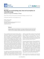
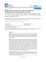
Figure 1
A schematic representation of retrovirus budding. (a) Gag proteins move to the plasma membrane and begin to associate with one another.
(b) Formation of electron-dense aggregates under a deforming plasma membrane follows. (c) Bud curvature steadily increases. (d) Membrane fusion
leads to pinching-off of the virion; (e) proteolytic processing of Gag leads to virion maturation and formation of an electron dense core. L-domain
mutants of most retroviruses arrest at a stage equivalent to (d) but with an extended stalk [3,4]; in other viruses, such as human T-lymphotropic
virus 1 (HTLV-1), arrest occurs at a stage roughly equivalent to (b) [38]. MA, matrix; CA, capsid; NC, nucleocapsid; Env, envelope proteins.
Out
Gag RNA Env
In
MA=
CA=
NC=
(a) (b) (c) (d) (e)
(MPMV) or simian immunodeficiency virus (SIV). MuLV
Gag could also interact with rat endophilin 1, another
member of the endophilin family [27].
The interaction between endophilin 2 and MuLV Gag was
confirmed using a fusion protein made up of glutathione-S-
transferase (GST) and endophilin 2, attaching this to beads
and using them to pull down Gag from MuLV-infected cells.
Significantly, 0.7% of the endophilin 2 present in MuLV-
producing cells became incorporated into the virions. Inter-
estingly, ␣-adaptin and clathrin, two other components of
the clathrin-mediated endocytic machinery [28], were also
found to be incorporated into MuLV virions. The region
required for binding to endophilin 2 was mapped to the
MA domain of the Gag protein. An intact endophilin 2
protein was required for Gag interaction, as determined in
the yeast two-hybrid system, but various fragments of
endophilin 2 could be incorporated into MuLV virions even
though they did not interact with Gag in this assay.
Overexpression of full-length endophilin 2 in MuLV-pro-
ducing cells resulted in a dose-dependent reduction in
virion production. Fragments of endophilin 2 were also
inhibitory, but to a somewhat lesser degree. In contrast, an
excess of endophilin 2 did not affect production of HIV-1
from cells, ruling out the idea that the effects on MuLV are
due to direct toxicity. This suggested that the specific
binding of endophilin 2 to MuLV Gag plays an important
role in MuLV production. It will be of considerable interest
to determine where virus production is arrested in over-
expressing cells. Inhibiting endophilin 2 levels by 80%
using a small interfering (si) RNA, however, did not seem to
affect viral production. This was attributed to the potentially
low levels of endophilin 2 required for virion production,
or the presence of other members of the endophilin family
that could make up for the reduction in endophilin 2.
Although perfectly plausible, these explanations do not
completely dispel the uncertainty introduced by the nega-
tive siRNA experiments. Hence, the conclusion that
endophilins are absolutely required for MuLV budding
remains to be confirmed by further experimentation.
Relatively little is known directly about the function of
endophilin 2, but endophilin 1 is a 40 kDa cytoplasmic
protein containing an amphipathic domain at the amino
terminus as well as a Src homology 3 (SH3) domain near
the carboxyl terminus [29]. It is a multifunctional protein
that is believed to participate in both early and late stages of
endocytosis [28], has lipid transferase activity [30] and is
considered capable of affecting membrane curvature [31] as
well as binding and deforming liposomes into tubules [32].
It can bind to proline-rich domains in multiple cellular pro-
teins, including dynamin and synaptojanin [33]. The closely
related endophilins 2 and 3, though less well characterized,
seem likely to possess similar properties [29].
Given the membrane-bending properties of endophilins, a
role for this family of proteins in virus budding seems, at
least superficially, an attractive hypothesis. But compared to
endocytosis, MVB formation and virus budding are topolog-
ically different processes, with endocytosis involving invagi-
nation into the cytoplasm whereas MVB formation and
virus budding involve evagination, away from the cyto-
plasm. It seems likely that much of the protein machinery
mediating these processes is fundamentally different (for
example, involving components of clathrin-coated pits
versus the ESCRT complex). It seems quite feasible,
however, that some proteins might be involved in both
processes, particularly those with the ability to bend and
fuse membranes. Certainly there is evidence for some cross-
talk, as shown by the interaction between endophilins and
ALIX, a key player in formation of ESCRT complexes and
virus release [17,34,35].
Although significant steps have been taken towards under-
standing virus budding during the past couple of years,
there are still a number of important issues that remain to
be addressed. How is the initial bud formed? It may be that
energetic requirements for membrane distortion can be met
simply by the I-domain-mediated assembly of Gag mole-
cules, resulting in movement of associated membrane lipid
molecules [36]. But what happens in the case of viruses like
MPMV that assemble in the cytoplasm? Is there a need for
cellular enzymes such as endophilin to introduce negative
curvature (bending towards the outside of the cell) by mod-
ifying the lipid composition of the membrane? How does
membrane pinching-off take place? The ESCRT complex is
intimately involved, but is the whole complex required and
what is the role of other factors such as the ubiquitin ligase,
Nedd4, that are clearly involved in the budding of certain
viruses [23,24]? How is the plasma membrane targeted for
budding? In macrophages HIV-1 can bud into vacuoles
[37], but what targets Gag and associated ESCRT complexes
to the cell surface in HIV-infected T cells? Given the pace of
progress in this area, driven in part by the urgency of devel-
oping novel antiretroviral drugs, we can be optimistic that
these and related questions will soon be answered, bringing
closer a detailed understanding of the mechanisms of virus
budding and membrane remodeling.
References
1. Swanstrom R, Wills JW: Synthesis, assembly, and processing
of viral proteins. In Retroviruses. Edited by Coffin JM, Hughes SH,
Varmus HE. Cold Spring Harbor, NY: Cold Spring Harbor Labora-
tory Press; 1997:263-334.
2. Hurley JH, Wendland B: Endocytosis: driving membranes
around the bend. Cell 2002, 111:143-146.
Journal of Biology 2003, Volume 3, Issue 1, Article 3 Yap and Stoye 3.3
Journal of Biology 2003, 3:3
3. Freed EO: Viral late domains. J Virol 2002, 76:4679-4687.
4. Göttlinger HG, Dorfman T, Sodroski JG, Haseltine WA: Effect of
mutations affecting the p6 gag protein on human
immunodeficiency virus particle release. Proc Natl Acad Sci
USA 1991, 88:3195-3199.
5. Yuan B, Campbell S, Bacharach E, Rein A, Goff SP: Infectivity of
Moloney murine leukemia virus defective in late assembly
events is restored by late assembly domains of other
retroviruses. J Virol 2000, 74:7250-7260.
6. Accola MA, Strack B, Göttlinger HG: Efficient particle produc-
tion by minimal Gag constructs which retain the carboxy-
terminal domain of human immunodeficiency virus type
1 capsid-p2 and a late assembly domain. J Virol 2000,
74:5395-5402.
7. Parent LJ, Bennett RP, Craven RC, Nelle TD, Krishna NK,
Bowzard JB, Wilson CB, Puffer BA, Montelaro RC, Wills JC:
Positionally independent and exchangeable late budding
functions of the Rous sarcoma virus and human
immunodeficiency virus Gag proteins. J Virol 1995,
69:5455-5460.
8. Garrus JE, von Schwedler UK, Pornillos OW, Morham SG, Zavitz
KH, Wang HE, Wettstein DA, Stray KM, Cote M, Rich RL, et al.:
Tsg101 and the vacuolar protein sorting pathway are
essential for HIV-1 budding. Cell 2001, 107:55-65.
9. Martin-Serrano J, Zang T, Bieniasz PD: HIV-1 and Ebola virus
encode small peptide motifs that recruit Tsg101 to sites
of particle assembly to facilitate egress. Nat Med 2001,
7:1313-1319.
10. VerPlank L, Bouamr F, LaGrassa TJ, Agresta B, Kikonyogo A, Leis J,
Carter CA: Tsg101, a homologue of ubiquitin-conjugating
(E2) enzymes, binds the L domain in HIV type 1 Pr55Gag.
Proc Natl Acad Sci USA 2001, 98:7724-7729.
11. Katzmann DJ, Odorizzi G, Emr SD: Receptor downregulation
and multivesicular-body sorting. Nat Rev Mol Cell Biol 2002,
3:893-905.
12. Raiborg C, Rusten TE, Stenmark H: Protein sorting into multi-
vesicular endosomes. Curr Opin Cell Biol 2003, 15:446-455.
13. Babst M, Katzmann DJ, Estepa-Sabal EJ, Meerloo T, Emr SD:
ESCRT-III, an endosome-associated heterooligomeric
protein complex required for MVB sorting. Dev Cell 2002,
3:271-282.
14. Babst M, Katzmann DJ, Synder WB, Wendland B, Emr SD: Endo-
some-associated complex, ESCRT-II, recruits transport
machinery for protein sorting at the multivesicular body.
Dev Cell 2002, 3:283-289.
15. Katzmann DJ, Babst M, Emr SD: Ubiquitin-dependent sorting
into the multivesicular body pathway requires the func-
tion of a conserved endosomal sorting complex, ESCRT-1.
Cell 2001, 106:145-155.
16. Martin-Serrano J, Yaravoy A, Perez-Caballero D, Bieniasz PD:
Divergent retroviral late-budding domains recruit vac-
uolar protein sorting factors by using alternative adaptor
proteins. Proc Natl Acad Sci USA 2003, 100:12414-12419.
17. von Schwedler UK, Stuchell M, Muller B, Ward DM, Chung HY,
Morita E, Wang HE, Davis T, He GP, Cimbora DM, et al.: The
protein network of HIV budding. Cell 2003, 114:701-713.
18. Babst M, Wendland B, Estepa EJ, Emr SD: The Vps4p AAA
ATPase regulates membrane association of a Vps protein
complex required for normal endosome function. EMBO J
1998, 17:2982-2993.
19. Ogura T, Wilkinson AJ: AAA+ superfamily ATPases: common
structure-diverse function. Genes Cells 2001, 6:575-597.
20. Goila-Gaur R, Demirov DG, Orenstein JM, Ono A, Freed EO:
Defects in human immunodeficiency virus budding and
endosomal sorting induced by TSG101 overexpression.
J Virol 2003, 77:6507-6519.
21. Martin-Serrano J, Zang T, Bieniasz PD: Role of ESCRT-I in
retroviral budding. J Virol 2003, 77:4794-4804.
22. Tanzi GO, Piefer AJ, Bates P: Equine infectious anemia virus
utilizes host vesicular protein sorting machinery during
particle release. J Virol 2003, 77:8440-8447.
23. Bouamr F, Melillo JA, Wang MQ, Nagashima K, De Los Santos
M, Rein A, Goff SP: PPPYEPTAP motif is the late domain
of human T-cell leukemia virus type 1 Gag and mediates
its functional interaction with cellular proteins Nedd4
and Tsg101. J Virol 2003, 77:11882-11895.
24. Gottwein E, Bodem J, Muller B, Schmechel A, Zentgraf H, Krausslich
HG: The Mason-Pfizer monkey virus PPPY and PSAP motifs
both contribute to virus release. J Virol 2003, 77:9474-9485.
25. Craven RC, Harty RN, Paragas J, Palese P, Wills JW: Late
domain function identified in the vesicular stomatitis virus
M protein by use of rhabdovirus-retrovirus chimeras. J Virol
1999, 73:3359-3365.
26. Hui EK, Barman S, Yang TY, Nayak DP: Basic residues of the
helix six domain of influenza virus M1 involved in nuclear
translocation of M1 can be replaced by PTAP and YPDL
late assembly domain motifs. J Virol 2003, 77:7078-7092.
27. Wang MQ, Kim W, Gao G, Torrey TA, Morse HC III, De Camilli
P, Goff SP: Endophilins interact with Moloney murine
leukemia virus Gag and modulate virion production. J Biol
2003, 3:4.
28. Mousavi SA, Malerod L, Berg T, Kjeken R: Clathrin-mediated
endocytosis. Biochem J 2004, 377:1-16.
29. Reutens AT, Begley CG: Endophilin-1: a multifunctional
protein. Int J Biochem Cell Biol 2002, 34:1173-1177.
30. Schmidt A, Wolde M, Thiele C, Fest W, Kratzin H, Podtelejnikov
AV, Witke W, Huttner WB, Soling HD: Endophilin I mediates
synaptic vesicle formation by transfer of arachidonate to
lysophosphatidic acid. Nature 1999, 401:133-141.
31. Huttner WB, Schmidt AA: Membrane curvature: a case of
endofeelin’. Trends Cell Biol 2002, 12:155-158.
32. Farsad K, Ringstad N, Takei K, Floyd SR, Rose K, De Camilli P:
Generation of high curvature membranes mediated by
direct endophilin bilayer interactions. J Cell Biol 2001,
155:193-200.
33. Ringstad N, Nemoto Y, De Camilli P: The SH3p4/Sh3p8/
SH3p13 protein family: binding partners for synaptojanin
and dynamin via a Grb2-like Src homology 3 domain. Proc
Natl Acad Sci USA 1997, 94:8569-8574.
34. Strack B, Calistri A, Craig S, Popova E, Gottlinger HG:
AIP1/ALIX is a binding partner for HIV-1 p6 and EIAV p9
functioning in virus budding. Cell 2003, 114:689-699.
35. Chatellard-Causse C, Blot B, Cristina N, Torch S, Missotten M,
Sadoul R: Alix (ALG-2-interacting protein X), a protein
involved in apoptosis, binds to endophilins and induces
cytoplasmic vacuolization. J Biol Chem 2002, 277:29108-29115.
36. Garoff H, Hewson R, Opstelten D-JE: Virus maturation by
budding. Microbiol Mol Biol Rev 1998, 62:1171-1190.
37. Amara A, Littman DR: After Hrs with HIV. J Cell Biol 2003,
162:371-375.
38. Le Blanc I, Prevost MC, Dokhelar MC, Rosenberg AR: The
PPPY motif of human T-cell leukemia virus type 1 Gag
protein is required early in the budding process. J Virol
2002, 76:10024-10029.
3.4 Journal of Biology 2003, Volume 3, Issue 1, Article 3 Yap and Stoye />Journal of Biology 2003, 3:3