Báo cáo sinh học: "Beyond toxicity: aryl hydrocarbon receptor-mediated functions in the immune system" docx
Bạn đang xem bản rút gọn của tài liệu. Xem và tải ngay bản đầy đủ của tài liệu tại đây (102.08 KB, 3 trang )
Stockinger: Journal of Biology 2009, 8:61
Abstract
The aryl hydrocarbon receptor is a ligand-activated trans crip-
tional regulator that binds dioxin and other exogenous contami-
nants and is responsible for their toxic effects, including immuno-
suppression. New evidence suggests, however, that the aryl
hydrocarbon receptor has a physiological role in the immune
system, and the immunosuppressive effects of dioxin may
reflect a more subtle disruption of the regulatory inter actions
between immune cells.
The aryl hydrocarbon receptor (AhR), also called the
dioxin receptor, is a transcriptional regulator best known
for mediating the toxicity of environmental contaminants,
most notably halogenated polycyclic aromatic hydro-
carbons such as dioxin. AhR has been studied extensively
for its pathological role in response to environmental
pollution, and there is a wealth of knowledge regarding its
signalling components as well as its structural features and
pharmacological effects. Although many aspects of AhR-
mediated toxicity have been described, the molecular
mechanisms underlying these are not well understood.
AhR is conserved across vertebrate and invertebrate
species, playing a role, for instance, in the development of
the nervous system in Caenorhabditis elegans, while in
Drosophila the AhR homolog spineless is involved in
development of antennae and legs as well as in aspects of
color vision [1].The intrinsic physiological functions of
AhR in mammals have been delineated from the phenotype
of the AhR knockout mouse [2-4]. These mice show
reduced fertility, smaller livers, possibly resulting from
vascular defects [1], and portal fibrosis. The strong conser-
vation of AhR in so many species as well as the mutant
phenotype suggest that it has roles beyond those of mediat-
ing toxicity of pollutants. More recently it has been
suggested that dioxin-mediated toxicity may, in fact, reflect
disruption of the endogenous function of this receptor by
inducing sustained and inappropriate AhR signaling due to
the stability of the toxin, and thus causing dysregulation of
physiological functions [5].
Toxic effects of dysregulation are particularly likely in the
immune system where highly complex interactions between
hematopoietic cells and their environment dictate the
outcome of challenge by pathogens, and indeed dioxin has
been known for decades to be immunotoxic, though
information on possible underlying mechanisms is sparse
[6]. Indications of immune defects have been described in
one of the three AhR knockout strains, but not the others,
prompting suggestions that differences in background or
infectious agents in the environment might have played a
role. However, immune challenges such as influenza or
Listeria monocytogenes applied to AhR knockout mice have
so far yielded little insight into the mechanism of changes
that have been reported in the responses of specific subsets
of immune cells, or the induction of specific subsets of
immune cells [7]. More recent experiments on the
expression of AhR in the lymphocytes of the immune
system have begun to suggest that ligands of AhR have
roles in the immune system that do not conform to the
notion of immunosuppression.
The lymphocytes of the adaptive immune system fall into
two major classes - B cells, which secrete antibodies, and
T cells, which act on other cells and, broadly speaking,
either activate other cells of the immune system (cells that
do this belong to a class known as CD4 T cells, or T helper
cells) or destroy infected cells (most cells that do this
belong to a class known as CD8 T cells). CD4 T cells are
further subdivided into four clearly defined subsets with
distinct functions that are mediated by the distinct
cytokines they secrete and that act on other immune cells
(Figure 1), including B cells, which they activate to secrete
antibody. All CD4 T cells to some extent regulate one
another’s activation, but regulatory T cells (T
REG
cells) are
specialized for suppressing the other subsets and are
thought to be essential for preventing autoimmunity.
Clearly, disruption of these regulatory interactions is likely
to have complex effects.
It has been assumed on the basis of global microarray
analysis of unseparated hematopoietic cell populations
that AhR expression is virtually ubiquitous in the immune
system [8]. We recently showed, however, that AhR is
differentially expressed in different lymphocyte subsets.
For instance, in the CD4 T cell lineage AhR expression is
Opinion
Beyond toxicity: aryl hydrocarbon receptor-mediated functions in
the immune system
Brigitta Stockinger
Address: Division of Molecular Immunology, MRC National Institute for Medical Research, Mill Hill, London NW7 1AA, UK.
Email:
61.2
Stockinger: Journal of Biology 2009, 8:61
restricted to T
H
17 cells [9], whereas it is absent from T
H
1
and T
H
2 cells and only marginally present in T
REG
cells.
AhR expression also varies in other immune cells,
including antigen-presenting cells such as dendritic cells
and macrophages, which are essential for the activation
and some of the effector functions of T cells (Figure 1),
although there is currently little information regarding
subset-specific expression.
Thus, the evaluation of micro array analysis or even
quantitative PCR of hetero genous cell populations can lead
to erroneous conclusions. For instance, expression of AhR
in unseparated total CD4 T cells, which are composed of
naïve cells, memory cells, T
H
1, T
H
2, T
H
17 and T
REG
cells
will be detectable at a very low level, and this has prompted
the conclusion that AhR is ubiquitously expressed in all
CD4 T cells when, in reality, the signal is caused by high
expression in the small subset of T
H
17 cells. Moreover,
although expression of AhR in T
REG
cells is detectable
when these cells are assayed on their own [10], by
comparison with expression in T
H
17 cells or hepatocytes,
this level of expression seems minimal [9], calling into
question its physiological relevance.
The differential expression of AhR in immune cells has
implications for the physiological functions of this trans-
criptional regulator. We now know that AhR plays a role in
promoting (though not in initiating) the differentiation of
T
H
17 cells, and more importantly, in inducing them to
secrete the cytokine interleukin (IL)-22 (a cytokine impli-
cated in the defense of mucosal barriers).
AhR-dependent induction of IL-22 is seen with several
distinct ligands, so it is not likely that a specific breakdown
product rather than the AhR trigger itself is responsible
for the effect. Assuming that this reflects a physiological
function of AhR, it is not easy to reconcile with the notion
that the role of AhR is to downregulate or suppress
immune responses [6]. To resolve this conflict, it has been
suggested that the immunosuppressive effects of aryl
hydrocarbons are due to the action of T
REG
cells, and that
some AhR ligands - for example, dioxin - induce T
REG
cells, whereas others induce T
H
17 cells [10]. I would argue
that there are alternative explanations for these findings.
There is a tendency in immunology to equate proportional
cell representation (given as percentage values) with
absolute numbers, and the contention that dioxin induces
T
REG
cells is based on this. It seems equally likely,
however, that dioxin kills cells - such as T
H
17 cells, as well
as other immune cells that may express the receptor at
high levels - whereas other AhR ligands do not. In that
case, T
REG
cells will be proportionally overrepresented in
the CD4 T cell population when exposed to dioxin because,
as a consequence of minimal receptor expression, they are
likely to escape death by overstimulation. This, however,
is far removed from induction, which would mean an
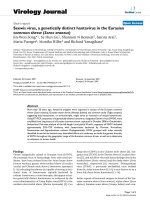
Figure 1
Functional subsets of CD4 T cells. Naïve CD4 T cells - that is,
T cells that have not yet been activated by antigen - circulate in the
blood and lymph until they are activated, usually by dendritic cells,
which are specialized for that function. They then proliferate and
differentiate into different functional subsets, distinguished by the
different cytokines they produce (indicated under each CD4 T cell
type): the cytokines act on other immune cells, activating them in
turn. The four known subsets of CD4 T cells are T
H
1 cells, which
induce infl ammatory responses that protect the tissues; T
H
2 cells,
which are largely responsible for protecting the epithelial surfaces of
the gut, lung and genitourinary system; T
H
17 cells, which produce
early infl ammatory responses; and T
REG
cells, which inhibit the
responses of the other cell types and are thought to provide
protection from autoimmune disease. IFN, interferon; IL, interleukin,
NK, natural killer; TGF, transforming growth factor. Modifi ed from
Figure 5-22 in DeFranco AL, Locksley RM, Robertson M: Immunity:
The Immune Response in Infectious and Infl ammatory Disease.
London: New Science Press; 2007.
T
H
1
IFN-γ
IL-22
IL-17
IL-10,
TGF-β
IL-4, IL-13,
IL-5
T
H
2T
H
17 T
REG
Naïve
CD4 T cell
Activated
macrophages,
NK cells,
CD8 T cells
Neutrophils Dendritic
cells
Eosinophils,
basophils,
mast cells,
alternatively
activated
macrophages
Activation of naïve
T cells by dendritic
cells
61.3
Stockinger: Journal of Biology 2009, 8:61
increase in absolute numbers (rather than percentage) of
T
REG
cells. Thus, it is not yet clear that different AhR
ligands actually induce different T cell types.
This issue is of some practical importance because
induction of T
REG
cells is a significant target of attempts to
suppress autoimmune diseases. For example, it has been
suggested that dioxin will suppress experimental allergic
encephalitis, a widely used model for multiple sclerosis,
through induction of T
REG
cells, a suggestion that is already
resulting in a drive to test AhR ligands for their suppressive
effect in this autoimmune model with a view to future
application in treatment of the disease.
While the role of AhR in the induction of the cytokine
IL-22 is now defined in mice as well as humans, this is
just the beginning. We still know little about the
physiological impact of AhR expression on other cells of
the immune system and the consequences of exposure to
AhR ligands for these. Given the complexity of cellular
interactions that underlie specific immune responses, the
global immune suppression diagnosed after dioxin
exposure is likely to hide a complicated network of
physiological dysregulation. While dioxin may be the
ligand of choice for toxicological studies because of its
potency and slow degradation, which reduces the
likelihood of ‘off-target’ effects, I would argue that off-
target effects may well be an integral feature of a tightly
orchestrated immune response. Immune responses
function in a tight interactive network of cells and media-
tors, and the behavior of dioxin in the immune system
may not reflect direct action on all the cell types that are
thought to be affected, and may indeed mask endogenous
functions of AhR that are prohibited or perturbed by
dioxin. There are multiple physiological ligands for AhR
and it is likely that these bind with varying affinities and
are rapidly degraded by induced metabolizing enzymes,
thus providing transient and tightly regulated stimuli.
Amongst these ligands are dietary components - flavones
and indoles, tryptophan metabolites as well as intrinsic
factors such as prostaglandin subtypes, lipoxin A4,
bilirubin and others [11]. It remains to be seen how such
endogenous AhR ligands influence various compo nents of
the immune system. Unraveling the complex functions of
AhR in the immune system, which may also give clues
towards crosstalk between the immune system, the
neuro endocrine system and the nervous system is more
likely to come from models that use physiological ligands
whose mode of action is under tight regulatory control.
Acknowledgements
I would like to thank Dr Alexandre Potocnik for critical comments on
this article.
References
1 McMillan BJ, Bradfield CA: The aryl hydrocarbon receptor
sans xenobiotics: endogenous function in genetic model
systems. Mol Pharmacol 2007, 72:487-498.
2 Schmidt JV, Su GH, Reddy JK, Simon MC, Bradfield CA:
Characterization of a murine Ahr null allele: involvement of
the Ah receptor in hepatic growth and development. Proc
Natl Acad Sci USA 1996, 93:6731-6736.
3 Fernandez-Salguero PM, Ward JM, Sundberg JP, Gonzalez FJ:
Lesions of aryl-hydrocarbon receptor-deficient mice. Vet
Pathol 1997, 34:605-614.
4 Mimura J, Yamashita K, Nakamura K, Morita M, Takagi TN,
Nakao K, Ema M, Sogawa K, Yasuda M, Katsuki M, Fujii-
Kuriyama Y: Loss of teratogenic response to 2,3,7,8-tetra-
chlorodibenzo-p-dioxin (TCDD) in mice lacking the Ah
(dioxin) receptor. Genes Cells 1997, 2:645-654.
5 Bock KW, Kohle C: Ah receptor: dioxin-mediated toxic
responses as hints to deregulated physiologic functions.
Biochem Pharmacol 2006, 72:393-404.
6 Kerkvliet NI: AHR-mediated immunomodulation: the role of
altered gene transcription. Biochem Pharmacol 2009, 77:
746-760.
7 Esser C: The immune phenotype of AhR null mouse
mutants: not a simple mirror of xenobiotic receptor over-
activation. Biochem Pharmacol 2009, 77:597-607.
8 Frericks M, Meissner M, Esser C: Microarray analysis of the
AHR system: tissue-specific flexibility in signal and target
genes. Toxicol Appl Pharmacol 2007, 220:320-332.
9 Veldhoen M, Hirota K, Westendorf AM, Buer J, Dumoutier L,
Renauld JC, Stockinger B: The aryl hydrocarbon receptor
links TH17-cell-mediated autoimmunity to environmental
toxins. Nature 2008, 453:106-109.
10 Quintana FJ, Basso AS, Iglesias AH, Korn T, Farez MF, Bettelli
E, Caccamo M, Oukka M, Weiner HL: Control of T(reg) and
T(H)17 cell differentiation by the aryl hydrocarbon recep-
tor. Nature 2008, 453:65-71.
11 Denison MS, Nagy SR: Activation of the aryl hydrocarbon
receptor by structurally diverse exogenous and endog-
enous chemicals. Annu Rev Pharmacol Toxicol 2003, 43:309-
334.
Published: 17 August 2009
doi:10.1186/jbiol170
© 2009 BioMed Central Ltd