Báo cáo sinh học: "TBP2 is a general transcription factor specialized for female germ cells" doc
Bạn đang xem bản rút gọn của tài liệu. Xem và tải ngay bản đầy đủ của tài liệu tại đây (416.28 KB, 4 trang )
Müller and Tora: Journal of Biology 2009, 8:97
Abstract
The complexity of the core promoter transcription machinery
has emerged as an additional level of transcription regulation
that is used during vertebrate development. Recent studies,
includ ing one published in BMC Biology, provide mechanistic
in sights into how the TATA binding protein (TBP) and its
vertebrate-specific paralog TBP2 (TRF3) switch function during
the transition from the oocyte to the embryo.
See research article />Regulation of initiation of transcription by RNA poly-
merase II (Pol II) is central to any developmental
process. A key regulatory step in eukaryotic transcription
initiation is the assembly of basal transcription
apparatus at the core promoter. This regulatory step has
been brought into the spotlight by the discovery of
multiple promoter binding factors that assemble into
different basal transcription factor complexes. These
complexes have to be matched in the future to the
diversity of core promoter types and features [1]. This
apparent diversity points towards a dynamic regulatory
role for this machinery [1], which is very poorly
understood.
The preinitiation complex includes the core promoter, Pol
II and the general transcription factors TFIIA, B, D, E, F
and H. Originally, the core promoter recognition factor
TFIID, which is composed of TATA-binding protein
(TBP) and 14 TBP-associated factors (TAFs), was thought
to be ubiquitous. Functional and genetic studies revealed
that TBP is not exclusively required for all protein-coding
gene transcrip tion in vertebrates [2]. In line with genetic
observations, biochemical analyses revealed the existence
of alternative initiation complexes that have been
suggested to replace TFIID in several in vivo and in vitro
systems [1-3]. The diversity in the components of
transcription initiation machinery prompts the questions
of why this diversity is present in metazoans and how the
various initiation complexes act in parallel in a cell or the
multicellular organism.
TBP has a crucial role in preinitiation complex assembly:
nucleating the binding of TFIID to promoters. However, it
is a member of a protein family, and other members of the
TBP family, such as TBP-like factor (TLF or TBPL1/TRF2/
TRP) and TBP2 (or TRF3/TBPL2), have been shown to
substitute for TBP to mediate Pol II and Pol III trans crip-
tion. TBP2 is a vertebrate-specific paralog of TBP, with
much higher similarity to TBP than TLF (TBP2 is about
90% similar to TBP in its core domain). Consistent with
this similarity, TBP2 can bind the TATA box, to interact
with the other general transcription factors TFIIA and
TFIIB and mediate Pol II transcription initiation in vitro,
just as TBP can [3,4]. These properties of TBP2 suggest a
function complementary to that of TBP and raise the
question of whether TBP and TBP2 are functional equiva-
lents or carry out specialized functions. Thus, given the
high level of similarity in biochemical properties between
TBP and TBP2, the cause and mechanism for the retention
of TBP2 following gene duplication remains to be explained.
Two recent publications studying TBP2 function in frogs
[5] and mice [6] provide some answers to this intriguing
problem.
Replacement of TBP by TBP2 in Xenopus
oocyte transcription
The transition from maternal to zygotic gene activation in
the embryo has been a tractable and informative model
system for studying the function of TBP family proteins in
vertebrate ontogeny. Knockdown studies in Xenopus and
zebrafish embryos showed that TBP and TBP2 are both
indispensable for embryonic development and are both
required for activation of zygotic genes [4,7]. Unexpectedly,
TBP2 was shown to have a specialized role restricted to the
ventral side of the embryo [4,7] and in hematopoiesis [8].
However, these results did not shed light on why TBP2
seemed to be mostly expressed in the female gonad in frogs
and why only a low level of expression was detected in frog
and fish embryos [4,5,7]. The apparent enrichment for
TBP2 in the female gonad contrasted with opposing
dynamics of TBP, suggesting a general feature for specific
activity of TBP2 in the ovary in anamniotes [4,7].
Minireview
TBP2 is a general transcription factor specialized for female germ
cells
Ferenc Müller* and Làszlò Tora
†
Addresses: *Department of Medical and Molecular Genetics, School of Clinical and Experimental Medicine, College of Medical and Dental
Sciences, University of Birmingham, Birmingham B15 2TT, UK.
†
Department of Functional Genomics, Institut de Génétique et de Biologie
Moléculaire et Cellulaire (IGBMC), UMR 7104 CNRS, UdS, INSERM U964, BP 10142, F-67404 Illkirch Cedex, France.
Correspondence: Ferenc Müller. Email: Làszlò Tora. Email:
97.2
Müller and Tora: Journal of Biology 2009, 8:97
To address the function of TBP2, in their recent BMC
Biology paper Akhtar and Veenstra [5] have investigated
the role of TBP2 in oocyte transcription and compared it
with that of TBP. They show that Xenopus oocytes lack
TBP protein, whereas TBP2 is the major TBP-type factor in
the germ cells. Later, in eggs and early embryos, TBP2
levels decrease, whereas TBP starts to accumulate after
meiotic maturation and during cleavage stages of develop-
ment (Figure 1). A major role for TBP2 in oocyte trans-
cription was suggested by the observation that TBP2 is
recruited to the transcriptionally active loops of the expanded
‘lampbrush’ chromosomes found in frog oocytes. The
authors [5] exploited an overexpression system to show
that in oocytes TBP2 is recruited to transcribed promoters
together with Pol II. In addition, the authors [5] show that
TBP2 is also recruited to Pol III promoters, further
suggesting that TBP2 probably replaces TBP in mediating
transcription by all three RNA polymerases.
The results described by Akhtar and Veenstra [5] highlight
the functional significance of the apparent differentially
available pool of TBP and TBP2 in oocytes and provide
mechanistic insights into the dynamics of TBP and TBP2
protein. At the end of oocyte maturation TBP2 is degraded
and transcription is globally repressed. By analyzing
whether TBP2 degradation is directly linked to this change
in general transcription, they show [5] that the repression
is established by the germinal vesicle breakdown stage of
oocyte development, a stage at which TBP2 degradation
has already started but has not reached its maximum.
Thus, it seems that it is not TBP2 degradation, but rather a
loss of association of TBP2 with promoters, that coincides
with transcriptional repression during meiotic maturation.
This argues against a direct role for TBP2 degradation in
the global shutdown of transcription during oocyte
matura tion. This conclusion, together with the observed
reduction of TBP2 and enrichment of TBP in embryos,
implies that the primary role of TBP2 degradation is to
facilitate factor switching and transcriptional regulation
during subsequent development (Figure 1).
The hypothesis of factor switching during the transition
from maternal gene activity is consistent with a series of
observations made in several vertebrate models ([2] and
references therein). The picture emerging from the study
by Akhtar and Veenstra [5] is that post-transcriptional
regulation of TBP and TBP2 is important for regulating
steady-state levels of TBP paralogs in frogs. This regulation
results in striking differences in protein availability, and
suggests a model for subfunctionalization (division of
functions) of TBP paralogs between oocytes and embryos.
A requirement for mouse TBP2 in female
germ cell development
The conclusions drawn from studies in frog oocytes [5]
match those stemming from expression analysis and recent
genetic loss-of-function studies carried out in mice
[6,9,10]. In mice, the expression of Tbp2 mRNA has been
detected specifically in the oocytes [4,9]. Although there is
controversy regarding the specificity of this expression, a
recent genetic study indicated that the main role of TBP2 is
restricted to the female germline ([3,10] and references
therein). TBP2 protein accumulates in the nuclei of
growing mouse oocytes during folliculogenesis, and its
level declines on ovulation to become undetectable after
fertilization [10] (Figure 1). In contrast, TBP is expressed
in the oocytes only at the beginning of folliculogenesis and
after fertilization, but not during oocyte growth [10]
(Figure 1).
Consistent with a specific expression in the ovary, Tbp2
-/-
mice are viable and show no obvious phenotype [6].
However, females lacking TBP2 are sterile as a result of
defective folliculogenesis. Tbp2
-/-
females lack fully grown
germinal-vesicle-stage oocytes and Pol II transcription is
perturbed mainly at the primary follicle stage, when wild-
type oocytes show extensive transcriptional activity. A
general decrease in transcription is indicated by the
reduced phosphorylation on serine 2 of Pol II and reduced
methylation of histone H3 lysine 4, which are markers of
active genes. Consequently, a significant number of oocyte-
specific genes are severely deregulated in Tbp2
-/-
females.
In agreement with the idea that TBP2 is the sole TBP-type
factor in oocytes, TBP is dispensable for correct oocyte
maturation and fertilization. In contrast, when TBP2 is
misexpressed in early mouse embryos, where it is normally
not expressed, it has a negative effect on cell proliferation,
leading to developmental arrest [6]. These data together
demonstrate that TBP2 is not required for mouse viability
but has a critical and specialized role in mammalian female
germ cell development, and they provide evidence for non-
redundant functions of TBP2 and TBP in vivo in the
mouse.
TBP2 as a vertebrate oocyte-specific TBP-
type factor
The observations from frogs and mice [5,6] clearly
establish TBP2 as an oocyte-specific TBP-type factor in
vertebrates. In both organisms during certain stages of
oocyte development, TBP is absent and dispensable (Figure 1).
Thus, the unique role of TBP2 in oocyte transcription, in a
highly specialized cell type, provides evidence that the
basal transcription machinery is highly flexible and can
switch factors depending on the cellular and ontogenic
requirements.
A common model for subfunctionalization of TBP and
TBP2 during the transition between oocyte and embryo is
thus emerging from two evolutionarily distant vertebrates,
although there remain important lineage-specific differ-
ences between them. In anamniotes, TBP2 proteins mostly
(although not completely) degrade before the embryo is
97.3
Müller and Tora: Journal of Biology 2009, 8:97
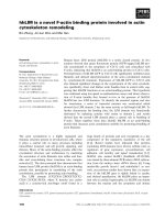
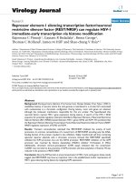
Figure 1
Regulation of TBP and TBP2 during oogenesis and the early stages of embryogenesis in vertebrates. Continuous line, frog; dashed line,
mouse; red, TBP2; blue, TBP; green, general transcription. Stages are represented at the top by light to dark shading, and at the bottom by
schematic representations. At most stages of oogenesis only TBP2 is expressed, which promotes oocyte-specific transcription during these
stages. Upon meiotic maturation, TBP2 is actively degraded following global repression of transcription in maturing oocytes (as has been
demonstrated in frogs). After fertilization and during the early stages of embryogenesis, TBP expression reaches the maximum levels that
are needed to start zygotic transcription. In frogs zygotic transcription is largely delayed until the mid blastula and this process is regulated by
late translation of maternal stores of tbp mRNA. In frog (and zebrafish) there are low levels of TBP2 during early stages of embryogenesis,
whereas in mice no TBP2 has been detected during embryogenesis. Global zygotic transcription initiation is delayed in both frog and mouse,
albeit to different developmental stages, and trace levels of zygotic transcription have been detected in both species before global genome
activation. The figure has been generated by summarizing experiments described in [4-7,9,10].
Frog
Mouse
Frog
Mouse
Fertilized egg/
zygote
Oogenesis Meiotic
maturation
Zygotic
gene expression
4000 cells
2 cells
Global activation
Translational
regulation
TBP2
TBP
Transcription
Global repression
Proteolytic
degradation
97.4
Müller and Tora: Journal of Biology 2009, 8:97
formed [5]. In contrast to mammals, a large amount of TBP
mRNA is produced maternally and seems to be prevented
from being translated in the oocyte and the early embryo.
To achieve factor switching, the maternal TBP mRNA
trans lation is activated before global zygotic gene activa-
tion to generate an abundant pool of TBP protein, thereby
becoming the dominant factor in the embryo.
The question remains: why is there a distinct requirement
for either of the two TBP paralogs in oocytes and embryos?
The high level of divergence of the amino termini between
TBP2 and TBP may hold the key to this question. One
possibility would be that the amino-terminal domain of
TBP2 could determine the association of TBP2 with a
special set of TAFs and/or other oocyte-specific factors
that, in turn, would confer the oocyte-specific core promoter
binding function to a non-canonical TFIID complex. Thus,
a specialized TBP2-containing TFIID-like complex could
act to mediate transcription from oocyte-specific genes
and, in contrast to TBP, could inhibit cell cycle regulatory
genes. Alternatively, the amino-terminal domain of TBP2
could function to regulate the DNA binding function of the
carboxy-terminal domain, or to regulate protein dynamics,
which as suggested by Akhtar and Veenstra [5] involves
regulation of protein degradation.
In summary, a protein very similar to TBP seems to have
evolved by gene duplication and has a non-redundant
regulatory function in transcription initiation in the verte-
brate oocyte. Further investigations are required to address
how TBP2 functions in the oocyte and what specific pro per-
ties and molecular mechanisms of transcription initiation
distinguish the oocyte from the soma and the embryo.
Acknowledgements
We thank S Bour for the illustration and ME Torres-Padilla for
critically reading the manuscript. We apologize to colleagues whose
work could not be cited owing to space and reference limitations
and was only covered by reviews instead. This work was supported
by a EUTRACC grant (LSHG-CT-2007-037445).
References
1. Juven-Gershon T, Kadonaga JT: Regulation of gene expres-
sion via the core promoter and the basal transcriptional
machinery. Dev Biol 2009, [Epub ahead of print].
2. Jones KA: Transcription strategies in terminally differenti-
ated cells: shaken to the core. Genes Dev 2007, 21:2113-
2117.
3. Deato MD, Marr MT, Sottero T, Inouye C, Hu P, Tjian R: MyoD
targets TAF3/TRF3 to activate myogenin transcription. Mol
Cell 2008, 32:96-105.
4. Bartfai R, Balduf C, Hilton T, Rathmann Y, Hadzhiev Y, Tora L,
Orban L, Muller F: TBP2, a vertebrate-specific member of
the TBP family, is required in embryonic development of
zebrafish. Curr Biol 2004, 14:593-598.
5. Akhtar W, Veenstra GJ: TBP2 is a substitute for TBP in
Xenopus oocyte transcription. BMC Biol 2009, 7:45.
6. Gazdag E, Santenard A, Ziegler-Birling C, Altobelli G, Poch O,
Tora L, Torres-Padilla ME: TBP2 is essential for germ cell
development by regulating transcription and chromatin
condensation in the oocyte. Genes Dev 2009, 23:2210-2223.
7. Jallow Z, Jacobi UG, Weeks DL, Dawid IB, Veenstra GJ:
Specialized and redundant roles of TBP and a vertebrate-
specific TBP paralog in embryonic gene regulation in
Xenopus. Proc Natl Acad Sci USA 2004, 101:13525-13530.
8. Hart DO, Raha T, Lawson ND, Green MR: Initiation of
zebrafish haematopoiesis by the TATA-box-binding pro-
tein-related factor Trf3. Nature 2007, 450:1082-1085.
9. Xiao L, Kim M, DeJong J: Developmental and cell type-spe-
cific regulation of core promoter transcription factors in
germ cells of frogs and mice. Gene Expr Patterns 2006, 6:
409-419.
10. Gazdag E, Rajkovic A, Torres-Padilla ME, Tora L: Analysis of
TATA-binding protein 2 (TBP2) and TBP expression sug-
gests different roles for the two proteins in regulation of
gene expression during oogenesis and early mouse devel-
opment. Reproduction 2007, 134:51-62.
Published: 30 November 2009
doi:10.1186/jbiol196
© 2009 BioMed Central Ltd