Báo cáo sinh học: "Adaptations of proteins to cellular and subcellular pH" pdf
Bạn đang xem bản rút gọn của tài liệu. Xem và tải ngay bản đầy đủ của tài liệu tại đây (215.94 KB, 4 trang )
Garcia-Moreno: Journal of Biology 2009, 8:98
Abstract
Bioinformatics-based searches for correlations between sub-
cellular localization and pI or charge distribution of proteins have
failed to detect meaningful correlations. Recent work pub lished
in BMC Biology finds that a physicochemical metric of charge
distribution correlates better with subcellular pH than does pI.
See research article />The need for tight regulation of intracellular pH is one of
the most important and constant organizing principles of
living systems. It is essential because the fundamental
energy transduction machinery of cells runs mainly on H
+
gradients and proton-coupled electron transfer reactions.
It is also necessary because pH determines the charge state
of weak acids and bases (for example, side chains of Asp,
Glu, His, Lys and Arg, and so on), and the charge state in
turn affects many physical and physiological properties of
bio logical molecules, especially proteins and RNAs (Figure 1).
Regulation of intracellular pH is so central to the living
state that even the most primitive cells capable of energy
transduction must have been able to control intracellular
pH by expelling H
+
produced from the hydrolysis of
organic compounds.
How tightly is pH regulated in cells and organisms? Consider
that the normal pH of human arterial blood is 7.40.
Depression below pH 7.35 leads to acidosis, a condition that
requires medical attention, and elevation to pH 7.50 results in
alkalosis, responsible for the unpleasant symp toms of altitude
sickness. Similarly small changes of 0.1 pH units in
intracellular pH or within subcellular compart ments can have
physiological consequences. What is remark able is that
despite the need for very tight regulation, the pH in various
cellular compartments varies significantly. In organs such as
the human stomach, it can be extreme.
In cells, pH is nearly neutral in the cytoplasm, in the endo-
plasmic reticulum and in mitochondria. It is more acidic in
vacuoles, lysosomes (as low as pH 5) and in the Golgi. It is
more basic in the nucleus and in peroxisomes (as high as
pH 8) [1]. Proteins can harness these differences in cellular
and subcellular pH for physiological purposes. For exam-
ple, the influenza virus requires exposure to the slightly
acidic conditions in the lysosome to become activated [2].
Human hemoglobin acts similarly as a pH sensor that
targets exercised tissue for delivery of oxygen by
responding to local acidity [2].
The tight regulation of cellular and subcellular pH might
imply that charged residues of proteins have been tailored
for structural or functional purposes under specific
conditions of pH. For example, the isoelectric point (pI) of
proteins, which describes the balance between acidic and
basic residues, might have co-evolved with the pH of the
organelles in which they exist to enable or to optimize
function [3]. Learning to recognize these adaptations
would be useful for the annotation of proteomes and for
understanding protein function and evolution. Previous
bioinformatics-based searches for correlations between
subcellular localization and pI or charge distribution have
yielded interesting nuggets but have failed to detect a
meaningful correlation [4]. The problem is that the pI is a
relatively insensitive global metric of the number and types
of ionizable groups; the distribution of pI values in a
proteome is inherently bimodal owing to the normal
differences in the pK
a
(the acid dissociation constant) of
the dominant acidic (Asp and Glu) and basic (Lys and Arg)
residues [4,5]. Now Chan and Warwicker [5] have
examined the correlation of intracellular pH with
calculated electrostatic contributions to stability. Using
this more physical metric of charge distribution, they
found that the average pH of maximal stability for proteins
in a subcellular compartment correlates better with sub-
cellular pH than does pI. The properties of histidine
residues underlie this correlation.
Effects of charge distribution on properties of
proteins
Identifying further meaningful correlations between
subcellular localization or subcellular pH with the number,
types, distribution and properties of ionizable groups in
proteins, starting from either sequence or structure, will be
interesting but challenging. The problem is that the charge
properties of proteins have evolved under pressure to
satisfy a large number of physical and biological con-
straints (Figure 1). Trying to identify trends or adaptations
by focus ing on one or two among the many constraints is
akin to examining a piece of music through statistical
analysis of musical notes or their values (that is, their
duration), ignoring all other attributes of the music.
Averaged over their entire oeuvre, the works of Bach and
Minireview
Adaptations of proteins to cellular and subcellular pH
Bertrand Garcia-Moreno
Address: Department of Biophysics, Johns Hopkins University, Baltimore, MD 21218, USA. Email:
98.2
Garcia-Moreno: Journal of Biology 2009, 8:98
Beethoven might indeed show statistically significant
differences in their predilection for some notes, but it is
unlikely that this will be reflected in any individual piece,
that the level of discrimination will be sufficient to identify
a composer from among a list of hundreds, or that this
approach will improve our understanding or appreciation
of music. A meaningful search for adaptations of proteins
to sub cellular pH should consider simultaneously some of
the following structural, biological and solution properties
of proteins, all of which are influenced by charged residues.
Spatial localization
The distribution of charges on a protein can be biased by
the protein’s location within a subcellular compartment.
For example, membrane proteins tend to be more basic
than cytosolic ones [4]. This might be an adaptation to
facilitate interactions between positive charges on the
protein and the predominantly negatively charged polar
head groups of membrane phospholipids. However, to
illustrate how deceptive general trends can be, consider
that some proteins that interact strongly and irreversibly
with membranes actually use Ca
2+
bridges that require
post-translational modifications that add negative charges
to the proteins [2].
Stability
The pH dependence of stability of proteins is governed by
differences in pK
a
values of ionizable groups in folded and
unfolded forms. Using structure-based continuum electro-
statics calculations, Alexov [6] showed previously that the
pI and the pH optimum for stability can be quite different.
Some proteins, such as proteases in the lysosome, are
clearly adapted for maximal stability and activity in the
relatively low pH of this compartment. Chan and Warwicker
[5] show that this correlation of the pH of maximal stability
with intracellular pH is only evident when the properties of
many proteins within a subcellular compartment are
averaged. Their study illustrates the power of quantitative
physicochemical approaches in the analysis of proteomes.
The effectiveness of this approach is also illustrated by
other studies of charge contributions to the stability of
thermophilic proteins. Proteins from thermophilic
organisms usually have a higher number of charges and
ion pairs than their mesophilic homologs [7]. An analysis
of the number and types of charges would conclude that
charges in thermophilic proteins were selected to enhance
the stability of proteins through Coulomb interactions.
However, a structure-based study of electrostatic contribu-
tions to stability using a physicochemical model failed to
detect any correlation between the excess charges in
thermophilic proteins and increased stability [7].
Solubility
Solubility is a critical factor in the evolution of protein
sequences and folds [8]. In general, charges in globular
proteins are surrounded by charges of the opposite sign
[9]. This would seem to reflect evolutionary tuning of
surface charges to maximize stabilizing Coulomb inter-
actions in the folded state, but it is more likely to be an
adaptation to enhance solubility. Charges affect the solu-
bility of proteins; therefore, the adaptation of charges to
tune any property of a protein for a specific subcellular pH
could affect solubility. Because there is no theory of protein
solubility, it is impossible to determine how a specific
distribution of charges affects solubility. In general,
solubility correlates with pI (proteins tend to be least
soluble at their pI); therefore, given the lack of correlation
between pI and subcellular pH, solubility probably does
not correlate well with subcellular pH. A large number of
ionizable residues, especially basic ones, can enhance the
solubility of a protein. However, unexpected factors can
also influence solubility. For example, the solubility of
folded proteins is affected by stability; a large energy gap
between the fully folded, soluble form and partially
unfolded, less soluble forms is needed to prevent popu-
lation of aggregation-prone states. Solubility can in fact be
affected by single mutations that do not alter the number
of charges in the protein.
Interactions
Charges are essential both to prevent and to stabilize
complexes of proteins with macromolecules and with small
molecules. A correlation has been found between the pH
optimum of stability of monomeric proteins and of their
complexes [10]. This suggests that pH-dependent proper-
ties of the monomers and of the complexes coevolved at
the same pH. The presence of a specific constellation of
charges for functional interactions can bias the distribution
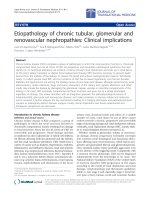
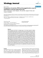
Figure 1
The distribution of ionizable residues (for example, the side chains
of Arg, Lys, His, Asp and Glu) in proteins might reflect adaptations
to cellular and intracellular pH. The search for these structural
adaptations should consider the important roles of charged residues
on the many different functional, structural, physical and biological
factors that influenced the evolution of proteins, as indicated here.
Arg
Localization
within cell
Stability
Solubility
Interactions
Lys
GluAsp
His
Minimizing local
fluctuations in pH
Enzymatic
activity
Sensitivity to
temperature,
pressure and ions
Buffering
98.3
Garcia-Moreno: Journal of Biology 2009, 8:98
and properties of charges in proteins. For example, regions
of high density of positive charge are usually found in the
faces of proteins that interact with nucleic acids. Any
correlation between nuclear pH and the distribution of
charges in nuclear proteins in a eukaryotic cell is likely to be
a secondary consequence of these functional adaptations.
Enzymatic activity
The ionizable groups that control enzymatic catalysis
usually titrate with highly perturbed pK
a
values tuned for
catalysis under the appropriate pH conditions. Just one of
these groups with perturbed pK
a
can have a dramatic
influence on the pH dependence of protein stability [11].
This might be the reason that the pH optimum of
enzymatic reactions is, in general, not correlated with the
pH of maximum stability. The adaptation of some proteins
to subcellular pH might involve tuning of the pK
a
values of
active-site residues. There are no computational tools that
reproduce the properties of internal ionizable groups
accurately, and so this essential adaptation to subcellular
pH cannot yet be examined quantitatively.
Environmental conditions
Previous attempts to identify protein adaptations to
subcellular pH have not considered that charges and their
contribution to stability, solubility, dynamics, conforma-
tion, function, and so on are sensitive to physical variables
(temperature, pressure), and to the chemical composition
of their milieu (osmolytes, ionic composition, metabolites).
Chan and Warwicker [5] point out that differences in the
distribution of charges in proteins from extremophiles and
mesophiles illustrate the important roles of charges in
proteins for adaptation to specific environmental
conditions. The influence of the ionic milieu on adaptations
to intracellular pH deserves special mention because
subcellular pH is coupled to ion homeostasis (that is,
changes in intracellular pH are coupled to changes in
concentration of other ions). Protein adaptations to unique
ionic environments are already known. For example,
hemoglobin, which is regulated physiologically by the large
anion 2,3-bis-phosphglycerate, has a cleft with a high
concentration of positively charged residues where the
di-anion binds [2]. Another example is that of extracellular
proteins or proteins that exist in vacuoles or other
compartments that can have high Ca
2+
concentrations.
These proteins might be expected to be more acidic to
maximize interactions of Asp and Glu residues with Ca
2+
to
enhance stability or for other purposes. Attempts to
identify protein adaptations to subcellular pH should
consider how the ionic milieu characteristic of a subcellular
compartment might have influenced these adaptations.
Buffer capacity
The regulation of subcellular pH is partly achieved through
the buffer capacity of metabolites and macromolecules.
This is the reason that a correlation of protein pI with
subcellular pH has been sought. However, the ability of a
protein to act as a buffer in a subcellular environment will
depend not just on the number of ionizable groups and
their pK
a
values, but also on the concentration of protein in
the compartment. A meaningful correlation between pI
and subcellular pH might yet be found if protein
concentration and compartment volume were taken into
consideration.
Biological considerations
The distribution of charges in proteins can be biased by a
number of biological factors that are not linked to intra-
cellular pH in an obvious way. Evolutionary history,
mutational bias, AT nucleotide bias, level of gene expres-
sion and optimization of translational efficiency are some
factors that will have to be examined closely.
Physicochemical considerations
Chan and Warwicker [5] have raised other interesting
issues that will have to be considered when searching for
protein adaptations to subcellular pH. The adaptations
might not be tuned for function in the steady state
intracellular pH, but rather to the transient changes in pH
that might be experienced in an intracellular compartment.
Chan and Warwicker also made the interesting suggestion
that the dependence of the pH of maximum stability on
subcellular pH might reflect the need to minimize
spontaneous fluctuations in H
+
concentration within a
compartment [5]. There is clear need for a more detailed
inventory of H
+
in subcellular compartments based on the
known number and calculated pK
a
of weak acids and bases,
and on emerging data of protein localization and concen-
trations. There is also need for improved understanding of
the diffusion properties of protons in the osmotically
complex intracellular environment, where there is no water
that is not under the influence of solute, and where,
formally speaking, the concept of pH is not valid.
Conclusions
Many examples of regulatory adaptations of proteins for
stability or function at a specific pH are known. If charges
in proteins have indeed been optimized for specific
functional purposes, quite possibly they also display struc-
tural adaptations to specific subcellular conditions of pH
and ionic composition. These adaptations will not be easily
identified from bioinformatics analysis of proteomes using
global metrics of charge distribution (such as pI). Although
in general, the mean properties of proteins might be less
informative than the deviations from the mean, in practice
the only correlations that have been found with intra-
cellular pH are with electrostatic properties calculated with
physical models, averaged over many different types of
proteins in a given subcellular compartment. To identify
further adaptations of proteins to subcellular pH it will be
useful to analyze proteomes with physicochemical models,
and to consider simultaneously many of the physical and
98.4
Garcia-Moreno: Journal of Biology 2009, 8:98
biological constraints that guided the evolution and the
adap tation of proteins to the pH and ionic properties of
their physiological milieu. This more integrative and
physical approach might begin to reveal how different
elements are combined in harmony to constitute the
symphony of the cell.
References
1. Demaurex N: pH homeostasis of cellular organelles. News
Physiol Sci 2002, 17:1-5.
2. Voet D, Voet JG: Biochemistry. 3rd edn. New York, Wiley; 2004.
3. Brett CL, Donowitz M, Rao R: Does the proteome encode
organelle pH. FEBS Lett 2006, 580:717-719.
4. Schwartz R, Ting CS, King J: Whole proteome pI values cor-
relate with subcellular localizations of protein for organ-
isms within the three domains of life. Genome Res 2001,
11: 703-709.
5. Chan P, Warwicker J: Evidence for the adaptation of protein
pH-dependence to subcellular pH. BMC Biol 2009, 7:69.
6. Alexov E: Numerical calculations of the pH of maximal
protein stability. Eur J Biochem 2004, 271:173-185.
7. Greaves RB, Warwicker J: Mechanisms of stabilization and
the maintenance of solubility in proteins from ther-
mophiles. BMC Struct Biol 2007, 7:18.
8. Niwa T, Ying B, Saito K, Jin W, Takada S, Ueda T, Taguchi H:
Bimodal protein solubility distribution revealed by an
aggregation analysis of the entire ensemble of Escherichia
coli proteins. Proc Natl Acad Sci USA 2009, 106:4201-4206.
9. Wada A, Nakamura H: Nature of the charge distribution in
proteins. Nature 1981, 293:757-758.
10. Kundrotas PJ, Alexov E: Electrostatic properties of protein-
protein complexes. Biophys J 2006, 91:1724-1736.
11. Isom, DG, Cannon BR, Castaneda CA, Robinson A, Garcia-
Moreno EB: High tolerance for ionizable groups in the
hydrophobic interior of proteins. Proc Natl Acad Sci USA
2008, 105:17784-17788.
Published: 2 December 2009
doi:10.1186/jbiol199
© 2009 BioMed Central Ltd