Báo cáo khoa học: "Translocational changes of localization of synapsin in axonal sprouts of regenerating rat sciatic nerves after ligation crush injury" pptx
Bạn đang xem bản rút gọn của tài liệu. Xem và tải ngay bản đầy đủ của tài liệu tại đây (234.87 KB, 9 trang )
-2851$/# 2)
9HWHULQDU\#
6FLHQFH
J. Vet. Sci. (2000),1(1), 1–9
Translocational changes of localization of synapsin in axonal sprouts of
regenerating rat sciatic nerves after ligation crush injury
Ku-birm Kwon, Jin-suk Kim
1
, Byung-joon Chang*
Department of Anatomy and Histology,
1
Department of Pharmacology and Toxicology, College of Veterinary Medicine, Konkuk
University, Seoul 143-701, Korea
Time-dependent translocational changes of Synapsin I
(SyI), a synaptic vesicle-associated phosphoprotein and its
involvement in the axonal transport were investigated in
the regenerating axonal sprouts. A weak SyI
immunoreactivity (IR) was found in the axoplasm of
normal axons. Rat sciatic nerves were crush-injured by
ligating with 1-0 silk thread at the mid-thigh level and
released from the ligation 24 h later. At various times after
release, immunocytochemistry was performed. SyI was
translocated from the proximal to the distal site of ligation
and also involved in the sprouting of regenerating axons.
The distribution patterns of SyI IR were changed in the
crush-injured nerves. SyI immunoreactive thin processes
were strongly appeared in the proximal region from 1 h
after release. After 3 h, a very strong IR was expressed.
The intense SyI immunoreactive thin processes were
elongated distally and were changed the distribution
pattern by time-lapse. After 12 h, strong immunoreactive
processes were extended to the ligation crush site. At 1
day, a very intense IR was expressed. At 2 days,
immunoreactive thin processes extended into the distal
region over the ligation crush site and strong IR was
observed after 3 days. SyI was accumulated in the
proximal region at the early phases after release. These
results suggest that SyI may be related to the
translocation of vesicles to the elongated membranes by a
fast axonal transport in the regenerating sprouts.
Key words:
sciatic nerve, regeneration, immunocytochemis-
try, Synapsin I.
Introduction
Illustration of the transported substances and the
mechanisms involved in the regeneration of the peripheral
nervous system (PNS) may be a great help to cure
peripheral nerve injuries and demyelinating diseases [26].
There is a dichotomy between the PNS and the central
nervous system (CNS) in their ability to regenerate
[1, 26, 27, 35]. The injured CNS neurons can not be
regenerated, whereas the injured PNS neurons can be
regenerated by reestablishing synaptic connections, thereby
resulting in recovering theis functions. The ability of nerve
regeneration may be attributed to the structural differences
in the cellular organization between the PNS and the CNS
[26-30, 34]. The CNS has no basal lamina of axons, while
the PNS has the basal lamina of Schwann cells which
plays an important role in the nerve regeneration [28].
Each fiber of both myelinated and unmyelinated peripheral
nerves is lying within a continuous basal lamina tube. A
sprout, the early axolemmal extension from the parent
axon, extends through the space between the basal lamina
and the myelin sheath. It has been reported that the sprout
formation was found as early as 5 h after injury [27]. The
node can produce multiple sprouts and these sprouts
appear to be regenerating axons in the proximal stump and
grow to the distal stump as a growth cone. Newly
synthesized membrane proteins were added to the axonal
growth cones. In the growth cone, materials and
membrane vesicles were preferentially provided for the
axonal growth [11].
SyI, the collective name for Synapsin Ia and Ib, is a
phosphoprotein associated with synaptic vesicles in the
nerve terminal [13, 15-17]. There are a slight difference in
molecular weight (MW) between synapsin Ia and Ib
[13,15-17]. The MW of Sy Ia is 86 kDa, and that of SyIb is
80 kDa. SyI is present only in the nerve terminals. Within
the terminals, it is associated with small synaptic vesicles
[5]. It is present virtually in all synapses [13, 15, 16] and
appears simultaneously with synapse formation during
development [23]. It is a peripheral protein of the
cytoplasmic surface of the vesicle. SyI acts as a link
protein between the vesicle and cytoskeletal matrix of the
terminal. Therefore, it seems like to connect synaptic
*Corresponding author
Phone: 82-2-450-3711; Fax: 82-2-3437-3661
E-mail:
2 Ku-birm Kwon et al.
vesicles and anchor them to the cytoskeletons in the
presynaptic terminals. SyI plays a regulatory role in
neurotransmitter release [14, 23]. In mature neurons, SyI is
concentrated almost exclusively in the presynaptic
terminals [9, 10, 23, 24] and colocalized with SV48 [9]
and synaptotagmin [10]. The essential function of the
synapsins is regulating the traffic of synaptic vesicles [33].
SyI and 200 kDa neurofilament protein, an axonal marker,
were colocalized. These indicate that SyI may be involved
in the axonal elongation [1, 2]. Immunocytochemical
studies on rat brain demonstrated that SyI IR is specifically
associated with the neuronal cytoskeleton as well as the
synaptic vesicles. Thus, SyI may play a critical role in the
dynamics of the cytoskeletal functions and the
cytoskeleton-membrane interactions [19].
Accumulation of transported materials can be studied at
a focal block of axonal transport caused by sever, crush
and cold block or ligature. Because we thought pools of
SyI may travel at a fast rate, the pattern of anterograde
transport of SyI was investigated in the regenerating
peripheral nerves. We investigated the time-dependent
translocational changes of SyI by axonal transport after
ligation crush injury and the involvement of SyI in
vesicular transport of membrane elongation in the sciatic
nerve regeneration.
Materials and Methods
Experimental animals
Forty-eight adult male Sprague-Dawley rats, weighing
250~300 g and aged 12~18 weeks, were used in this study.
Feed and water were provided ad libitum.
Operation procedures
The animals were slightly anesthetized with diethyl ether
and then deeply anesthetized by a mixture of Ketamine
(Ketara
®
: 50 mg/kg I.M.) and Xylazine (Rompun
®
: 10 mg/
kg I.M.) or pentobarbital sodium (Entobar
®
: 60 mg/kg I.P.).
Left sciatic nerves were exposed at the mid-thigh level
through the sciatic notch and simply crushed by a strong
ligation with 1-0 silk thread [1, 7, 8, 21, 30, 34]. No
ribbon-shaped ligation was performed to avoid rejection
reaction of surrounding tissues [8]. After 1 day, the rats
were anesthetized again as the saml way and the nerves
were released from the ligation. Skin was resutured
without sideways tearing. No infectious signs were found
in the animals operated by this way. Preliminary
experiments [28] showed that there was no difference in
the results obtained from animals operated either sterilely
or non-sterilely.
Preparation of tissues
Immediately, at 1h, 2h, 3h, 5h, 6h, 8h, 12h, 18h, 1 day,
2 days, 3 days, 4 days, 5 days, 7 days, and 14 days after
release, the animals were deeply anesthetized with diethyl
ether. Using a probe-ended stainless steel gastric tube, the
rats were slowly perfused transcardially with vascular
rinse solution, followed by 4% paraformaldehyde in 0.1 M
PBS (pH 7.4) [32]. After perfusion, the left sciatic nerve
segments including ligated area were excised and
postfixed in the same fixative for 4 h at 4
o
C. After washing
in 0.1 M PBS for 1 h, the nerve segments were infiltrated
by increasing phosphate buffered sucrose solution for
cryoprotection; 10% for 1 day, 20% for 1 day, and finally
30% for 3 days at 4
o
C. The sunk nerve segments were
excised and quickly frozen by a snap-freezing in the
isopentane inside of the liquid nitrogen bottle for 10 min.
The nerve segments were embedded in the tissue freezing
embedding medium, frozen rapidly, and sectioned
longitudinally with a 10-
µ
m thickness by cryosection. The
sections were thaw-mounted on the prepared gelatin-
coated slide glasses [32] and air-dried for 1 day at room
temperature.
Light microscopic ABC immunocytochemistry
After air-dried for 1 day at room temperature, sections
were washed three times in 0.1 M PBS for 10 min. The
sciatic nerve segment sections were transferred into buffer
A (0.1 M PBS with 1% BSA and 0.2% saponin) including
0.5% H
2
O
2
for 30 min to block endogenous peroxidase
activity. The sections were rinsed with 0.05 M glycine in
buffer A for 10 min. After washing with buffer A, sections
were incubated in 1.5% normal goat serum (Serotec) in
0.1 M PBS with 10% BSA and 0.2% saponin (buffer B)
for 1 h. Sections were sequentially incubated for 1 day at
4
o
C with rabbit polyclonal anti-synapsin I (Calbiochem-
Novabiochem), which cross-reacts with rat SyI and was
diluted to 1 : 100 with antibody dilution solution [1]. After
washing 3 times with buffer A for 10 min, sections were
incubated for 1 h with biotinylated secondary goat anti-
rabbit IgG antibody working solution (Vector) with a
dilution of 1 : 400 at room temperature by the method as
described by Hsu et al. [25].
After washing with buffer A, sections were incubated for
1 h with avidin-biotinylated horseradish peroxidase complex
(Vectastain
®
Elite ABC HRP-conjugated reagent, Vector)
in buffer A at room temperature [32]. The ABC complex
was prepared by the mixture of 1 : 100 dilution of
Vectastain A and B reagents in buffer A 30 min prior to
use at room temperature. After washing with buffer A,
sections were reacted with 0.02% 3,3'-DAB 4 HCl (Sigma)
in 0.05 M Tris-buffered saline (TBS) for 30 min. Then the
sections were reacted with 0.05% H
2
O
2
in 0.02% DAB in
0.05 M TBS for 10 min. After washing with 0.1 M PBS
for 10 min, the sections were mounted with a Histotec
permanent aqueous mountant (Serotec). Counterstain was
performed separately using 1% cresyl violet. The changes
in the distribution of SyI were observed and evaluated. The
Translocational changes of localization of synapsin I in axonal sprouts of regenerating rat sciatic nerves agter ligation crush injury 3
immunocytochemical staining procedure for appropriate
negative controls was performed with the omission of the
primary antibody or the omission of the goat anti-rabbit
IgG.
Results
Normal sciatic nerve (SCN) fibers
In normal rats, SCN fibers immunostained with anti-
Synapsin I (SyI) antibody (Ab) showed a weak
immunoreactivity (IR) (Fig. 1). A slight immunoreaction
appeared throughout the axoplasm in some fibers. IR was
also found in the contact site between the axolemma and
the myelin sheath. Both the myelinated and unmyelinated
nerve fibers showed a weak immunoreactivity.
Ligation crush-injured SCN fibers
No IR was found in the crush, just proximal, and distal
regions in the perfused rat SCN segment after immediate
release from ligation (Fig. 2). However, the distribution
patterns of IR were changed at 1 h after release. In the
SCN segments at 1 h after release from ligation, IR was
appeared at the proximal region of the ligation crush site.
Moderate immunoreactive thin processes were detected in
the proximal regions about 2 mm proximal to the site of
ligation crush (Fig. 3A). No morphological changes were
observed in the more than 4 mm proximal region to the
crush site. However, within 2 mm proximal region to the
crush site, myelin sheaths were degraded and nerve fibers
were remained clearly. Many immunoreactive thin
processes were extended along the outer surface of the
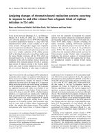
Fig. 1.
Normal rat SCN fibers immunostained with anti-SyI Ab.
Weak immunoreactivity (IR) was found. Slight immunoreaction
(arrowheads) was expressed throughout the axoplasm.
120
Fig. 2.
Immediately perfused SCN immunostained with anti-SyI
Ab. No IR was found.
120
Fig. 3.
SCN segment at 1 h after release. (A) IR was appeared
proximal to the crush (arrowheads).
50 (B) Immunoreactive
thin processes (arrowheads) were shown in the proximal region.
475 (C) IR was extended from the nodal region (large
arrowhead) of axon. IR was expressed in the axolemma (small
arrowheads) and throughout the axoplasm (arrows). IR was
exhibited to both proximal and distal directions from the node in
the proximal region (arrowheads).
475
4 Ku-birm Kwon et al.
damaged myelinated nerve fibers in this region (Fig. 3B).
IR was extended from the node of Ranvier (Fig. 3C). IR
was observed in the axolemma and throughout the
axoplasm. IR was exhibited to both proximal and distal
directions from the node in the proximal region. Similar
patterns were observed in the SCN at 2 h after release.
In the SCN segment at 3 h after release, many strongly
immunoreactive thin processes were shown just in the
proximal to the ligation crush site (Fig. 4A & 4B). Using
the counter cresyl violet staining (Fig. 4C), the strongly
immunoreactive thin processes were exhibited, assumed
them as regenerating axonal sprouts (Fig. 4D). In contrast,
there was no IR in the crush and distal regions (Fig. 4B &
4E). In the SCN segment at 5 h (Fig. 5), 6 h, and 8 h (Fig.
6) after release from the ligation, a similar pattern of IR
was observed. Strong SyI immunoreactive thin processes
were shown in the proximal region.
In the SCN segment at 12 h after release from the
ligation, SyI immunoreactive thin processes were extended
to the ligation crush site (Fig. 7A). Strong immunoreactive
thin processes were exhibited in the proximal region (Fig.
7B). Regenerating axonal sprouts were extended to the
crush region. A distinct SyI IR was seen in the proximal
region. In the SCN segment at 1 day after release from the
ligation, very strong SyI immunoreactive thin processes
were shown in the proximal region and extended to the
crush region (Fig. 8A). Many strong immunoreactive thin
processes were shown in the proximal region (Fig. 8B).
However, no IR was shown in the distal region (Fig. 8C &
8E). The IR at 1 day was much intenser than the former
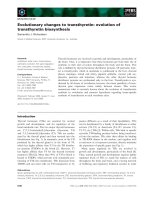
Fig. 4.
SCN segment at 3 h after release. (A and B) Strong immunoreactive thin processes (arrowheads) were shown proximal to the
crush. No immunoreactivity were shown in the crush and distal regions. (A)
50 (B) Higher magnification of Fig 4A.
120 (C)
Counter cresyl violet staining.
120 (D) Strong immunoreactive thin processes were exhibited in the proximal region (arrowheads). IR
was expressed throughout the axoplasm.
475 (E) No IR was shown in the distal region.
475
Fig. 5 and 6.
SCN segment at 5 h (Fig. 5) and 8 h (Fig. 6) after release. Immunoreactive thin processes (arrowheads) were shown the
proximal to the crush.
50
Translocational changes of localization of synapsin I in axonal sprouts of regenerating rat sciatic nerves agter ligation crush injury 5
Fig. 7.
SCN segment at 12 h after release. (A) Strong immunoreactive thin processes (arrowheads) were shown in the proximal region
and extended to the crush site (arrows).
50 (B) Strong immunoreactive thin processes were exhibited in the proximal region
(arrowheads).
120
Fig. 8.
SCN segment at 1 day after release. (A) Strong immunoreactive thin processes were shown in the proximal region (arrowheads)
and extended to the crush region (arrows).
50 (B) Many strong immunoreactive thin processes were shown in the proximal region
(arrowheads).
120 (C) No IR was shown in the distal region.
120 (D) Strong immunoreactive thin processes were shown in the
proximal region and extended from the node of Ranvier (large arrowhead). IR was expressed in the axolemma (arrows) and throughout
the axoplasm (arrowheads).
475 (E) No IR was shown in the distal region.
475
Fig. 9.
SCN segment at 2 days after release. (A) Strong immunoreactive thin processes were shown in the proximal region (arrowheads)
and extended to the crush region(arrows).
50 (B) Strong immunoreactive thin processes were shown in the proximal region. IR was
expressed in the periphery of the axoplasm (arrowheads).
475
6 Ku-birm Kwon et al.
groups. The IR was strong especially in the proximal
region to the ligation crush site. The IR was strongly
expressed in the axolemma and throughout the axoplasm
in the proximal region. Very strong immunoreactive thin
processes were extended from the node of Ranvier within
2 mm proximal to the ligation site (Fig. 8D). A similar
distribution pattern of IR was shown in the SCN segment
at 2 days after release (Fig. 9A). A weak IR was expressed
in the distal region. A strong IR was expressed in the
periphery of the axoplasm in the proximal and the crush
regions (Fig. 9B).
In the SCN sections at 3 days after release, strong SyI
immunoreactive thin processes were extended over the site
of ligation crush and most of sprouts were extended into
the distal region of the crush (Fig. 10A & 10B). Strong
immunoreactive thin processes were also shown in the
distal to the crush site. No distinct SyI IR was seen in the
degenerating parent axons in the distal region (Fig. 10B).
Similar distribution patterns were observed in the SCN
segment at 7 days (Fig. 11), 14 days, and 28 days after
release from the ligation. But after 7 days of release, the
crush site was slightly swelled. IRs at the various time
intervals were weaker than the previous groups. The
negative control sections reacted with normal serum were
not stained.
Discussion
Significant amounts of proteins and materials are
transported from the site of their synthesis for axonal
regeneration since the axon itself is unable to synthesize
them [20]. In addition, axonal lipids and proteins are
produced in the neurons for the regeneration of injured
nerves. These materials are transported to the distal site of
injury over the injured part by a slow or fast axonal
transport [26]. The axoplasmic transport and the molecular
mechanisms by which the synapsins are conveyed from
cell bodies to nerve terminals still remain to be elucidated.
The fast axonal transport in the nerve regeneration
contributes to the insertion of the regenerating sprout of
glycoprotein into the axolemma. SyI is synthesized in the
neuronal cell bodies and conveyed to the synaptic
terminals by the process of axonal transport together with
most axonal and synaptic proteins. The normally
transported SyI accumulates at the nerve endings [31].
Recently it has been demonstrated that the transport
mechanism of synaptic vesicles in the presynaptic terminal
can be applied to the regeneration [1, 2]. Slow axonal
transport provides the bulk of the axoplasmic and cytoskeletal
proteins, whereas fast axonal transport contributes to the
conveyance of elements for the axolemma. Because SyI is
a surface membrane protein, it is likely related to vesicular
accumulation and fast axonal transport [20].
SyI is one of the proteins that are highly specific to the
nerve terminals. SyI had been referred to for several years
as protein I [6, 18], until its virtually ubiquitous and
specific localization at synapses was known [16]. The SyI
binds to neurofilament1,2, small synaptic vesicles [5],
actin [22], and tubulin [3]. The colocalization of SyI and
Fig. 10.
SCN segment at 3 days after release. (A) Strong immunoreactive thin processes were shown in the proximal (arrowheads),
crush region (arrows) and extended to the distal region (arrows).
50 (B) Strong immunoreactive thin processes were shown in the
proximal and crush region (arrowheads), and extended to the distal region (arrows).
120
Fig. 11.
SCN segment at 7 days after release. Immunoreactive
thin processes were shown in the proximal (arrowheads) and
crush region (arrows), and extended to the distal region (arrows).
Crush part was slightly swelled.
50
Translocational changes of localization of synapsin I in axonal sprouts of regenerating rat sciatic nerves agter ligation crush injury 7
neurofilament has implicated that SyI-immunoreactive pro-
cesses occur in the axons but not in the Schwann cells and
other non-neural cells [35]. Ca
2+
influx through the
presynaptic Ca
2+
channel activates Ca
2+
-calmodulin-
dependent protein kinase, which phosphorylates SyI, then
detaches from synaptic vesicles, and is released from the
actin, microtubules, and other synaptic vesicles [24]. SyI
plays an important role in the movement of vesicles to the
active sites in the presynaptic membrane, thus plays a
regulatory role for neurotransmitter release [12]. In
addition, SyI may be involved in the elongation of
regenerating axons in the PNS regeneration [1, 7, 8, 12].
However, the involvement of SyI in the PNS regeneration
is still controversial.
In this study, we elucidated the involvement of SyI in the
PNS regeneration by immunocytochemistry with special
emphasis on a fast axonal transport. SyI has not previously
been detected immunocytochemically in the axons of
normal nerves [16] until Akagi et al. [1] have
demonstrated the presence of SyI in both the normal
myelinated and unmyelinated axons. Batinger et al. [4]
have shown that the bulk of SyI is transported at a velocity
of 6 mm/day, while a small amount of SyI is transported at
a more rapid velocity up to 240 mm/day. In the normal
nerve fibers, the morphological result from the present
study is corresponded to the biochemical data. Synapsin I-
like immunoreactive materials were accumulated only in
the proximal to the crush site, while SV2 and p38-like
materials were accumulated bidirectionally in the axons
with all sizes. The transmembrane components, SV2 and
p38, were retrogradely transported, while SyI was not
retrogradely transported. SyI is also known to be trans-
ported with the fast axonal transport in the non-autonomic
axons like rat sciatic nerve [8].
In this study, the changes in the distribution of SyI in the
injured peripheral nerve were observed using an experi-
mental animal model for PNS regeneration. Although the
axons and myelin sheaths were injured by a ligation crush,
the continuity of axons remained. Therefore, the transport
of SyI in the regenerating axons were observed more in
detail by immunocytochemistry [21]. The ligation crush
method is better than the forcep or hemostat crush method
in confirming an exact crush site and observing the
transported distribution of SyI [13].
In the kinetics of the axonal transport, three pools of SyI
present biochemically. The first pool of newly synthesized
SyI departs from the cell body immediately after synthesis.
The second and third pools enter the axon after delay of
more than one day. We performed ligation of the nerve by
1-0 silk thread and released it after 1 day to observe the
pools of SyI in vivo. Booj et al. [7] have reported that SyI
rapidly accumulates in parallel with synaptic vesicle-
specific integral membrane proteins proximal to the crush
site. The integral membrane proteins of synaptic vesicles,
but not SyI, accumulate distally to the crush [7, 13]. These
indicate that the synaptic vesicle membranes moving
retrogradely from the nerve terminal to cell bodies do not
carry appreciable amounts of SyI. SyI travels down the
axon only anterogradely. Therefore, the translocation of
SyI can be observed in the longitudinal section.
Previous studies have shown that some synaptic vesicle-
associated proteins like synaptophysin [30] and synapto-
tagmin [34] were localized in the regenerating axonal
sprouts emanating from the nodes of Ranvier. Synap-
tophysin and synaptotagmin were localized in the
proximal region at 1 day after release. Recently, SyI has
been reported to express in the regenerating axonal sprouts
and growth cones. The immunoreactive regenerating
sprouts appeared in the proximal region at 1 day and in the
distal region at 3 days. This result suggests that SyI travels
along the axon by a slow axonal transport [1]. In contrast,
our study showed that SyI immunoreactive processes
appeared at very early stages. The result indicates that SyI
may be involved in the PNS regeneration and that the
changes in the early accumulation of SyI may be related to
the fast axonal transport. Dahlstr m et al. [12] have
reported that four different synapsins, SyIa, Ib, IIa, and
IIb33, are accumulated in the crushed nerve. A large
amount of Sy Ib and a small amount of Sy Ia are
accumulated in the parent axons proximal to the crush site
up to 8 h after crushing. They concluded that Sy Ib may be
transported rapidly in association with membranous
organelles, while Sy Ia may be carried slowly in the
axoplasm. Akagi et al. [1] have found that SyI IR in the
regenerating axons was mainly associated with vesicular
organelles. They suggested that SyI IR found on the
vesicular organelles might represent Sy Ib in the early
sprouts and growth cones of the regenerating axons.
In this study, the SyI, including both Sy Ia and Ib,
accumulates at very early stages after release. The material
localized at the early stages may represent mainly Sy Ib.
De Camilli et al. [13] insisted that the effect of SyI on the
axonal transport was not likely to occur in vivo since it
would require concentrations of SyI normally present only
in the nerve terminals but not in axons. However, the result
is not consistent with ours. SyI is likely to be axonally
transported from the cell body to the terminals. It is
strongly expressed especially in the regenerating axonal
sprouts. In our study, the distribution of SyI supports the
results done by Akagi et al. [1] and Booj et al. [21]. SyI
immunoreactive thin processes appeared from the proximal
region to the crush site and extended into the distal region
after time-lapse. SyI immunoreactive processes were ex-
pressed in the proximal region until 8 h after release. This
result is consistent with that of Booj et al. [7], but not with
that of Akagi et al. [1]. They showed that SyI IR was
expressed in the proximal region at 1 day after release on
the vesicular organelles. In this study, SyI IR was strongly
8 Ku-birm Kwon et al.
expressed from proximal to crush region at 1 day after
release. An electron microscopic study may be necessary
to elucidate the involvement of SyI on the vesicular
organelles on the ultrastructural level.
In conclusion, the distribution patterns of SyI IR were
changed. SyI was accumulated in the proximal region at
very early stages after release. SyI was translocated from
the proximal to distal site of ligation by the time lapse.
These results suggest that SyI may be involved in the PNS
regeneration in addition to a role as a regulator of neuro-
transmitter release. In addition, the early accumulation of
SyI suggests that SyI may be related to the translocation of
vesicles to elongated membrane by a fast axonal transport
in the regenerating sprouts.
References
1.
Akagi S., Mizoguchi A., Sobue K., Nakamura H., Ide C.
Localization of Synapsin I in normal fibers and regenerating
axonal sprouts of the rat sciatic nerve. Histochem. Cell Biol.
1996,
105
, 365-373.
2.
Anderton B. H., Breinburg D., Downes M. J., Green P. J.,
Tomlinson B. E., Ulrich J., Wood J. N., Kahn J.
Monoclonal antibodies show that neurofibrillary tangles and
neurofilaments share antigenic determinants. Nature. 1982,
298
, 84-86.
3.
Baines A. J., Bennett V.
Synapsin I is a microtubule-
bundling protein. Nature. 1986,
319
, 145-147.
4.
Batinger C., Willard M.
Axonal transport of Synapsin I-
like proteins in rabbit retinal ganglion cells. J. Neurosci.
1987,
7
, 3723-3735.
5.
Benfenati F., B hler M., Jahn R., Greengard P.
Interactions of Synapsin I with small synaptic vesicles :
Distinct sites in Synapsin I bind to vesicle phospholipids and
vesicle proteins. J. Cell Biol. 1989,
108
, 1863-1872.
6.
Bloom F. E., Ueda T., Battenberg E., Greengard P.
Immunocytochemical localization, in synapses, of protein I,
an endogenous substrate for protein kinases in mammalian
brain. Proc. Natl. Acad. Sci. USA. 1979,
76(11)
, 5982-5986.
7.
B j S., Goldstein M., Fischer-Colbrie R., Dahlstr m A.
Calcitonin gene-related peptide and chromogen A : presence
and intra-axonal transport in lumbar motor neurons in the
rat, a comparison with synaptic vesicle antigen in
immunohistochemical studies. Neurosci. 1989.
30
, 479-501.
8.
B j S., Larsson P. A., Dahll f A. G., Dahlstr m A.
Axonal
transport of Synapsin I- and cholinergic synaptic vesicle-like
material ; further immunohistochemical evidence for
transport of axonal cholinergic transmitter vesicles in motor
neurons. Acta Physiol. Scand. 1985,
128
, 155-165.
9.
Burry R. W., Ho R. H., Matthew W. D.
Presynaptic
elements formed on polylysine-coated beads contain
synaptic vesicle antigens. J. Neurocytol. 1986,
15
, 409-419.
10.
Chun J. JM., Shatz C. J.
Redistribution of synaptic vesicle
antigens is correlated with the disappearance of a transient
synaptic zone in the developing cerebral cortex. Neuron.
1988,
1
, 297-310.
11.
Craig A. M., Wyborski R. J., Banker G.
Preferential
addition of newly synthesized membrane proteins at axonal
growth cones. Nature. 1995,
375
, 592-594.
12.
Dahlstr m A. B., Czernik A. J., Li J. Y.
Organelles in fast
axonal transport. What molecules do they carry in
anterograde vs retrograde directions, as observed in
mammalian systems? Mol. neurobiol. 1992,
6
, 157-177.
13.
De Camilli P., Greengard P
. The synapsins. Annu. Rev.
Cell Biol. 1990,
6
, 433-460.
14.
De Camilli P., Vitadello M., Canevini M. P., Zanoni R.,
Jahn R., Gorio A.
The synaptic vesicle proteins Synapsin I
and synaptophysin(protein 38) are concentrated both in
efferent and afferent nerve endings of the skeletal muscle. J.
Neurosci. 1988,
8
, 1625-1631.
15.
De Camilli P., Greengard P.
Synapsin I : A synaptic
vesicle-associated neuronal phosphoprotein. Biochem.
Pharmacol. 1986,
35(24)
, 4349-4357.
16.
De Camilli P., Cameron R., Greengard P.
Synapsin
I(Protein I), a nerve terminal-specific phosphoprotein. I. Its
general distribution in synapses of the central and peripheral
nervous system demonstrated by immunofluorescence in
frozen and plastic sections. J. Cell Biol. 1983a,
96
, 1337-
1354.
17.
De Camilli P., Harris S. M. Jr., Huttner W. B., Greengard
P.
Synapsin I(Protein I), a nerve terminal-specific
phosphoprotein. II. Its specific association with synaptic
vesicles demonstrated by immunocytochemistry in agarose-
embedding synaptosomes. J. Cell Biol. 1983b,
96
, 1355-
1373.
18.
De Camilli P., Ueda T., Bloom F. E., Battenberg E.,
Greengard P.
Widespread distribution of protein I in the
central and peripheral nervous system. Proc. Natl. Acad. Sci.
USA. 1979,
76
, 5977-5981.
19.
Goldenring J. R, Lasher R. S., Lou Vallano M., Ueda T.,
Naito S., Sternberger N. H., Sternberger L. A.,
DeLorenzo R. J.
Association of Synapsin I with neuronal
cytoskeleton. J. Biol. Chem. 1986,
261(18)
, 8495-8504.
20.
Griffin J. W., Price D. L., Drachman D. B., Morris J.
Incorporation of axonally transported glycoproteins into
axolemma during nerve regeneration. J. Cell Biol. 1981,
88
,
205-214.
21.
Haftek J., Thomas P. K.
Electron-microscopic observation
on the effects of localized crush injuries on the connective
tissues of peripheral nerves. J. Anat. 1968,
103(2)
, 233-243.
22.
Han H. Q., Greengard P.
Remodeling of cytoskeletal
architecture of nonneuronal cells induced by Synapsin. Proc.
Natl. Acad. Sci. USA. 1994,
91
, 8557-8561.
23.
Harada A., Sobue K., Hirokawa N.
Developmental
changes of Synapsin I subcellular localization in rat
cerebellar neurons. Cell Struct. Funct. 1990,
15
, 329-342.
24.
Hirokawa N., Sobue K., Kanda K., Harada A., Yorifuji
H.
The cytoskeletal architecture of the presynaptic terminal
and molecular structure of Synapsin I. J. Cell Biol. 1989,
108
, 111-126.
25.
Hsu S. M., Raine L., Fanger H.
Use of Avidin-Biotin-
Peroxidase Complex(ABC) in immunoperoxidase techniques:
A comparison between ABC and unlabeled antibody(PAP)
procedures. J. Histochem. Cytochem. 1981,
29(4)
, 577-580.
Translocational changes of localization of synapsin I in axonal sprouts of regenerating rat sciatic nerves agter ligation crush injury 9
26.
Ide C.
Peripheral nerve regeneration. Neurosci. Res. 1996,
25
, 101-121.
27.
Ide C., Kato S.
Peripheral nerve regeneration. Neurosci.
Res. 1990, Suppl. 13, S157-S164.
28.
Ide C., Tohyama K., Yokota R., Nitatori T., Onodera S.
Schwann cell basal lamina and nerve regeneration. Brain
Res. 1983,
288
, 61-75.
29.
Nah J. W., Chung I. H., Lee W. T., Shin T. S.
Morphometrical study of regenerating peripheral nerve in
young and adult rats. Yonsei J. Med. Sci. 1985,
18(2)
, 423-
436.
30.
Okajima S., Mizoguchi A., Masutani M., Tomatsuri M.,
Tamai K., Hirasawa Y., Ide C.
Synaptophysin
immunocytochemistry in the regenerating sprouts from the
nodes of Ranvier in injured rat sciatic nerve. Brain Res.
1993,
631
, 133-136.
31.
Petrucci T. C., Macioce P., Paggi P.
Axonal transport
kinetics and posttranslocational modification of Synapsin I
in mouse retinal ganglion cells. J. Neurosci. 1991,
11(9)
,
2938-2946.
32.
Priestley J. V.
Neurochemistry, Ch. 3. Immunocytochemical
techniques for the study of the nervous system(ed. Turner
AJ, Bachelard HS). pp.71-120. IRL press, Oxford, 1997.
33.
S dhof T. C.
Synaptic vesicle cycle : a cascade of protein-
protein interactions. Nature. 1995,
375
, 645-653.
34.
Uehara K., Mizoguchi A., Mizuno K., Ide C.
Localization
of synaptotagmin in the regenerating sprouts emanating
from the nodes of Ranvier. Acta Histochem. Cytochem.
1995,
28(5)
, 401-407, 1995.
35.
Wiklund P., Ekstr m Per A. R., Edbladh M., Tonge D.,
Edstr m A.
Protein kinase C and mouse sciatic nerve
regeneration. Brain Res. 1996,
715
, 145-154.