Báo cáo khoa học: "Prevalence and Clinical Characterization of Gastric Helicobacter Species Infection of Dogs and Cats in Korea" pps
Bạn đang xem bản rút gọn của tài liệu. Xem và tải ngay bản đầy đủ của tài liệu tại đây (280.08 KB, 11 trang )
J O U R N A L O F
Veterinary
Science
J. Vet. Sci. (2002), 3(2), 123-133
ABSTRACT
10)
This study w as carried out to evaluate the
prevalence and clinical characterizations of gastric
Helicobacter
spp. infection of dogs and cats in Korea.
The prevalence of
Helicobacter
spp. infection of dogs
and cats determ ined by urease test w as 78.4% and
64%, respectively, although
Helicobacter
genus-specific
PCR assay showed that it was 82.3% and 84%. U rease
mapping results based on urease te st showed that
total positive rate of tested tissues from clinically
abnormal dogs was significantly higher than that
from clinically normal dogs (p=0.0018; Odds ratio =
6.118; 95% Confidence Interval = 1.96~19.103). These
findings were consistent with the results of
Helicobacter
genus-specific PCR assay w hich show ed that positive
rate of the fundus (100%) and the antrum (100%) of
clinically abnormal dogs w as significantly higher
than that of same gastric regions of clinically normal
dogs (77.5 and 67.5% respectively). In comparison of
gastric regions betwee n clinically normal dogs and
abnormal dogs, positive rate of ure ase test for the
fundus (100%) and body (90.9%) in clinically abnormal
dogs w as significantly higher than that of abnormal
dogs (72.5% and 57.5% respectively; p<0.05). The
results of urease mapping in dogs and cats also
indicated that
Helicobacter
colonization in the fundus
w as more dense compared w ith the density in the
body and antrum. In
Helicobacter
species-spe cific
PCR assay for dogs, 32 of 42 fundic tissues (76.2%)
w ere positive for
H. heilmannii
and tw o (4.8%) were
positive for
H. felis
. In cats, 18 of 21 fundic tissues
(85.7%) w ere positive for
H. heilmannii
and 2 (9.5%)
w ere positive for
H. felis
. Gastritis scores of fundic
tissues from clinically abnormal infected dogs w ere
similar to that from noninfected dogs and evidence of
upregulation of IL-1
β
, IL-8, and TNF-
α
mRNA w as
*
Corresponding author: Hong-Ryul Han, DVM, Ph.D.
Department of Veterinary Internal Medicine, College of Veterinary
Medicine, Seoul National University. Republic of Korea
Tel: +82-2-880-8683, Fax: +82-2-875-5588
E-mail:
not detected in gastric fundic tissues from clinically
abnormal infe cted dogs. This study suggested that
Helicobacter
spp. infection in domestic dogs including
private ow ned pet dogs and cats is highly prevalent
usually w ith no clinical sign but high density of
colonization can be related to gastrointestinal signs
Key w ords:
Helicobacter spp., Prevalence, PCR, Dog, Cat
Introduction
Helicobacter species are spiral-shape or curved gram-
negative bacteria inhabit the mucus, glands and parietal
cells of the stomach. Since the initial isolation of Helicobacter
pylori (H. pylori) from human gastric tissue in 1983 by
Warren and Marshall, evidence implicating the bacterium
as the causative agent of gastritis and duodenal ulcer has
been established [16,34,53]. More recently, the bacterium
also known to be a cofactor in the development of gastric
adenocarcinoma and gastric lymphoma in humans [40,56].
Gastric spiral organisms in various animals, including
pigs with gastric ulcers (Helicobacter heimannii), cheetahs
with severe gastritis (Helicobacter acinonychis), ferrets with
gastritis and peptic ulcers (Helicobacter mustelae), monkeys
(Helicobacter nemestrinae), rodents (Helicobacter muridarum)
and dolphins have been also described [2,7,12,22,32,42]. In
dogs and cats, several gastric spiral organisms also have
been reported since the turn over the century, however, their
presence has been largely ignored and even understanded as
gastric commensals [13,54]. In now, these gastric organisms
have received renewed attention because of the H. pylori
has been proved as a strong gastric pathogen in humans.
Research works for gastric spiral organisms in domestic
pets, especially dogs and cats was initially focused on the
development of suitable animal model for studying H. pylori
infection and extended to evaluate their clinical significance
in these animals [32,43,44,46].
The main gastric spiral organisms described in dogs and
cats are morphologically distinct from H. pylori with their
more tightly coiled body shape and larger size [24,31].
Because these organisms cannot be distinguished when they
are examined in gastric tissue by light microscopy, they are
Prevalence and Clinical Characterization of Gastric Helicobacter Species Infection of
Dogs and Cats in Korea
Cheol-Yong Hwang, Hong-Ryul Han* and Hwa-Young Youn
Department of Veterinary Internal Medicine, College of Veterinary Medicine, Seoul National University, Seoul 151-742, Korea
Received Dec. 16, 2001 / Accepted May 16, 2002
124 Cheol-Yong Hwang, Hong-Ryul Han and Hwa-Young Youn
collectively referred to as "gastiric Helicobacter-like organisms
(GHLO)" [31]. To date, Helicobacter felis (H. felis),
Helicobacter heilmannii (H. heilmannii), Helicobacter pametensis,
Helicobacter bizzozeronii, Helicobacter salomonis, Helicobacter
bilis, and Flexispira rappini have been identified on the
basis of 16S ribosomal RNA sequencing, DNA hybridization,
species specific polymerase chain reaction (PCR) analysis,
electron microscopic appearance, and protein profiling
analysis [5,9,20,27,28,31,36,37].
Infection of GHLO is highly prevalent in dogs and cats.
The prevalence of GHLO in dogs has been reported to be
between 61 to 100 %; it is seen in 61-82% of dogs with
recurrent vomiting [15,25,57], 67-86% of clinically healthy
pet dogs [8,57], and almost 100% of laboratory beagles and
shelter dogs [6,8,24]. In cats, the prevalence of GHLO has
been reported to be similar to that of the dogs between 41
to 100 % [15,18,21,25,30,36,38]. The observations that the
high prevalence of GHLO in closely contacted cat groups
like as research colonies and animal shelters have been
identical with that of the dogs. H. pylori infection has been
observed in group of laboratory cats and commercial vendor,
but not in private owned pet cats [13,18,19].
In Korea, some studies for GHLO infection in dogs were
also previously reported and showed that the infection rate
was similar to that of foreign studies. These studies
however, were only based on closely contacted healthy
laboratory beagle colony and kennel dogs [1,35,39]. To date,
the prevalence survey study for GHLO infection in private
owned pet dogs and any group of cats in Korea has not been
reported yet.
Despite the frequent occurrence of GHLO in dogs and
cats, the relationships between these bacteria and clinical
manifestation have not been clearly understood with
gastritis accompanying infection in some but not all infected
dogs and cats [8,21,25,57].
Several reports of GHLO infections in humans have lead
to speculation that animals especially dogs and cats may
serve as a source for human infection [6,23,50,51]. There is
therefore a need to determine the prevalence of Helicobacter
species in domestic pets in order to evaluate the possible
risk to human health, and also to that of the host animals,
in which gastritis and related complains can occur.
Therefore, this study was carried out to evaluate the
prevalence and clinical characterizations of gastric
Helicobacter spp. infection of dogs including private owned
pet dogs and cats in Korea.
Materials and Methods
Experimental animals and gastric tissue sam pling
Canine gastric tissues were collected at necropsy from 4
euthanized pet dogs having gastrointestinal signs (like
intermittent vomiting with or without blood in vomitus and
inappetence or anorexia which were not related with other
non-gastric causes), 24 euthanized pet dogs had no
gastrointestinal signs and 12 laboratory beagle dogs being
used in another toxicological study. Feline gastric tissues
were taken at necropsy from 24 cats being used in terminal
procedure part of one research project. Up to 5 full-
thickness gastric tissue samples were collected from the
fudus, the cardia and the antrum of these animals with a
sterile 6 mm skin biopsy punch. Collecting tissue samples
for PCR analysis were frozen at -80
℃
pending analysis and
samples for histopathologic examinations were fixed in 10%
buffered formalin. Samples from the fundus for cytokine
analysis were collected, snap-frozen in liquid nitrogen and
stored at
–
80
℃
pending analysis. The remnants were used
for urease mapping, direct smear examination and culture.
Endoscopic biopsies obtained from 4 clinically healthy pet
dogs, 7 patient dogs with gastrointestinal signs and one
clinically healthy pet cat using a pediatric endoscope and
biopsy forceps. Endoscopic biopsies procured from the
fundus (near cardia), the body (greater curvature), and the
antrum (near pylorus). Two biopsies were taken from each
site for urease testing, PCR analysis, direct smear
examination and histopatologic examination. Additional
fundus site biopsy for cytokine analysis was snap-frozen in
liquid nitrogen and stored at
–
80
℃
pending analysis.
Direct gastric tissue smear test
Gastric tissue samples were impressed on slide glass and
were stained with Diff-Quick (International Reagent Co.,
Japan) and evaluated by light microscopy for the presence
of Helicobacter spp.
Urease m apping
Urease mapping was performed to determine semi-
quantitatively the density of colonization by Helicobacter
spp. in different regions of the stomach. Gastric tissue
samples were places in sterile tubes containing 200
㎕
of a
solution composed of urea, sodium azide, phenol red, and
phosphate-buffered saline (pH 6.5). Samples were incubated
at 37
℃
for 24 hours and observed at 1, 3, 12, and 24 hours
for a change in the color of the indicator medium. A change
from orange-red to bright pink was considered a positive
results, and the time of color changes were recorded.
Culture of gastric tissue samples
Gastric fundic tissue samples were directly smeared on
trypicase soy agar base (DIFCO, U.S.A.) supplemented with
5% bovine blood and containing trimethoprim, vancomycin,
and polymyxin B. Culture plates incubated for 7 days at 37
℃
in a moist microaerophilic atmosphere provided by
anaerobic jar with a Campypak system (Campypak Plus;
Beckton Dickinson Microbiology system, U.S.A.).
Bacterial strains
For evaluating the sensitivity of Helicobacter-genus and
species-specific PCR assay, H. felis (ATCC 51211) purchased
from the American Type Culture Collection (ATCC; U.S.A.)
Prevalence and Clinical Characterization of Gastric Helicobacter Species Infection of Dogs and Cats in Korea 125
and H. pylori (KCTC 2948) purchased from the Korean
Collection for Type Cultures (KCTC; Korea) were used as
standard Helicobacter strains.
PCR
DNAs from gastric tissue samples and standard bacteria
were extracted with a Wizard genomic DNA purification
kit (Promega, U.S.A.) according to the manufacturer's
instructions.
Helicobacter genus specific PCR assay was performed
with C97 and C98 primers which amplify the 16S rRNA
gene of Helicobacter species (Table 1.). DNA samples (5
㎕
)
were added to a reaction mixture containing 400
μ
M
dNTPs, 1X PCR buffer, 2.5 U of Taq DNA polymerase
(ABgene, U.K.), 0.6
μ
M of each primer, and distilled water
in a total volume of 50
㎕
. PCR samples were heated to 9
4
℃
for 2.5 min once, followed by 40 cycles of denaturation
at 94
℃
for 1 min, primer annealing at 50
℃
for 1 min, and
extension at 72
℃
for 1 min, with a final extension at 72
℃
for 15 min in a Biometra (U.S.A.) personal thermocycler.
PCR products were subjected to electrophoresis on a 2%
agarose gel containing 0.5
㎍
of ethidium bromide per
㎖
and visualized over UV light.
Helicobacter species-specific PCR assay was performed
with fundic tissue samples as follows. H. felis and H. pylori
specific PCR was performed with primers which amplify the
urease B gene of H. felis and H. pylori (Table 1.). The PCR
mixture and the cycle was the same as the Helicobacter
genus specific PCR assay. H. heilmannii specific PCR assay
was performed with primers which amplify the urease B
gene of H. heilmannii (Table 1.). Two microliters of DNA
was added to the PCR mixture described above in a total
volume of 50
㎕
. The temperature and time schedule was as
follows: 1 cycle of denaturation at 94
℃
for 3 min, annealing
at 52
℃
for 2 min, and extension at 72
℃
for 3 min followed
by 30 cycles at 94
℃
for 30 s, 52
℃
for 30 s, and 72
℃
for 1
min. Following completion of the 30 cycles, additional one
cycle at 94
℃
for 20 s, 52
℃
for 20 s, and 72
℃
5 min was
performed.
Table 1.
Primers used for PCR
Primer Primer directiona and sequence product size (bp)
C97
5
′
-GCTATGACGGGTATCC- 3
′
400
C98
5
′
-GATTTTACCCCTACACCA- 3
′
H. felis
F, 5
′
-ATGAAACTAACGCCTAAAGAACTAG
1,150
R, 5
′
-GGAGAGATAAAGTGAATATGCGT
H. pylori
F, 5
′
-GGAATTCCAGATCTATGAAAAAGATTAGCAGAAAAG- 3
′
1,707
F, 5
′
-GGAATTCGTCGACCTAGAAAATGCTAAAGAGTTG- 3
′
H. heilmannii
F, 5
′
-GGGCGATAAAGTGCGCTTG- 3
′
580
R, 5' -CTGGTCAATGAGAGG- 3
′
a F, forward; R, reverse.
Cloning and nucleotide sequence analysis of PCR
products
To confirm the identity of the Helicobacter species-specific
PCR assay products with target genes, the PCR products
were cloned with Topo TA Cloning Kit (Invitrogen, U.S.A.)
and the plasmids containing correct inserts were analysed.
Histopathologic examination
For evaluating the relation of Helicobacter spp. infection
and clinical gastritis in dogs, formalin fixed gastric fundic
tissue samples from 9 clinically abnormal infected dogs
(Urease test positive, PCR assay positive and had clinically
abnormal gastrointestinal signs), and 8 clinically healthy
uninfected dogs (Urease test negative, PCR positive and had
not clinically abnormal gastrointestinal signs) were embedded
in paraffin, sectioned and stained with hematoxylin and
eosin.
For the histopathological assessment, the presence of
lymphocyte aggregates, and the mean numbers of leukocytes
at X400 fields were recorded under microscopy. Gastritis
was grade as follows: "no gastritis", no lymphocyte aggregate
or leukocytes; "mild gastritis", no lymphocyte aggregates
and < 10 leukocytes per field; "moderate gastritis",
lymphocyte aggreates present and/or 10 to 50 leukocytes per
field; "severe gastritis". lymphocyte aggreates present and
>50 leukocytes per field [37].
Analysis of cytokine gene expression in gastric
fundic tissues
Gastric tissue samples from the fundus were collected
from 9 clinically abnormal infected dogs and 8 uninfected
healthy dogs described previously, snap-frozen in liquid
nitrogen, and stored at -80
℃
pending analysis. RNA was
extracted from the tissues with an RNA extraction kit
(NucleoSpin RNA II; Macherey-Nagel, Germany) according
to the manufacturer's instruction. mRNA expression for
TNF-
α
, IL-1
β
, IL-8 was determined by reverse
transcription (RT)-PCR. The RNA was reverse transcribed
126 Cheol-Yong Hwang, Hong-Ryul Han and Hwa-Young Youn
with cDNA synthesis kit (First Strand cDNA Synthesis Kit;
MBI Fermentas, Lithuania) and resulting cDNA served as
a template for PCR assay. PCR primers for canine TNF-
α
,
IL-1
β
, and IL-8 were used to amplify their respective
cDNAs (Table 2.). The PCR reaction was run for 94
℃
for 5
min one time, followed by 35 cycles of 94
℃
for 1 min, 45
℃
for 1 min, and 72
℃
for 1 min with a final extension at 72
℃
for 10 min in a Biometra (U.S.A.) personal thermocycler.
PCR products were subjected to electrophoresis on a 2%
agarose gel containing 0.5
㎍
of ethidium bromide per
㎖
and visualized over UV light. For the positive control, canine
leukocyte-derived RNA from concanavalin A stimulated canine
peripheral blood leukocytes was used. The RT-PCR results
were judged as negative or positive only regardless of
staining intensity.
Statistical analysis
Deferences in total positive rate of urease activity and
total positive rate of Helicobacter genus-specific PCR assay
of gastric region between clinically normal and abnormal
dogs were evaluated by Bonferroni t-test. Deferences in
these variables in cats and between gastric region in each
dog groups were also evaluated by Bonferroni t-test.
Deferences in total positive rate of urease activity between
clinically normal and abnormal dogs and between gastric
region in dogs were evaluated by logistic regression. All
statistical analyses were performed with software package
SAS (release 8.0, SAS Institute, Cary, NC, U.S.A.). A
statistical significance level of 0.05 was used for analyses.
Results
Urease m apping
1) Dogs
Urease mapping results of clinically normal and
abnormal dogs were summarized in Table 3. Most of
positive results were detected within 12 hours incubation,
but 6 of 11 fundic tissue samples from clinically abnormal
dogs showed positive results within 1 hour. Forty of 51
tested dogs showed positive results at least one gastric
region and the detection rate of Helicobacter spp. in dogs
determined by urease test was 78.4%.
Total positive rate of tested tissues from clinically
abnormal dogs was significantly higher than that from
clinically normal dogs (p=0.0018; Odds ratio = 6.118; 95%
Confidence Interval = 1.96~19.103). In comparison of gastric
regions, total positive rate of the fundus was higher than
that of other gastric regions and the deference between the
antrum and the fundus was statistically significant
(p=0.0013; Odds ratio = 4.4438; 95% Confidence Interval =
1.791~10.997). Positive rate of the fundus (100%) and the
body (90.9%) of clinically abnormal dogs was significantly
higher than that of same gastric regions from clinically
normal dogs (72.5%, 57.5% respectively; p<0.05). In
clinically normal dogs, positive rate of the fundus (72.5%)
was significantly higher than that of the antrum (40%;
Table. 2.
Primers used in the RT-PCR to detect mRNA of cytokines
Primer Primer directiona and sequence product size (bp)
IL-1
β
F, 5
′
-GAGGTTCCAATGTGAAGTGC- 3
′
291
R, 5
′
-CCTGTAACTTGCAGTCCACC- 3
′
IL-8
F, 5
′
-ACTTCCAAGCTGGCTGTTGC- 3
′
172
R, 5
′
-GGCCACTGTCAATCACTCTC- 3
′
TNF-
α
F, 5
′
-CCAAGTGACAAGCCAGTAGC- 3
′
274
R, 5
′
-TCTTGATGGCAGAGAGTAGG- 3
′
a F, forward; R, reverse.
Table 3.
Urease mapping results of clinically normal and abnormal dogs
Group (No. of dogs)
Site
No. of urease activity (%)
< 1hr 1 - <3hr 3 -<12h 12-24h Total positive
Clinically normal dogs
(40)
Fundus
3 (7.5) 18 (45) 7 (17.5) 1 (2.5) 29 (72.5)
Body
0 (0) 9 (22.5) 13 (32.5) 1 (2.5) 23 (57.5)
Antrum
0 (0) 8 (20) 7 (17.5) 1 (2.5) 16 (40)
Clinically abnormal dogs
(11)
Fundus
6 (54.5) 3 (27.3) 1 (9.1) 1 (9.1) 11 (100)
Body
1 (9.1) 4 (36.4) 5 (45.5) 0 (0) 10 (90.9)
Antrum
1 (9.1) 2 (18.2) 5 (45.5) 0 (0) 8 (72.7)
Prevalence and Clinical Characterization of Gastric Helicobacter Species Infection of Dogs and Cats in Korea 127
p<0.05) although there were no differences between gastric
regions in clinically abnormal dogs.
2) Cats
Many samples showed positive results within 12 hours.
Total positive rate of the fundus was 64% and this rate was
higher than that of the body (32%) and the antrum (28%),
but statistically significant difference (P<0.05) was only
detected between the fundus and the antrum (Table 4).
Table 4.
Urease mapping results of cats
Site
(No. of
samples)
No. of urease activity (%)
< 1hr 1 - <3hr 3 - <12h 12-24h
Total
positive
Fundus
(25)
2 (8) 3 (12) 8 (32) 3 (12) 16 (64)
Body
(25)
1 (4) 2 (8) 3 (12) 2 (8) 8 (32)
Antrum
(25)
1 (4) 4 (16) 2 (8) 0 (0) 7 (28)
PCR assay
1)
Helicobacter
genus-specific PCR assay in dogs
Helicobacter genus-specific PCR assay (Fig. 2) in dogs
showed that positive rate of the fundus (100%) and the
antrum (100%) of clinically abnormal dogs was significantly
higher than that of same gastric region of clinically normal
dogs (77.5% and 67.5%; P<0.05). Total positive rate of the
fundus (82.3%) was highest but was not statistically
significant compared with the other regions (P>0.05). There
were also no significant deferences of positive rate between
gastric region in each dog groups (P>0.05) (Table 5).
Table 5.
Results of Helicobacter genus-specific PCR assay
in dogs
No. of Positive (%)
Group
(No. of dogs)
Fundus Body Antrum
Clinically normal
dogs (40)
31 (77.5) 31 (77.5) 27 (67.5)
Clinically
abnormal dogs (11)
11 (100) 10 (90.9) 11 (100)
Total (51)
42 (82.3) 41 (78.8) 38 (74.5)
2) Helicobacter
genus-specific PCR assay in cats
Positive rate of the fundus, the body and the antrum of
cat was 84%, 80% and 79% respectively, but these
deferences were not statistically significant (p>0.05).
Table 6.
Results of Helicobacter genus-specific PCR assay
in cats
(No. of cats) Site No. of positive (%)
Cats (25)
Fundus
21 (84)
Body
20 (80)
Antrum
19 (79)
3)
Helicobacter
species-specific PCR assay in dogs
Each set of primers was shown to amplify the gene from
which it was derived, without cross-hybridizing with the
corresponding gene of the two other species (Fig. 3, Fig. 4).
Thirty-two of 42 fundic tissue samples tested (76.2%) were
positive for H. heilmannii and two samples (4.8%) from
clinically normal dogs were positive for H. felis (Table 7). No
amplification products corresponding to H. pylori were
detected and 8 samples (19%) were negative for all species-
specific PCR assay (Table 7). There were no significant
differences of the results between clinically normal and
abnormal dogs.
Table 7.
Results of Helicobacter species-specific PCR assay
in dogs
No. of Species identified (%)
Group
(No. of dogs)
H.
heilmannii
H. felis H. pylori
non-ide ntified
Clinically normal
dogs (31)
23 (74.2) 2 (6,5) 0 (0) 6 (19.4)
Clinically abnormal
dogs (11)
9 (81.8) 0 (0) 0 (0) 2 (18.1)
Total (42)
32 (76.2) 2 (4.8) 0 (0) 8 (19)
4) Helicobacter
species-specific PCR assay results in
cats
Eighteen of 21 fundic tissue samples (85.7%) tested were
positive for H. heilmannii and 2 (9.5%) were positive for H.
felis. H. pylori was not detected and 1 sample were not
amplified by all species-specific PCR assay (Table 8, Fig. 3,
Fig. 4).
Table 8.
Results of Helicobacter species-specific PCR
assay in cats
(No. of Cats)
No. of Species identifie d (%)
H.
heilmannii
H. felis H. pylori non-identified
Cats (21) 18 (85.7) 2 (9.5) 0 (0) 1 (4.7)
128 Cheol-Yong Hwang, Hong-Ryul Han and Hwa-Young Youn
5) Nucleotide homology of the Helicobacte r species-
specific PCR products
There was greater than 97% identity between the
sequences of H. heilmannii specific PCR products and
GeneBank sequences of the urease B gene of H. heilmannii
(Accession No. L25079). In comparision of H. felis PCR
products, greater than 98% identity was detected (Accession
No. X69080).
Evaluation of
Helicobacter
spp. infection state by
different detection methods
Thirty-nine of 51 (76.5%) dogs and 16 (64%) of 25 cats
were positive for all test performed with gastric fundus
tissues. Eight (15.7%) dogs and 4 (16%) cats showed
negative in all tests. In direct tissue smear test, the results
were concordant with the results of other tests in all dogs
and cats but 4 dogs and 5 cats (2 dogs which were negative
in direct tissue smear test were positive in other tests; 2
dogs and 5 cats which were positive in direct smear test
were negative in urease test; Table 9).
Table 9.
Evaluation of Helicobacter infection state in
gastric fundic tissues by different detection
methods
Smeara Ureaseb PCRc
No. of dogs
with pattern
No. of cats
with pattern
+ + +
39 16
+ - +
2 5
- + +
2 0
- - -
8 4
a: Direct tissue smear test : + = positive,
–
= negative.
b: Urease test; + = positive,
–
= negative.
c: PCR assay; + = positive,
–
= negative.
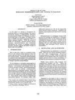
Fig. 3.
Detection of H. heilmannii DNA (580bp) in gastric
tissues by PCR assay. Lanes: M, DNA ladder; 1, H.
heilmannii infected dog; 2, H. heilmannii infected cat; 3,
DNA from H. pylori (KCTC 2948); 4, DNA from H. felis
(ATCC 51211).
Fig. 4.
Detection of H. felis DNA (580bp) in gastric tissues
by PCR assay. Lanes: M, DNA ladder; 1, H. felis infected
dog; 2, H. felis infected cat; 3, DNA from H. felis (ATCC
51211); 4, DNA from H. pylori (KCTC 2948).
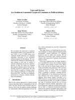
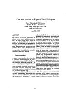
Fig. 1.
Direct impression smear of gastric tissue from
Helicobacter spp. infected dogs. Arrows indicate spiral
shaped Helicobacter organisms.
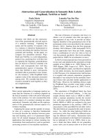
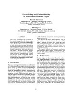
Fig. 2.
Detection of Helicobacter spp. DNA (400bp) in
gastric tissues by Helicobacter genus-specific PCR assay.
Lanes: M, DNA ladder; 1, infected dogs; 2, infected cat; 3,
DNA from H. pylori (KCTC 2948); 4, DNA from H. felis
(ATCC 51211).
Prevalence and Clinical Characterization of Gastric Helicobacter Species Infection of Dogs and Cats in Korea 129
Culture result
Helicobacter organisms were not cultured from all gastric
fundic tissue samples of dogs and cats.
Histopathologic findings
Most of clinically abnormal infected dogs had mild to
moderate gastritis consisting of scattered leukocytes but
similar degree of gastritis also was detected in clinically
normal uninfected dogs. Whatever severe gastritis was only
detected in one clinically abnormal infected dogs, there were
no correlation between the presence of the bacteria with
clinical signs and the gastritis score.
Table 10.
Gastritis results for clinically abnormal Helicobacter
spp. infected and noninfected normal dogs
No. of dogs w ith result
Degree of
gastritis
Clinically abnorm al
infected (n = 8)
Clinically normal
noninfected (n = 7)
None
1 2
Mild
2 1
Moderate
4 4
Severe
1 0
Analysis of cytokine gene expression in gastric
fundic tissues
RT-PCR assay of gastric fundic tissues from dogs for
detecting existence of cytokines did not show the evidence of
upregulation of IL-1
β
, IL-8, or TNF-
α
mRNA in ether
clinically abnormal infected or uninfected dogs. Appropriated
reactions were only detected positive control samples (Fig. 5).
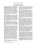
Fig. 5.
Detection of mRNA for IL-1
β
, IL-8, and TNF-
α
in
gastric tissues by RT-PCR. Agarose gel electrophoresis of
DNA products. Lanes: M, DNA ladder; 1 to 7, Helicobacter
spp. infected dogs; +, positive control (peripheral blood of dog).
Discussion
Since the discovery that H. pylori is a pathogen in
humans, many studies have been evaluated the prevalence
of Helicobacter infection and the relationship between
infection and gastric pathology in other animals. This study
was carried out with purpose of evaluating the prevalence
and characterization of Helicobacter infection in domestic
dogs and cats. In the present study, the prevalence of
Helicobacter spp. in dogs and cats were evaluated with
direct gastric tissue smear test, urease test which is
commonly used in detecting H. pylori infection in humans
and PCR assay. These test results in the present study
showed that the prevalence of Helicobacter spp. in dogs (>
78.4%) and cats (> 64%) was as high as in previous reports
[1,8,15,21,25,30,35,36,38,57].
Urease mapping based on urease test showed that total
positive rate of tested tissues from clinically abnormal dogs
was significantly higher than that from clinically normal
dogs (p=0.0018; Odds ratio = 6.118; 95% Confidence Interval
= 1.96~19.103). These findings were consistent with the
results of Helicobacter genus specific PCR assay which
showed that positive rate of the fundus (100%) and the
antrum (90.9%) of clinically abnormal dogs was significantly
higher than that of same gastric regions of clinically normal
dogs (77.5 and 67.5% respectively). However, a previous
report showed that there was no difference of the prevalence
between clinically normal and abnormal dogs [57]. In spite
of higher prevalence in clinically abnormal dogs, rate of
showing positive urease activity within one hour in
clinically abnormal dogs also higher than that in clinically
normal dogs. It suggested that the density of Helicobacter
colonization in clinically abnormal dogs was higher than
that in clinically normal dogs based on the fact that urease
activity is depended on the density of Helicobacter
colonization.
The results of urease mapping in dogs also indicated that
Helicobacter colonization in the fundus was more dense
compared with the density in the antrum. These pattern of
colonization was similar to that observed in previous reports
conducted with naturally acquired helicobacteriasis and
experimentally infected dogs and cats [5,21,38,46,57]. In
comparison of gastric regions between clinically normal and
abnormal dogs, positive rate of urease test for the fundus
and body in clinically abnormal dogs was significantly
higher than that in normal dogs (p<0.05). These results
combined with the higher degree of colonization in clinically
abnormal dogs may consider the possibility that high degree
of Helicobacter spp colonization in the fundus and body can
arise gastrointestinal signs in dogs.
To the best of our knowledge, this is the first report of
evaluating the Helicobacter spp. infection of cats in Korea
130 Cheol-Yong Hwang, Hong-Ryul Han and Hwa-Young Youn
although the number of cats evaluated was so small and
limited to only one laboratory colony. The pattern of urease
mapping results of cats was similar to that of dogs, which
showed that colonization was less dense in the antrum of
the stomach compared with the density in the fundus and
body. Therefore, for reducing the possibility of false negative
result in urease test, using biopsy tissues from the fundus
and body rather than from the antrum is recommended. All
of cats investigated in the present study had no
gastrointestinal signs and the rate of showing positive
urease activity within one hour was low. This result also
supported the possibility as previously mentioned that high
degree of Helicobacter spp. colonization might induce
gastrointestinal signs.
In several recently developed PCR assays for detecting
Helicobacter infection, two targets the urease and 16S rRNA
genes, were appeared promising because partial or whole
sequence information is available for both [3,4,14,17,26,
52,55]. In the present study, Helicobacter-specific primer
pair C97 and C98 [14] which generate 16S rRNA amplicons
of approximately 400 bases was used for Helicobacter
genus-specific PCR assay. In Helicobacter genus-specific
PCR assay in dogs and cats, Helicobacter spp. detecting rate
(dog= 82.3%, cat = 84%) was slightly higher than that of
urease test (dog = 78.4, cat = 64%). There were no significant
deference of positive rate between gastric regions in dogs
and cats. These results combined with the results of urease
mapping indicated that Helicobacter spp. infection rate
between gastric regions were not different but truly in
colonization density.
For detecting Helicobacter spp. infection in gastric
tissues, direct tissue smear test, urease test and PCR assay
was used and each test results of fundic tissues were
compared. Concordant results among the different diagnostic
tests were reached for 92% of the dogs and 80 % of the cats
evaluated. These results were similar to that of one previous
report conducted in cats [36]. In direct tissue smear test,
results were concordant with the results of other tests in all
dogs and cats but 2 dogs which were negative although
urease test and PCR asssy test showed positive. Two dogs
and 5 cats were positive for direct tissue smear test and
PCR assay but showed negative result in urease test.
According to these results, direct tissue smear test and PCR
assay appeared more sensitive than urease test. These
observations concur with those in studies of experimentally
H. felis infected cats and of dogs with naturally acquired
helicobacteriasis [45,46]. The results of the present study
also suggested that PCR assay is the most sensitive test for
the detection of Helicobacter infection in dogs and cats. This
observation is agreement with results obtained in studies of
mice and dogs experimentally infected with H. felis and of
humans and cats infected with H. pylori, which showed that
PCR assay was more sensitive than histology, bacterial
culture, and urease test [10,29,41,45].
PCR assay also has been known to be a specific methods
to distinguish between Helicobacter species [36]. Primer
pairs used for Helicobacter species-specific PCR assay in the
present study were designed for amplifing urease B gene of
H. heilmannii, H. pylori and H. felis. Each set of primers
was shown to amplify the gene from which it was derived
without cross-reaction with the corresponding gene of the
two other species.
In Helicobacter species-specific PCR assay for dogs, 32 of
42 fundic tissues (76.2%) were positive for H. heilmannii
and two (4.8%) were positive for H. felis (Table 7). In cats,
18 of 21 fundic tissues (85.7%) were positive for H.
heilmannii and 2 (9.5%) were positive for H. felis. Observation
that high prevalence of H. heilmannii in domestic dogs and
cats is agreement with results obtained in previous foreign
studies [36,37] and one study of domestic dogs [35]. H.
Pylori infection in cats has been observed in group of
laboratory cats and commercial vendor, but not in private
owned pet cats [13,18,19]. In the present study, No
amplification products corresponding to H. pylori were
detected in both dogs and cats. This finding is important,
because this may indicated that dogs and cats do not
represent a source of H. pylori for the human population, at
least in Korea. Eight fundic tissues (19%) from dogs and 1
tissue (4.7%) from cat were negative for all species-specific
PCR assays although positive for genus-specific PCR assay.
Possibly these negative results were due to yet another
Helicobacter spp. with a urease that primer sets used in the
present study were unable to amplify.
H. heimannii detected most frequently in this study is
generally known to be unculturable by standard methods
that have been successful with other Helicobacter species
[47]. Similarly, all gastric fundic tissue samples from dogs
and cats in this study were negative in culture although
some H. felis which is usually culturable were only detected
on H. felis specific PCR assay. The main problem was that
contaminations with other bacteria were occurred frequently
although some antibiotics were inserted in culture medium.
These contaminations might prevent the growth of Helicobacter
spp. or make the growing colonies of Helicobacter spp. to be
undetectable by covering whole agar medium.
The relationship between Helicobacter spp. infection and
clinical manifestation have not been identified in dogs and
cats, because Helicobacter spp. infection have been found in
clinically normal and abnormal dogs and cats [11,21,25,57].
This study and another previous studies of dogs and cats
found no correlation between the severity of mucosal lesions
of noninfected cases and that of infected cases [8,37,48].
Moreover, evidence of upregulation of IL-1
β
, IL-8, and
TNF-
α
mRNA which is highly expressed in H. pylori
infected human gastric tissues were not detected in gastric
fundic tissues from clinically abnormal infected dogs even in
one dogs showed severe gastritis.
This study suggested that Helicobacter spp. infection in
domestic dogs including private owned pet dogs and cats is
highly prevalent with no clinical sign but high density of
Prevalence and Clinical Characterization of Gastric Helicobacter Species Infection of Dogs and Cats in Korea 131
colonization can be related to gastrointestinal signs.
Therefore, diagnostic tests for detecting Helicobacter spp.
infection like PCR for gastric biopsies are highly
recommended in dogs and cats having chronic gastritis signs
(usually intermittent vomiting) and effective treatment for
eradicating the organism should be applied if the animals
were proved to be infected.
Acknowledgments
This study was supported by the 2000 SNU Research
Fund.
References
1.
An, J. H., Nam, H. W., Han, J. H., and Kim, D
. The
detection of Helicobacter-like organisms in dogs. Korean
J. Vet. Clin. Med. 1999,
16
, 281-288.
2.
Bronsdon, M. A., Goodw in, C. S., Sly, L. I., Chilvers,
T., and Schoenknecht, F. D
. Helicobacter nemestrinae
sp. nov., a spiral bacterium found in the stomach of a
pig tailed macaque (Macaca nemestrina). Int. J. Syst.
Bacteriol. 1991,
41
, 148-153.
3.
Chuanfu, L. I., Tuanzuh, H. A., Donald, A.,
Ferguson, D. A., Chi, D. S., Zhao, R., Patel, N. R.,
Krishnaswamy, G., and Tomas, E.
A newly developed
PCR assay of H. pylori in gastric biopsy, saliva, and
feces; Evidence of high prevalence of H. pylori in saliva
supports oral transmission. Dig. Dis. Sci. 1996,
41
,
2142-2149.
4.
Clayton, C. L., Kleanthous, H., Coates, P. J., Morgan,
D. D., and Tabaqchali, S.
Sensitive detection of
Helicobacter pylori by using polymerase chain reaction.
J. Clin. Microbiol. 1992,
30
, 192-200.
5.
De Majo, M. M., Pennisis, M. G., Carbone, M., Fe ra,
M. T., Masucci, M., Meil, F., and Cavallari, V.
Occurence of Helicobacter spp. in gastric biopsies of cats
living in different kinds of colonies. Eur. J. Comp.
Gastroenterol. 1998,
3
, 13-18.
6.
Eaton, K. A., Paola, J. P., and Johnson, S. E.
Gastritis associated with gastric bacteria in asymptomatic,
random source dogs. Vet. Pathol. 1992,
29
, 454.
7.
Eaton, K. A., Dew hirst, F. E., Radin, M. J., Fox, J.
G., Paster, B. J., Krakowka, S., and Morgan, D. R.
Helicobacter acnonyx sp. nov., isolated from cheetahs
with gastritis. Int. J. Syst. Bacteriol. 1993,
43
, 99-106.
8.
Eaton, K. A., Dew hirst, F. E., P aster, B. J., Tzellas,
N., Coleman, B. E., Paola, J ., and Sherding, R.
Prevalence and varieties of Helicobacter species in dogs
from random sources and pet dogs: Animal and public
health implications. J. Clin. Microbiol. 1996,
34
,
3165-3170.
9.
El-Zaatari, F. A. K., Woo, J. S., Badr, A., Osato, M.
S., Serna, H., Lichtenberger, L. M., Genta, R. M.,
and Graham, D. Y.
Failure to isolate Helicobacter
pylori from stray cats indicates that H. pylori in cats
may be an anthroponosis-an animal infection with a
human pathogen. J. Med. Microbiol. 1997,
46
, 372-376.
10.
Fabre, R., Sobhani, I., Laurent-Puig, P., Hedef, N.,
Yazigi, N., Vissuzaine, C., Rodde, I., P otter, F.,
Mignon, M., Etienne, J. P., and Braquet, M.
Polymerase
chain reaction assay for the detection of Helicobacter
pylori in gastric biopsy specimens: comparison with
culture, rapid urease test, and histological tests. Gut
1994,
35
, 905-908.
11.
Feinstein, R. E., and Olsson, E.
Chronic gastro-
enterocolitis in nine cats. J. Vet. Diagn. Invest. 1992,
4
,
293-298, 1992.
12.
Fox, J. G., Taylor, N. S., Edmonds, P., and Brenner,
D. J.
Campylobacter pylori subsp. mustelae subsp. nov.
isolated from the gastric mucosa of ferrets (Mustela
putorius furo), and an emended description of Camphy-
lobacter pylori. Int. J. Syst. Bacteriol. 1988,
38
, 367-370.
13.
Fox, J. G., Batcheder, M., Marini, R., Yan, L.,
Handt, L., Li, X., Shames, B., Hayward, A.,
Campbell, J., and Murphy, J. C.
Helicobacter
pylori-induced gastritis in the domestic cat. Infect.
Immun. 1995,
63
, 2674-2680.
14.
Fox, J. G., D ewhirst, F. E., Shen, Z., Feng, Y.,
Taylor, N. S., Paster, B. J., Ericson, R. L., Lau, C.
N., Correa, P., Araya, J. C., and Roa, I.
Hepatic
Helicobacter species identified in bile and gallbladder
tissue from Chileans with chronic cholecystitis.
Gastroenterology 1998,
114
, 755-763.
15.
Geyer, C., Colbatzky, F., Lechner, J., and Hermanns,
W.
Occurrence of spiral-shaped bacteria in gastric
biopsies of dogs and cats. Vet. Rec. 1993,
133
, 18-19.
16.
Graham, D.
Campylobacter pylori and peptic ulcer
disease. Gastroenterology 1989,
96
, 615-625.
17.
Hammar, M., Tyszkiewicz, T., Wadstrom, T. and
O'Toole, P. W.
Rapid detection of Helicobacter pylori in
gastric biopsy material by polymerase chain reaction. J.
Clin. Microbiol. 1992,
30
, 54-58.
18.
Handt, L. K., Fox, J. G., Dewhirst, F. E., Fraser, G.
J., Paster, B. J., Yan, L. L., Rozm iarek, H., Rufo, R.
and Stalis, I. H.
: Helicobacter pylori isolated from
domestic cat: public health implications. Infect. Immun.
1994,
62
, 2367-2374.
19.
Handt, L. K., Fox, J. G. Stalis, I. H., Rosemarie, R.,
Lee, G., Lin, J ., Li, X. and Kleanthous, H.
Characterization of feline Helicobacter pylori strains
and associated gastritis in a colony of domestic cats. J.
Clin. Microbiol. 1995,
33
, 2280-2289.
20.
Hänninen, M. L., Happonen, I., Saari, S. and
Jalava, K.
Culture and characteristics of Helicobacter
bizzozeronii, a new canine gastric Helicobacter sp. Int. J.
Syst. Bacteriol. 1996,
46
, 160-166.
21.
Happonen, I., Saari, S., Castren, L., Tyni, O.,
Hanninen, M. L. and Westerm arck, E.
Occurrence
and topographical mapping of gastric Helicobacter-like
132 Cheol-Yong Hwang, Hong-Ryul Han and Hwa-Young Youn
organisms and their association with histological
changes in apparently healthy dogs and cats. J. Vet.
Med. Asso. 1996,
43
, 305-315.
22.
Harper, C. M., Dangler, C. A., Xu, S., Feng, Y.,
Shen, Z., Sheppard, B., Stamper, A., Dewhirst, F.
E., P aster, B. J. and Fox, J. G.
: Isolation and
characterization of Helicobacter sp. from the gastric
mucosa of dolphins, lagenorhynchus actus and delphinus
delphis. Appl. Environ Microbiol. 2000,
66
, 4751-4757.
23.
Heilmann, K. L. and Borchard, F.
Gastritis due to
spiral shaped bacteria other than H. pylori: clinical,
hostological, and ultrastructural findings. Gut, 1991,
32
,
137-140.
24.
Henry, G. A., Long, P. H., Burns, J. L. and
Charbonneau, D. L.
Gastric spirillosis in beagles. Am.
J. Vet. Res. 1987,
48
, 831-836.
25.
Hermanns, W., Kregel, K., Breuer, W. and Le chner,
J
. Helicobacter-like organisms: Histological examination
of gastric biopsies from dogs and cats. J. Comp. Pathol.
1995,
112
, 307-318.
26.
Hoshina, S., Kahn, S. M., Jiang, W., Green, P. H. R.,
Neu, H. C., Chin, N., Morotami, M., LoGerfo, P. and
Weinstein, I. B.
Direct detection and amplification of
Helicobacter pylori ribosomal 16S gene segment from
gastric endoscopic biopsies. Diagn. Microbiol. Infect. Dis.
1990,
13
, 473-479.
27.
Jalava, K., Kaartinen, M., Utriainen, M., Happonen,
I. and Hänninen, M. L.
Helicobacter salomonis sp.
nov., a canine gastric Helicobacter sp. related to
Helicobacter felis and Helicobacter bizzozeronii. Int. J.
Syst. Bacteriol. 1997,
47
, 975-982.
28.
Jalava, K., On, S. L. W., Vandamm e, P. A. R.,
Happonen, I., SuKura, A. and Hännine n, M. L.
Isolation and identification of Helicobacter spp. from
canine and feline gastric mucosa. Appl. Environ.
Microbiol. 1998,
64
, 3998-4006.
29.
Kong, L., Smith, J. G., Bramhill, D ., Abruzzo, G. K.,
Bonfiglo, C., Cioffe, C., Flattery, A. M., Gill, C. J.,
Lynch, L., Scott, P. M., Silver, L., Thompson, C.,
Kropp, H. and Bartizal, K.
A sensitive and specific
PCR method to detect Helicobacter felis in a conventional
mouse model. Clin. Diagn. Lab. Immunol. 1996,
3
,
73-78.
30.
Lavelle, J. P., Landas, S., Mitros, F. A., and
Conklin, J . L.
Acute gastritis associated with spiral
organisms from cats. Dig. Dis. Sci. 1994,
39
, 744-750.
31.
Lee, A., Hazell, S. L., O'Rourke, J . L. and
Kouprach, S.
Isolation of spiral-shaped bacterium from
the cat stomach. Infect. Immun. 1988,
56
, 2843-2850.
32.
Lee, A., P hillips, M. W., O'rourke, J . L., Paster, B.,
J., Dew hirst, F. E., Fraser, G. J., Fox, J. G., Sly, L.
I., Rom aniuk, P. J., Trust, T. J. and Kouprach, S.
Helicobacter muridarum sp. nov., a microaerophilic
helical bacterium with a novel ultrastructure isolated
from the intestinal mucosa of rodents. Int. J. Syst.
Bacteriol. 1992,
42
, 27-36.
33.
Lee, A., Karakowka, S., Fox, J. G., Otto, G., Eaton,
K. A. and Murphy, J. C.
Role of Helicobacter felis on
chronic canine gastritis. Vet. Pathol. 1992,
29
, 487-494.
34.
Lee, A., Fox, J. G. and Hazell, S.
: Pathogenicity of
Helicobacter pylori : a perspective. Infect. Immun. 1993,
61
, 1601-1610.
35.
Nam, H. W. and Kim, D.
Prevalence of Helicobacter
species infection in dogs. Korea J. Vet. Res. 2000,
40
,
747-753.
36.
Neiger, R., Dieterich, C., Burnens, A., Waldvogel,
A., Corthesy-Theulaz, I., Halter, F., Lauerburg, B.
and Schmassmann, A.
Detection and prevalence of
Helicobacter infection in pet cats. J. Clin. Microbiol.
1998, 36, 634-637.
37.
Norris, C. R., Marks, S. L., Eaton, K. A., Torabian,
S. Z., Munn, R. J. and Solnick, J. V.
Healthy cats are
commonly colonized with "Helicobacter heilmannii" that
is associated with minimal gastritis. J. Clinic. Microbiol.
1999,
37
, 189-194.
38.
Otto, G., Hazell, S. H., Fox, J. G., How lett, C. R.,
Murphy, J. C., O'Rourke, J. L. and Lee, A.
Animal
and public health implications of gastric colonization of
cats by Helicobacter-like organisms. J. Clin. Microbiol.
1994,
32
, 1043-1049.
39.
Park, J . H., Lee, B. J., Kim, C. K., Park, T. K., Park,
J. H., Kim , C. H., Li, C. H. and Lee, Y. S.
Pathologic
examination of stomach from beagle dogs spontaneously
infected with Gastrospirillum sp. Korean. J. of the Lab.
Anim. Sci. 1998,
14
, 121-126.
40.
Parsonnet, J., Friedman, G. D.,Vandersteen, D. P.,
Chang, Y., Vogelm an, J. H., Orentreich, N. and
Sibley, R. K.
Helicobacter pylori infection and the risk
of gastric calcinoma. N. Eng. J. Med. 1991,
325
,
1127-1131.
41.
Perkins, S. E., Yan, L. L., Shen, Z., Hayward, A.,
Murphy, J. C. and Fox, J. G.
Use of PCR and culture
to detect Helicobacter pylori in naturally infected cats
following triple antimicrobial therapy. Antimicrob.
Agents Chemother. 1996,
40
, 1486-1490.
42.
Queiroz, D. M. M, Rocha, G. A., Mendes, E. N.,
Moura, S. B., Oliveira, A. M. R. and Miranda, D.
Association between Helicobacter and gastric ulcers
disease of pars esophagea in swine. Gastroenterology.
1996,
111
, 19-27.
43.
Radin, M. J., Eaton, K. A., Krakowka, S., Morgan,
D. R., Lee, A., Otto, G. and Fox, J.
Helicobacter pylori
gastric infection in gnotobiotic beagle dogs. Infect.
Immun. 1990,
58
, 2606-2612.
44.
Rossi, G., Rossi, M., Vitali, C. G., Fortuna, D.,
Burroni, D., Pancotto, L., Capecchi, S., Sozzi, S.,
Rezoni, G., Ghiara, P. and Taccini, E.
A
conventional beagle dog model for acute and chronic
infection with Helicobacter pylori. Infect. Immun. 1999,
67
, 3112-3120.
Prevalence and Clinical Characterization of Gastric Helicobacter Species Infection of Dogs and Cats in Korea 133
45.
Simpson, K. W., McDonough, P . L., Strauss-Ayali,
D., Chang, Y. F., Harpending, P. and Valentine, B.
A.
Helicobacter felis infection in dogs: effect on gastric
structure and function. Vet. Pathol. 1999,
36
, 237-248.
46.
Simpson, K. W., Strauss-Ayali, D., Scanziani, E.,
Straubinger, R. K., Mcdonough, P. L., Straubinger,
A. F., Chang, Y., Dom eneghini, C., Arebi, N. and
Calam, J.
Helicobacter felis infection is associated with
lymphoid follicular hyperplasia and mild gastritis but
normal gastric secretory function in cats. Infect. Immun.
2000,
68
, 779-790.
47.
Solnick, J. V. and Tompkins. L. S.
Helicobacter
pylori
and gastroduodenal disease: pathoge nesis
and host-parasite interaction.
Infect. Agents Dis.
1992,
1
, 294-309.
48.
Solnick, J. V., O'Rourke, J. L., Lee, A., Paster, B. J.,
Dew hirst, F. E. and Tompkins, L. S.
An uncultured
gastric spiral organism is a newly identified Helicobacter
in humans. J. Infect. Dis. 1993,
168
, 379-385.
49.
Solnick, J . V., Canfield, D. R. and Parsonnet, J.
Seroprevalence of Helicobacter pylori infection in rhesus
monkeys. Gastroenterology 1996,
110 (Suppl.)
, A261.
50.
Stolte, M., Wellens, E., Bethke, B., Ritter, M. and
Eidt, H.
Helicobacter heilmannii (formerly Gastrospirillum
hominis) gastritis: an infection transmitted by animals?
Scand. J. Gastroenterol. 1994, 29, 1061-1064.
51.
Tomson, M. A., Storey, P., Greer, R. and Cleghorn,
G. J.
Canine-human transmission of Gastrospillium
hominis. Lancet 1994,
343
, 1605-1607.
52.
Van Zw et, A. A., Thijs, J. C., Kooistra-Smid, A. M.
D., Schirm, J. and Snijider, J. A. M.
Senstivity and
culture compared with that of polymerase chain
reaction for detection of Helicobacter pylori from antral
biopsy samples. J. Clin. Microbiol. 1993,
31
, 1918-1920.
53.
Warren, J. R. and Marshall, B. J.
Unidentified
curved bacilli on gastric epithelium in active gastritis.
Lancet 1983,
1
, 1273-1275.
54.
Weber, A. F., Hasa, O. and Sautter J. H.
Some
observations concerning the presence of spirilla in the
fundic glands of dogs and cats. Am. J. Vet. Res. 1958,
19
, 677-680.
55.
Weiss, J., Elvira, J. M., Silva, D. A. and Gassner, D.
Comparisom of PCR and other diagnostic techniques for
detection of Helicobacter pylori infection in dyspeptic
patients. J. Clin. Microbiol. 1994, 32, 1663-1668.
56.
Wotherspoon, A. C., Ortiz-Hidalgo, C., Falzon, M.
R. and Isaacson, P. G.
Helicobacter pylori-associated
gastritis and primary B-cell gastric lymphoma. Lancet
1991,
338
, 1175-1176.
57.
Yamasaki, K., Suematsu, H. and Takahashi, T.
Comparison of gastric lesions in dogs and cats with and
without gastric spiral organisms. J. Am. Vet. Med.
Assoc. 1998,
212,
529-533.