Báo cáo khoa học: "Immunohistochemical Study of the Pancreatic Endocrine Cells inthe BALB/c mice: An Unique Distributional Pattern of Glucagon" pptx
Bạn đang xem bản rút gọn của tài liệu. Xem và tải ngay bản đầy đủ của tài liệu tại đây (220.26 KB, 7 trang )
JOURNAL OF
Veterinary
Science
J. Vet. Sci. (2002), 3(3), 167-173
Abstract
4)
The regional distribution and relative frequency of
insulin-, glucagon-, somatostatin- and pancreatic
polypeptide (PP)-producing endocrine cells in the
pancreas of BALB/c mouse were investigated by
immunohistochemical method. The pancreas of mice
was divided into two portions; pancreatic islets and
exocrine portions, and pancreatic islets were further
subdivided into two regions (central and peripheral
regions) and the relative frequency and regional
distribution of immunoreactive cells against insulin,
glucagon, somatostatin and PP antisera were monitored.
In the pancreatic islet portions, insulin-immunoreactive
cells were located in the central regions and they
were randomly dispersed in the whole pancreatic
islets in some case of the small islets. Quite different
from t hose of other mammals, gl uc ag on- immunore ac t i ve
cells were dispersed throughout central to peripheral
regions in case of large islets and in the smaller ones,
most of these cells were situated in the peripheral
regions. Somatostatin-immunoreactive cells were detected
in the peripheral regions with various frequencies.
Although some cells were demonstrated in the central
regions of pancreatic islets, most of PP-immunoreactive
cells were located in the peripheral regions. In the
exocrine portions, all four types of immunoreactive
cells were demonstrated in the BALB/c mouse. Some
peculiar distributional patterns of pancreatic endocrine
cells were found in BALB/c mouse, especially in case
of glucagon-immunoreactive cells.
Key words : BALB/c mouse, pancreatic endocrine cell,
immunohistochemistry
*
Corresponding author: Dr. Hyeung-sik Lee
*
Department of Biology, Faculty of Natural Sciences, Kyungsan
University, Kyungsan, Kyungpook, 712-240, Republic of KOREA
*
Tel : +82-53-819-1436, Fax : +82-53-819-1558
*
E-mail :
Introduction
BALB/c mouse is an inbred albino (A,b,c.) mouse. Now it
is widely distributed and one of the most widely used inbred
mouse strains. This strain is particularly well known for the
production of plasmacytomas on injection with mineral oil.
These tumours form the basis for the production of
monoclonal antibodies. So it is used as a general-purpose
strain in many different disciplines. Besides, it has good
breeding performance and long reproductive life span.
Normally this strain has low mammary tumor incidence but
canbeinfectedwiththemammarytumorvirusbyfostering
C3H (which carries the virus), and it then gets a high
incidence of mammary tumors
1, 2
. In addition, this strain
shows high sensitivity to X-irradiation
3, 4
and low LD
50
to
X-irradiation
5
and recommended host for transplantable
tumours: melanoma HP and pleomorphic sarcoma 5180.
It is generally known that pancreas of vertebrates is
subdivided into two portions, one is exocrine portions where
digestive enzymes are released and the other is endocrine
portions where regulatory hormones such as insulin, glucagon,
somatostatin and pancreatic polypeptide (PP) are released
into blood circulation. The appearance, regional distribution
and relative frequency of these regulatory hormones secreted
by endocrine cells in the pancreas were well recognized by
histochemistry
6
, immunofluorescence method
7
and immuno-
histochemistry
8
. Except above regulatory hormones, peptide
YY-, neuropeptide YY-
9
, motilin-
10
and chromogranin family-
11,
12
immunoreactive cells were also demonstrated in the
vertebrate pancreas. The pancreas has been treated as a
valuable organ for endocrine studies and endocrine pancreas
has been extensively studied, associated with diabetes
13
.In
addition, the investigations of gastroenteropancreatic endocrine
cells have been considered as an important part of a
phylogenetic studies
14
.
Until now, the regional distribution and relative frequency
of major four types of endocrine cells, insulin, glucagon,
somatostatin and PP, were reported in the pancreas of the
hamster
15
,woodmouse
16
,C57BL/6mouse
17
, preobese and
obese yellow Avy/- mouse
18
,vole
19
, obese ob+/ob+ mouse
20
,
Immunohistochemical Study of the Pancreatic Endocrine Cells in the BALB/c mice:
An Unique Distributional Pattern of Glucagon
Sae-Kwang Ku, Hyeung-Sik Lee
1*
and Jae-Hyun Lee
2
Pharmacology & Toxicology Laboratory, Central Research Laboratories, Dong-Wha Pharm. Ind. Co.
1
Department of Biology, Faculty of Natural Sciences, Kyungsan University
2
Department of Histology, College of Veterinary Medicine, Kyungpook National University
Received May 29, 2002 / Accepted July 15, 2002
168 Sae-Kwang Ku, Hyeung-Sik Lee and Jae-Hyun Lee
sand rat
21
, Japanese field vole
22
,guineapig
23
and ICR
mouse
24
. In addition, angiotensin Ⅱ-immunoreactive cells
were found in the pancreas of mouse
25
and appearances of
calcitonin gene-related peptide- and cholecystokinin- immu-
noreactive cells in the rat pancreas were also reported
26, 27
.
With the increasing demands of diabetic animal models and
usefulness of irradiation in many fields, the regional distribution
and relative frequency of pancreatic endocrine cells, especially
insulin- and glucagon-producing cells in the laboratory
animals have been concerned in recent years
17, 18, 28
.Many
researchers suggested that species-dependent characteristic
distribution of cells producing different hormones in the
pancreas of each species of animals might be due to feeding
habits and now it is are generally accepted
29
.Inaddition,it
was also reported that different regional distribution and
relative frequency of endocrine cells in the pancreatic islets
were demonstrated in different portions of the pancreas
even if they were same pancreas of same animal
16
.And
strain-dependent characteristic distribution of these immu-
noreactive cells was also detected with the increase of
producing genetically mutated laboratory animals and breeding
of specific laboratory animals having specific disease or
unique nature, especially in rat and mouse
16-18, 20, 24
.
Although many studies have elucidated the regional
distribution and relative frequency of different endocrine
cells in the pancreas of the various vertebrates including
various species and strains of rodents, the reports were
seldom dealing with the endocrine cells in the pancreatic
islets of BALB/c mouse in spite of its biological, physiological
and anatomical differences from the other rodents.
Theobjectofthisstudywastoclarifytheregional
distribution and relative frequency of the endocrine cells in
the pancreas of BALB/c mouse by specific immuno-
histochemistry using four types of specific antisera against
insulin, glucagon, somatostatin and PP.
Materials and Methods
Five adult BALB/c mice (7-wk old, 26-38 body weight
upon receipt) were acquired from the Charles River
Laboratories (Yokohama, Japan) and they were used in this
study without sexual distinction. After phlebotomized under
anesthetizing with ethyl ether, samples from the pancreas
were fixed in Bouin's solution. After paraffin embedding, 3-4
㎛ serial sections were prepared. Representative sections of
each tissue were stained with hematoxylin and eosin for
light microscopic examination of the normal pancreatic
architecture.
Each representative section was deparaffinized, rehydrated
and immunostained with the peroxidase anti-peroxidase
(PAP) method
30
. Blocking of nonspecific reaction was performed
with normal goat serum prior to incubation with the specific
antisera (Table 1). After rinsed in phosphate buffered saline
(PBS; 0.01M, pH 7.4), the sections were incubated in
secondary antiserum. They were then washed in PBS buffer
and finally the PAP complex was prepared. The peroxidase
reaction was carried out in a solution 3,3'- diaminobenzidine
tetrahydrochloride containing 0.01% H
2
O
2
in Tris-HCl buffer
(0.05M, pH 7.6). After immunostained, the sections were
lightly counterstained with Mayer's hematoxylin and the
immunoreactive cells were observed under light microscope.
The specificity of each immunohistochemical reaction was
determined as recommended by Sternberger
30
, including the
replacement of specific antiserum by the same antiserum,
which had been preincubated with its corresponding antigen
and the relative frequency of occurrence of each type of
immunoreactive cells was placed into one of five categories
according to their observed numbers as seen using light
microscopy. The relative frequency of occurrence of each
type of IR cell was placed into one of five categories, not
detected (-), rare (; mean values were below 2/one filed), a
few (+; mean values were below 5/one filed), moderate (++;
mean values were below 10/one filed) and numerous (+++;
mean values were up to 20/one filed), according to their
observed mean numbers as seen under one filed of light
microscope (×200).
The local animal research committee approved the
experimental protocol. The animals used were cared for in
accordance with the principles of the "Guide for the Care
and Use of Laboratory Animals" prepared by the National
Academy of Sciences (NIH publication 86-23 revised 1985).
Table 1. Antisera used in this study
Antisera raised
*
Code Source Diluton
Insulin 842613 Diasorin, Stillwater, Minnesota 1 : 2000
Glucagon 927604 Diasorin, Stillwater, Minnesota 1 : 2000
Somatostatin 917600 Diasorin, Stillwater, Minnesota 1 : 1000
PP
1)
A619 DAKO Corp., Carpinteria, California 1:600
*All antisera were raised in rabbits,
1)
PP: human pancreatic polypeptide
Immunohistochemical Study of the Pancreatic Endocrine cells in the BALB/c mice: An Unique Distributional Pattern of Glucagon 169
Results
In this study, all four kinds of the immunoreactive
endocrine cells were detected with the antisera against
insulin, glucagon, somatostatin and PP in the pancreas of
BALB/c mice. The pancreatic islets of this study were
distinguished into two distinct layers, central and
peripheral regions with their composition of immunoreactive
cells. According to the regions of the pancreas, different
regional distribution and relative frequency of these
immunoreactive cells were observed and these differences
are shown in Table 2. Spherical to spindle or occasionally
oval to round-shaped immunoreactive cells were located in
the pancreas.
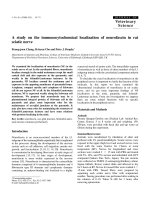
Insulin-immunoreactive cells
These cells were located in the central regions with
numerous frequency. In addition, cells showing a few
frequency were also demonstrated in the peripheral regions
(Fig. 1a). However, some insulin-immunoreactive cells were
randomly distributed throughout the whole pancreatic islets
in a case of relatively small islets, which were mainly
located between pancreatic duct and exocrine portions (Fig.
1b, c). In the exocrine portion, they were randomly scattered
between pancreatic acinar cells or interlobular connective
tissues with rare frequency (Fig. 1d). No insulin-
immunoreactive cells were detected in the pancreatic duct
portions.
Glucagon-immunoreactive cells
In case of relatively large pancreatic islets, they were
dispersed throughout central to peripheral regions intermingled
with other 3 types of immunoreactive cells and showed
regular relative frequencies between central and peripheral
regions, with moderate frequency (Fig. 2a-c). In case of
relatively small pancreatic islets, most of glucagon-
immunoreactive cells were situated in the peripheral
regions but no cells were found in the central regions (Fig.
2d). In the exocrine portion, they were randomly scattered
between pancreatic acinar cells or interlobular connective
tissues with a few frequency (Fig. 2e). No glucagon-
immunoreactive cells were detected in the pancreatic duct
portions.
Somatostatin-immunoreactive cells
These immunoreactive cells were located in the
peripheral regions with moderate frequency. However, no
somatostatin-immunoreactive cells were demonstrated in
the central regions where numerous insulin-immunoreactive
cells were found (Fig. 3a). In the exocrine portion, they were
randomly scattered between pancreatic acinar cells or
interlobular connective tissues with moderate frequency
(Fig. 3b). No somatostatin-immunoreactive cells were
detected in the pancreatic duct portions in this study.
PP-immunoreactive cells
Although some cells were demonstrated in the central
regions of pancreatic islets with rare frequency, most of
these cells were located in the peripheral regions with a few
frequency (Fig. 3c, d). In the exocrine portion, they were
randomly scattered between pancreatic acinar cells or
interlobular connective tissues with moderate frequency
(Fig. 3e). However, no cells were detected in the pancreatic
duct portions in this study.
DISCUSSION
Insulin is synthesized in the B cells of the pancreatic
islets and regulates the serum glucose levels
31
. In the mammals,
the regional distribution and relative frequency of
insulin-immunoreactive cells in the pancreas were reported in
the wood mouse
16
,hamster
15
,C57BL/6mouse
17
,voles
19
,ICR
mouse
24
, three-toed sloth
32
, Australian brush-tailed possum
33
,
opossum
34
and laboratory animals
29
.Fromtheseprevious
reports, it is well recognized that insulin cells are situated
in the central regions of pancreatic islets and other cells,
such as glucagon-, somatostatin- and PP-immunoreactive
cells, surround them. And they were also demonstrated
frequently associated with acinar cells and pancreatic duct.
However, somewhat different from other researchers, Reddy
Table 2. Regional distributions and relative frequencies of the endocrine cells in the pancreas of BALB/c mouse
Immunoreactive cells
Pancreatic islets portion
Exocrine portion Pancreatic duct portion
Central Peripheral
Insulin +++ + ± -
Glucagon ++ ++ + -
Somatostatin -++++ -
PP
1)
± ± ++ -
* Relative frequencies; +++: numerous, ++: moderate, +: a few, ±:rare,-: not detected
1)
PP: human pancreatic polypeptide.
170 Sae-Kwang Ku, Hyeung-Sik Lee and Jae-Hyun Lee
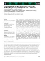
Fig. 1. Insulin-immunoreactive cells in the pancreas of th
e
BALB/c mice; Most of immunoreactive cells were situated i
n
the central regions of pancreatic islets (a) and they were
randomly dispersed in the whole pancreatic islets in som
e
case of the small islets (b; c is high magnification of b). I
n
addition, insulin-immunoreactive cells were also detected i
n
the exocrine portions (d). a, b: ×150; c, d: ×300, PA
P
method.
Fig. 2. Glucagon-immunoreactive cells in the pancreas o
f
the BALB/c mice; They were dispersed throughout central t
o
peripheral regions in a case of large islets (a ~ c) and in th
e
smaller ones, most of immunoreactive cells were situated i
n
the peripheral regions (d). In addition, some cells were
demonstrated in the exocrine portions (e). a ~ d: ×150; e:
×300, PAP method.
Immunohistochemical Study of the Pancreatic Endocrine cells in the BALB/c mice: An Unique Distributional Pattern of Glucagon 171
Fig. 3. Somatostatin- and PP-immunoreactive cells in the
pancreas of the BALB/c mice; Somatostatin- immunoreactiv
e
cells were located in the peripheral regions of the pancreati
c
islets regardless of their size (a) and some cells were also
demonstrated in the exocrine portions (b). Although some
cells were demonstrated in the central regions of pancreati
c
islets (c), most of PP-immunoreactive cells were located i
n
the peripheral regions (d). In addition, some PP-
immunoreactive cells were also detected in the exocrine
portions (e) a, c, d: ×150; b, e: ×300, PAP method.
et al
35
reported that they were observed in most islets
where they occurred as groups of cells peripherally and
within the pancreatic islets of several marsupial species. In
the present study, most of insulin-immunoreactive cells
were restricted to the central regions of islets similar to that
of previous rodents
15-19, 24, 28
. Different from other rodents,
where these cells were found in the lining epithelium of
pancreatic duct, no insulin-immunoreactive cells were
situated in the pancreatic duct portions. In addition, some
cells were randomly distributed in case of relatively small
islets of BALB/c mouse. According to the location of these
smaller islets, which were correlated with pancreatic duct in
this study and the previous reports that showed possibility
of insulin cells originated from pancreatic duct stem cells
36,
37
, these smaller ones were regarded as a juvenile or infant
typed cell clusters composed of insulin-immunoreactive cells.
Anyway, these differences compared to that of other rodents
were considered as peculiar distributional patterns of
BALB/c mouse.
Glucagon is synthesized in the A cells of the pancreas and
regulates glucose levels in blood
31
. Morphologically similar
cellsarealsoobservedinthedigestivetractofthedog.
Although glucagon-immunoreactive cells were located in the
mantle and peripheral regions of mammalian pancreatic
islets, exocrine portions and pancreatic duct
15-19, 24, 29, 32-34
,
species-dependent variations were also reported. In the
equine pancreas, A-cells demonstrated by anti-glucagon
were found in the center of pancreatic islets where in most
vertebrates, insulin-immunoreactive cells were numerously
found
38
. In addition, it was also reported that under specific
disease conditions such as obese (diabetic condition) mouse,
glucagon-immunoreactive cells were intermingled with
insulin-immunoreactive cells in the central regions of pancreatic
islets, in contrast, normal non-obese littermates showed a
peripheral localization of these immunoreactive cells
20
. Although
glucagon-immunoreactive cells were situated in the peripheral
regions in case of relatively small islets, they were randomly
distributed in the central to peripheral regions in case of
relatively large ones where numerous insulin-immunoreactive
cells were located and these results were different from
those of other mammals. In addition, they were not demon-
strated in the pancreatic duct portions. These distributional
patterns were considered as peculiar patterns of BALB/c
mouse.
Somatostatin, which consisted of 14 amino acids, was
isolated from hypothalamus of sheep for the first time. It
could be divided into straight form and cyclic form
39
.This
substance inhibited the secretion of the gastrin, cholecystokinin,
secretin, glucagon, insulin, motilin and gastric acid
40
and
the absorption of amino acid, glucose and fatty acid in the
gastrointestinal tract
41
. So far as investigated, somatostatin-
immunoreactive cells are located in the peripheral regions of
mammalian pancreatic islets and exocrine portions
15-19, 24, 29,
32-34
. Well corresponding to these previous studies, most of
somatostatin cells were found in the peripheral regions
172 Sae-Kwang Ku, Hyeung-Sik Lee and Jae-Hyun Lee
where intermingled with glucagon- and PP-immunoreactive
cells and they occupied outmost regions of pancreatic islets.
PP is a peptide hormone containing 36 amino acids,
which is synthesized by F cells in the pancreatic islets
31
.
The specific function of this peptide is not clear, however,
inhibition of food intake has been postulated as a possible
function of this peptide
31
.AndPolaket al
41
reported that it
promoted the secretion of gastric acid and stimulated the
glycolysis of liver in avian species. It has been revealed that
PP-immunoreactive cells were conspicuously distributed in
the peripheral regions of pancreatic islets and exocrine
portions in mammalian species, if they occurred
15-19, 24, 29, 33, 34
.
In addition, colocalization with serotonin in the pancreatic
islets of the opossum
34
and cattle
42
was also demonstrated.
Anyway, da Mota et al
32
reported that PP-immunoreactive
cells were not found in the pancreas of the three-toed sloth.
In the present study, well corresponding to previous
studies
15-19, 24, 29, 33, 34
, PP-immunoreactive cells were detected
in the outmost regions of pancreatic islets, although rare
frequencied cells were intermingled with other immunoreactive
cells in the central regions where insulin-immunoreactive
cells were most predominant.
In conclusion, some peculiar distributional patterns of
pancreatic endocrine cells, especially, glucagon-immunoreactive
cells, were demonstrated in the BALB/c mice.
Reference
1. Hilgers J, Arends JWA. A series of recombinant inbred
strains between BALB/cHeA and STS/A strains. Curr
Top Microbiol Immunol. 122:31-37, 1985.
2. Potter M. The BALB/c mouse. Current Topics in Microbiol
Immunol.122, Berin: Springer-Verlag, 1985.
3. Roderick TH. The response of twenty-seven inbred strains
of mice to daily doses of whole body X-irradiation.
Radiation Res. 20:631-639, 1963.
4. Storer JB. Longevity and gross pathology at death in 22
inbred strains of mice. J Gerontol. 21:404-409, 1966.
5. Yuhas JM, Storer JB. On mouse strain differences in
radiation resistance: hematopoietic death and endogenous
colony-forming unit. Radiation Res. 39:608-622. 1969.
6. Kobayashi K, Syed Ali S. Cell types of the endocrine
pancreas in the shark, Scylliorhinus stellaris as
revealed by correlative light and electron microscopy.
Cell Tissue Res. 215:475-490, 1981.
7. Orci L. Macro- and micro-domains in the endocrine
pancreas. Diabetes. 31:538-564, 1982.
8. Sternberger LA, Hardy PH, Cuculis JJ, et al.The
unlabeled antibody enzyme method of immunocy-
tochemistry: Preparation and properties of soluble
antigen-antibody complex (Horseradish peroxidase-
antihorseradish peroxidase) and use in identification of
spirochetes. J Histochem Cytochem. 18:315-333, 1970.
9. Ali-Rachedi A, Varndell IM, Adrian TE, et al.Peptide
YY (PYY) immunoreactivity is co-stored with glucagon-
related immunoreactants in endocrine cells of the gut
and pancreas. Histochemistry. 80:487-491, 1984.
10. Yamada J, Campos VJM, Kitamura N, et al.An
immunohistochemical study of endocrine cells in the
pancreas of Caiman latirostris (Alligatorinae), with
special reference to pancreatic motilin cells. Biomed
Res. 7:199-208, 1986.
11. Rindi G, Buffa R, Sessa F, et al.ChromograninA,B
and C immunoreactivities of mammalian emdocrine
cells: Distribution from costored hormones/prohormones
and relationship with argyrophil component of secretory
granules. Histochemistry. 85:19-28, 1986.
12. Ito H, Hashimoto Y, Kitagawa H, et al. Distribution of
chromogranin containing cells in the porcine gastroen-
teropancreatic endocrine system. Jpn J Vet Sci. 50:
395-404, 1987.
13. Jansson L, Sandler S. The influence of cyclosporin A on
the vascular permeability of the pancreatic islets and on
diabetes induced by multiple low dose of streptozotocin
in the mouse. Virchows Archiv A Pathol Anat
Histopathol. 412:225-230, 1988.
14. D'Este L, Buffa R, Pelagi M, et al. Immunohistochemical
localization of chromogranin A and B in the endocrine
cells of the alimentary tract of the green frog, Rana
esculanta. Cell Tissue Res. 277:341-349, 1994.
15. Camihort G, Del Zotto H, Gomez-Dumm CL, et al.
Quantitative ultrastructural changes induced by sucrose
administration in the pancreatic B cells of normal
hamsters. Biocell. 24:31-37, 2000.
16. Yukawa M, Takeuchi T, Watanabe T, et al.Proportions
of various endocrine cells in the pancreatic islets of
wood mice (Apodemus speciosus). Anat Histol Embryol.
28:13-16, 1999.
17. Gomez-Dumm CL, Console GM, Lunna GC, et al.
Quantitative immunohistochemical changes in the
endocrine pancreas of nonobese diabetic (NOD) mice.
Pancreas. 11:396-401, 1995.
18. Warbritton A, Gill AM, Yen TT, et al. Pancreatic islet
cells in preobese yellow Avy/- mice: relation to adult
hyperinsulinemia and obesity. Proc Soc Exp Biol Med.
206:145-151, 1994.
19.SasakiM,AraiT,UsuiT,et al. Immunohistochemical,
ultrastructural, and hormonal studies on the endocrine
pancreas of voles (Microtus arvalis) with monosodium
aspartate-induced diabetes. Vet Pathol. 28:497-505,
1991.
20. Starich GH, Zafirova M, Jabelenska R, et al.A
morphological and immunohistochemical investigation
of endocrine pancreas from obese ob+/ob+ mice. Acta
Histochem. 90:93-101, 1991.
21. Donev S, Petkov P, Marquie G, et al. Immuno-
histochemical investigations of the endocrine pancreas
in normoglycemic sand rat (Psammomys obesus). Acta
Diabetol Lat. 26:309-313, 1989.
22. Ohara N, Kitamura N, Yamada J, et al. Immuno-
Immunohistochemical Study of the Pancreatic Endocrine cells in the BALB/c mice: An Unique Distributional Pattern of Glucagon 173
histochemical study of gastroenteropancreatic endocrine
cells of the herbivorous Japanese field vole, Microtus
montebelli. Res Vet Sci. 41:21-27, 1986.
23. Reddy SN, Bibby NJ, Elliott RB. Cellular distribution of
insulin, glucagon, pancreatic polypeptide hormone and
somatostatin in the fetal and adult pancreas of the
guinea pig: a comparative immunohistochemical study.
Eur J Cell Biol. 38:301-305, 1985.
24. Ku SK, Lee HS, Lee JH. Immunohistochemical study of
the pancreatic endocrine cells in the ICR mice. Korean
JVetRes. 42:21-28, 2002.
25. Leung PS, Chan HC, Wong PY. Immunohistochemical
localization of angiotensin Ⅱ in the mouse pancreas.
Histochem J. 30:21-25, 1998.26. Ding WG, Guo LD,
Kitasato H, et al. Phylogenic study of calcitonin gene-
related peptide-immunoreactive structures in the pancreas.
Histochem Cell Biol. 109:103-109, 1998.
27. Shimizu K, Kato Y, Shiratori K, et al.Evidencefor
existence of CCK-producing cells in rat pancreatic islets.
Endocrinology. 139:389-396, 1998.28.FuQ,HondaM,
Ohgawara H, et al. Morphological analysis of pancreatic
endocrine cells in newborn animals delivered by
experimental diabetic rats. Diabetes Res Clin Pract.
31:57-62, 1996.
29. Wieczorek G, Pospischil A, Perentes EA. Comparative
immunohistochemical study of pancreatic islets in
laboratory animals (rats, dogs, minipigs, nonhuman
primates). Exp Toxicol Pathol. 50:151-172, 1998.
30. Sternberger LA. The unlabeled antibody peroxidase-
antiperoxidase (PAP) method. In: Sternberger LA (ed).
Immunocytochemistry, New York: John Wiley & Sons,
pp. 104-169, 1979.
31. Hsu WH, Crump MH. The endocrine pancreas. In:
McDonald LE, Pineda MH (eds). Veterinary endocrinology
and reproduction, Philadelphia: Lea & Febiger, pp.
186-201, 1989.
32.daMotaDL,YamadaJ,GergeLL,et al. An immuno-
histochemical study on the pancreatic endocrine cells of
the three-toed sloth, Bradypus variegatus. Arch Histol
Cytol. 55:203-209, 1992.
33. Leigh CM, Edwin NA. Light-microscopic immuno-
cytochemical study of the endocrine pancreas in the
Australian brush-tailed possum (Trichosurus vulpecula).
Eur J Histochem. 36:237-241, 1992.
34. Krause WJ, Cutts JH 3rd, Cutts JH, et al.Immuno-
histochemical study of the developing endocrine pancreas
of the opossum (Didelphis virginiana). Acta Anat
(Basel). 135:84-96, 1989.
35. Reddy S, Bibby NJ, Fisher SL, et al. Immunolocalization
of insulin, glucagon, pancreatic polypeptide and soma-
tostatin in the pancreatic islets of the possum, Trichosurus
vulpecula. Gen Comp Endocrinol. 64:157-162, 1986.
36. Slack JM. Developmental biology of the pancreas.
Development. 121:1569-1580, 1995.
37. Regitnig P, Spuller E, Denk H. Insuloma of the pancreas
with isular-ductular differentiation in its liver metastasis
- idication of a common stem-cell origin of the exocrine
and endocrine components. Virchows Arch. 438:624-
628, 2001.
38. Helmstaedter V, Feurle GE, Forssmann WG. Insulin-,
glucagon- and somatostatin-immunoreactive cells in the
equine pancreas. Cell Tissue Res. 172:447-454, 1976.
39. Brazeau P, Vale W, Burgurs R, et al.Hypothalamic
polypeptide that inhibits the secretion of immunoreactive
pituitary growth hormone. Science. 179:77-79, 1973.
40. Kitamura N, Yamada J, Calingasan NY, et al. Immuno-
cytochemical distribution of endocrine cells in the
gastrointestinal tract of the horse. Equine Vet J. 16:
103-107, 1984.
41. Polak JM, Adrian TE, Bryant MG, et al.Pancreatic
polypeptide in the insulomas, gastrinomas and gluca-
gonomas. Lancet. 1:328-330, 1976.
42. Nakajima S, Kitamura N, Yamada J, et al.Immuno-
histochemical study on the endocrine pancreas of cattle
with special reference to coexistence of serotonin and
glucagon or bovine pancreatic polypeptide. Acta Anat
(Basel); 131:235-240, 1988.