Báo cáo khoa học: " Influence of Isoflurane Anesthesia on Pulsatility Index and Peak Systolic Velocity of Basilar Artery in Dogs by Doppler Ultrasonography" pdf
Bạn đang xem bản rút gọn của tài liệu. Xem và tải ngay bản đầy đủ của tài liệu tại đây (143.51 KB, 3 trang )
JOURNAL OF
Veterinary
Science
J. Vet. Sci. (2002), 3(3), 203-205
Abstract
9)
This study was performed to examine the influence
of isoflurane anesthesia on the pulsatility index (PI)
and the peak systolic velocity (PSV) of the blood flow
in the basilar artery of dogs by duplex Doppler
ultrasonography. Twelve healthy dogs were used to
measure the PI and the PSV under the conscious
state and isoflurane anesthesia. The pulsatility index
(PI) and the peak systolic velocity (PSV) in the basilar
artery were measured five times with random intervals.
The blood pressure was measured. The PI and PSV
values in dogs under isoflurane anesthesia were 1.37
±
0.32 and 72
±
19 cm/sec, whereas those in the
consciousdogswere1.37
±
0.13 and 81
±
16 cm/sec,
respectively. The indirect mean arterial systolic and
diastolic pressures under isoflurane anesthesia were
107 and 51 mmHg, whereas those in the conscious
dogs were 133 and 74 mmHg. Though the isoflurane is
generally known to induce hypotension, there were
no significant differences in the PI and PSV between
the isoflurane-anesthetized and the conscious dogs. In
conclusion, the isoflurane anesthesia did not influence
the PI and PSV in the basilar artery of dogs.
Key words: Dog, Doppler ultrasonography, isoflurane,
peak systolic velocity, pulsatility index
Introduction
It has been reported that the pulsatility index (PI) and
the peak systolic velocity in the basilar artery measured to
examinebraindamagecanbeusedforthepredictionand
the diagnosis of brain damage in the dog by Doppler
ultrasonography [4]. Concerning the isoflurane anesthesia,
it has been known that the arterial pressure and vascular
*
Corresponding author:
Department of Radiology, College of Veterinary Medicine, Seoul
National University San 56-1 Shillim-dong, Kwanak-gu, Seoul
151-742, Korea
Tel: +82-2-880-8692, Fax: +82-2-880-8662,
E-mail:
resistance, which is reflected in the PI, were decreased in
dogs [9, 11]. In order to examine the abnormal velocity and
pulsatility index in the basilar, knowledge of baseline values
are of important. However, only a few studies were performed
to establish the normal major blood flow profile in dogs by
Doppler ultrasonography [12, 13]. Besides, the normal range
and anesthetic influence on the peak systolic velocity and
the pulsatility index in the basilar artery have not even
been established.
The aim of this study is to examine how the isoflurane
anesthesia influences on the pulsatility index and the peak
systolic velocity of the blood flow in the basilar artery of the
dogs by Doppler ultrasonography.
Materials and Methods
Experiment Animals
Twelve healthy one-year-old beagle dogs, weighing 6.4~10
kg, were used without sex discrimination. All of the dogs
were considered to be normal following physical and
hemodiagnostic (complete blood count) examinations and a
Dirofilaria immitis immunodiagnostic test (Snap; IDEXX
Laboratories Co., USA).
Doppler Ultrasound
Doppler ultrasonography was performed using a Toshiba
260A with a 3.5 MHz sector transducer (5.66-25.00 kHz
pulse repetition frequencies, 100 Hz wall filter). Doppler
waveforms were recorded at gains in which noises first
became apparent and at pulse repetition frequencies that
were sufficient to prevent aliasing.
BasilarArtery(BA)
Dogs were placed in right lateral recumbency. The basilar
artery was examined through the foramen magnum acoustic
window. Following an initial B-mode examination, color flow
Doppler was performed to identify the vessels of interest.
Once identified, pulsed wave Doppler was initiated and the
waveform analysis was performed after freezing the image.
The Doppler angle was maintained between 30-40 degrees.
Measurements such as PSV, mean velocity and end diastolic
velocity (EDV) were made on a representative spectral
Influence of Isoflurane Anesthesia on Pulsatility Index and Peak SystolicVelocityof
Basilar Artery in Dogs by Doppler Ultrasonography
Ki-Chang Lee, Min-Cheol Choi
*
and Jung-Hee Yoon
College of Veterinary Medicine, Seoul National University Seoul 151-742,Korea
Received July 25, 2002 / Accepted September 2, 2002
204 Kichang Lee, Mincheol Choi and Junghee Yoon
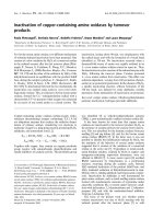
waveform (Fig. 1). The waveform analyses were performed
5 times at random intervals. The PI value was calculated by
the equation, PI = (PSV - EDV)/ mean velocity.
Fig. 1. Pulsatility index (PI) measurement at basilar artery.
Drawing of calipers from the peak (a) to the end diastolic
spectrum(c) to measure the PI of the blood flow in the
basilar artery is shown.
a: peak systolic velocity, b: mean velocity, c: end diastolic
velocity
Inhalation Anesthesia
Twelve beagle dogs were anesthetized with isoflurane.
Before performing experiments, feed was withheld for 12
hours. For the inhalation anesthesia, a semi-open circle
anesthetic system (Anesthesia Apparatus FO-20S, Acoma
Medical Industry Co., Tokyo, Japan), with Tec-type vaporizer
for isoflurane (Acoma Vaporizer 1 MK- Ⅲ,AcomaMedical
Industry Co., Tokyo, Japan), out of circle, was used for the
whole period of experiment. Induction was produced by 4%
isoflurane (Aerane
®
, Choongwae medical Co. Ltd., Seoul, Korea)
in oxygen via facemask without any preanesthetics. After
induction of anesthesia, endotracheal tube was inserted and
the dog was placed in the right lateral recumbency.
Lactated Ringer s solution was administered intravenously
at a rate of 5 ㎖/㎏/h. Body temperature was maintained at
approximately 38 ±0.5℃ with a water pad and blanket.
Two percent of isoflurane in oxygen was delivered via
endotracheal tube for at least 60 minutes.Determination of
baseline MAC was initiated at 1.5% isoflurane and was
duplicated following the method of Eger et al. (1980).
Respiratory gases were monitored continuously using a gas
analysis module (M-CaiOV, Datex-Ohmeda, Helsinki, Finland)
connected to an anesthetic patient monitoring system (S-3,
Datex-Ohmeda, Helsinki, Finland).
Blood Pressure
Blood pressure was measured by an indirect (i.e.,
oscillometric) blood pressure monitor [10]. Dogs were placed
in right lateral recumbency. Limb circumference over the
left dorsal pedal artery was measured, and a cuff width of
approximately 40% to 60% of the limb circumference was
chosen. Three readings were taken at 5-minute intervals.
Systolic and diastolic blood pressures were recorded. One of
the authors (Lee) took all the measurements to obtain all
values in an identical fashion.
Data Analysis
Statistical evaluation was performed using the SPSS
statistical computer program. A one-way ANOVA (post-hoc
scheffe) was applied to data analyses.
Results
Pulsatility Index and Peak Systolic Velocity of
Blood Flow in Basilar Artery
The mean PI and PSV values under isoflurane anesthesia
(1.5 %) were 1.37 0.32 (mean SD) and 72 19 (mean SD)
cm/sec and those in the conscious dogs were 1.370.13 and
8116 cm/sec, respectively (Fig. 2). The Doppler angle had
the range of 30~40°.Therearenosignificantdifferencesof
PI and PSV values between isoflurane-anesthetized and the
conscious dogs.
Fig. 2. Duplex Doppler image of the basilar artery
A spectral waveform for velocity measurement in basilar
artery (empty arrow) is shown on the left in figure 2. Real
time image and sample gate (arrow) are on right side of the
each image. The basilar artery showed typical parabolic
velocity profile and low resistance flow pattern.
Blood Pressure
Mean values of blood pressure under isoflurane anesthesia
were 107 ± 15 (mean ± SD) mmHg in systole and 51 ±
12 (mean ± SD) mmHg in diastole, respectively. In
conscious dogs, the values were 137 ± 13 (mean ± SD)
mmHg in systole and 78 ± 15 (mean ± SD) mmHg in
diastole. The systolic and diastolic blood pressures decreased
significantly in isoflurane group (p<0.05) compared to the
normal value.
Discussion
It has been generally known that isoflurane anesthesia
induced hypotension and decreased vascular resistance [5, 7,
9, 11]. This study showed the significantly decreased blood
pressures in the isoflurane-anesthetized dogs, which are
Influence of Isoflurane Anesthesia on Pulsatility Index and Peak Systolic Velocity of Basilar Artery in Dogs by Doppler Ultrasonography
205
accordant with the results of the other reports.
Evans et al. and Blohme et al. reported that the PI is one
of the indicators of peripheral resistance [1, 3]. Theoretically,
it is known that the PI value is inversely proportional to the
vascular resistance. That is to say, when the vascular
resistance is decreased, the PI value is increased, and vice
versa. The PI of the basilar artery under isoflurane anesthesia
was expected to increase, but it showed the negligible
difference in anesthetized dogs when compared to normal
value. Though there are some controversies concerning the
PI in intra- and/or extra-cranial arteries, it is important to
know the normal value in the basilar artery for examining
the cerebral blood flow indirectly. In this study, the mean PI
and peak velocity in the basilar artery were 1.37 ±0.13 and
72 ± 19 cm/sec, respectively. And these values under
anesthesia were not significantly different from those of the
normal values.
Many controversies have been reported about isoflurane
anesthetic influences on the cerebral blood flow and velocity.
Jones et al. reported that isoflurane caused cerebral
vasodilatation and an increase in cerebral perfusion in dogs
[8]. Meanwhile, Thiel et al. reported isoflurane caused little
change on the blood flow and the velocity in the middle
cerebral artery of humans [14]. To the contrary, Holzer et
al.andNewberget al. stated that isoflurane decreased the
cerebral blood flow in human [6,11].
Though intracranial cerebral blood flow velocity was not
measured in this study, it could be deduced that isoflurane
did not affect the PSV as well as the PI in the basilar artery
when compared to those in the normal one. This means the
blood flow of the extra cranial artery like basilar artery was
not affected under isoflurane anesthesia.
Conclusively, the decreased blood pressure under isoflurane
anesthesia did not influence on the PI and PSV in the
basilar artery. Therefore isoflurane anesthesia can be used
safely without any changes of blood flow.
References
1.Blohme,L.,Pagani,M.,Parra-Hoyos,H.,Olofsson,
P., Takolander, R., and Swedenborg, J. Changes in
middle cerebral artery flow velocity and pulsatility
index after carotid endarterectomy. Eur. J. Vasc. Surg.
1991, 5,659-663.
2. Cheung, A.T., Levy, W.J., Weiss, S.J., Barclay, D.K.,
and Stecker, M.M. Relationships between cerebral
blood flow velocities and arterial pressures during
intra-aortic counterpulsation. Cardiothorac Vasc Anesth.
1998, 12(1), 51-57.
3. Evans, D.H., Barrie, W.W., Asher, M.J., Bentley, S.,
and Bell, P.R. The relationship between ultrasonic
pulsatility index and proximal arterial stenosis in a
canine model. Circ. Res. 1980, 46, 470-475.
4. Fukushima, U., Sasaki, S., Okano, S., Oyamada, T.,
Yoshikawa, T., Hagio, M., and Takase, K. Non-
invasive diagnosis of ischemic brain damage after
cardiopulmonary resuscitation in dogs by using transcranial
Doppler ultrasonography. Vet. Radiol. Ultrasound. 2000,
41(2), 172-177.
5.Hellyer,P.,Muir,W.W.3rd,Hubbell,J.A.,and
Sally, J. Cardiorespiratory effects of the intravenous
administration of tiletamine-zolazepam to dogs. Vet.
Surg. 1989, 18(2), 160-165.
6. Holzer, A., Greher, M., Hetz, H., Standhardt, H.,
Donner, A., Heinzl, H., Zimpfer, M., and Illievich,
U.M. Influence of aortic blood flow velocity on changes
of middle cerebral artery blood flow velocity during
isoflurane and sevoflurane anesthesia. Eur. J. Anaesthesiol.
2001, 18(4), 238-244.
7. Hysing, E.S., Chelly, J.E., Doursout, M.F., and
Merin, R.G. Comparative effects of halothane, enflurane,
and isoflurane at equihypotensive doses on cardiac
performance and coronary and renal blood flows in
chronically instrumented dogs. Anesthesiology. 1992, 76(6),
979~984.
8. Jones, R.S. and Snowdon, S.L. Experimental inve-
stigation of the cardiovascular and respiratory effects of
increasing concentrations of isoflurane in the dog. Res.
Vet. Sci. 1986, 40(1), 89-93.
9. Klide, A.M. Cardiopulmonary effects of enflurane and
isoflurane in the dog. Am. J. Vet. Res. 1976, 37(2),
127-131.
10. Meurs, K.M., Miller M.W., Slater, M.R., and Glaze
K. Arterial Blood pressure measurement in a population
of healthy geriatric dogs. Am. Anim Hosp Assoc. 2000,
36, 497-500.
11. Newberg, L.A., Milde, J.H., and Michenfelder, J.D.
Systemic and cerebral effects of isoflurane-induced
hypotension in dogs. Anesthesiology. 1984, 60(6), 541-
546.
12. Spaulding K.A. A review of sonographic identification
of abdominal blood vessels and juxtavascular organs.
Vet. Radiol. Ultrasound. 1997, 38(1), 4-23.
13. Szatmari, V., Sotonyi, P., and Voros, K. Normal
duplex Doppler waveforms of major abdominal blood
vessels in dogs: a review. Vet. Radiol. Ultrasound. 2001,
42(2), 93-107.
14. Thiel, A., Zickmann, B., Zimmermann, R., and Hem-
pelmann, G. Transcranial Doppler sonography: effects
of halothane, enflurane and isoflurane on blood flow
velocity in the middle cerebral artery. Br .J .Anaesth.
1992, 68(4), 388-393.