Báo cáo khoa học: " Identification of a putative cellular receptor 150 kDa polypeptide for porcine epidemic diarrhea virus in porcine enterocytes" pps
Bạn đang xem bản rút gọn của tài liệu. Xem và tải ngay bản đầy đủ của tài liệu tại đây (583.69 KB, 7 trang )
-2851$/ 2)
9H W H U L Q D U \
6FLHQFH
J. Vet. Sci.
(2003),
/
4
(3), 269–275
Short Communication
Identification of a putative cellular receptor 150 kDa polypeptide for
porcine epidemic diarrhea virus in porcine enterocytes
Jin Sik Oh, Dae Sub Song and Bong Kyun Park*
Department of Microbiology, Virology Lab, College of Veterinary Medicine and School of Agricultural Biotechnology,
Seoul National University, Seoul 151-742, Korea
Porcine epidemic diarrhea virus (PEDV) causes an
acute enteritis in pigs of all ages, often fatality for
neonates. PEDV occupies an intermediate position
between two well characterized members of the
coronavirus group I, human coronavirus (HCoV-229E)
and transmissible gastroenteritis virus (TGEV) which
uses aminopeptidase N (APN), a 150 kDa protein, as their
receptors. However, the receptor of the PEDV has not
been identified yet. A virus overlay protein binding assay
(VOPBA) was used to identify PEDV binding protein in
permissive cells. The binding ability of PEDV to porcine
APN (pAPN) and the effects of pAPN on infectivity of
PEDV in Vero cells were also investigated. VOPBA
identified a 150 kDa protein, as a putative PEDV receptor
in enterocytes and swine testicle (ST) cells. Further the
PEDV binding to pAPN was blocked by anti-pAPN and
pAPN enhanced PEDV infectivity in Vero cells. In
conclusion, these results suggested that pAPN may act as
a receptor of PEDV.
Key words:
PEDV, cellular receptor, porcine aminopeptidase N
Porcine epidemic diarrhea virus (PEDV), a member of
the family
Coronaviridae
is an enveloped and single-
stranded RNA virus [10]. It causes severe diarrhea in pigs,
especially in newborn pigs. PEDV and transmissible
gastroenteritis virus (TGEV) are not serologically related
to each other, though both infect digestive tract and induce
very similar clinical signs [5].
6HYHUDOPHPEHUVRIFRURQDYLUXVVHURJURXS,LQFOXGLQJ
KXPDQ FRURQDYLUXV +&R9( 7*(9 DQG IHOLQH
LQIHFWLRXVSHULWRQLWLVYLUXV),39XVHDPLQRSHSWLGDVH1
$31 DV WKHLU FHOOXODU UHFHSWRU >@ 7KH KLJKHVW $31
DFWLYLW\ LV GHWHFWHG LQ WKH EUXVK ERUGHU PHPEUDQH RI WKH
HQWHURF\WHV DQG WKH UHQDO SUR[LPDO WXEXOH FHOOV >@
3UHYLRXVO\WKHVHTXHQFLQJRIWKH0V025)DQG1JHQH
RI&9VWUDLQRI3('9ZDVFRPSOHWHG7KH60V0
25)DQG1JHQHVRID%ULWLVKVWUDLQRI3('9DOVREHHQ
VHTXHQFHG >@ 7KHVH GDWD LQGLFDWHG WKDW 3('9
RFFXSLHV DQ LQWHUPHGLDWH SRVLWLRQ EHWZHHQ WZR ZHOO
FKDUDFWHUL]HGPHPEHUVRIWKHJURXS,FRURQDYLUXV+&R9
(DQG7*(9%RWK+&R9(DQG7*(9XVH$31
DVWKHLUUHFHSWRU>@
A virus overlay protein binding assay (VOPBA) was
used for identifying the putative cellular receptor in several
viruses [14]. And Schenten
et al
. reported that the soluble
form receptor could enhance the infection of HIV (human
immunodeficiency virus) [26].
The objectives of this study were to identify a cellular
receptor in permissive cells using VOPBA and to
determine whether the PEDV infectivity would be
enhanced by soluble porcine APN treatment on Vero cells.
The continuous Vero cell line (ATCC, CCL-81) was
regularly maintained in
α
-MEM (minimal essential
medium) supplemented with 5% fetal bovine serum (FBS),
and 2% antibiotic-antimycotic agent mixture (penicillin,
10,000 IU/ml; streptomycin, 10,000
µ
g/ml; and
amphotericin B, 25
µ
g/ml; Invitrogen, Grand Island, N.Y.).
PEDV strain KPEDV-9 which was used for this study has
been endorsed to the Green Cross Veterinary Product Co.,
Ltd. (Suwon, Korea) for manufacturing PEDV live vaccine
by the National Veterinary Research and Quarantine
Service (Anyang, Korea). KPEDV-9 was propagated in
Vero cells with virus replication medium (VM),
α
-MEM
supplemented 0.02% yeast extract, 0.3% tryptose
phosphate broth and 2
µ
g of trypsin (T-VM), as described
previously [22]. And KPEDV-9 was propagated in Vero
cells with VM containing pAPN (A-VM) instead of
trypsin. ST (swine testicle) and PK-15 (porcine kidney)
cells were grown in MEM supplemented with 5% FBS,
and 2% antibiotic-antimycotic agent mixture. TGEV,
Pyungtak 45 strain was cultured in ST cells.
7KH PRQRFORQDO DQWLERGLHV IRU YLUXV RYHUOD\ SURWHLQ
ELQGLQJDVVD\923%$RI7*(9DQG3('96SURWHLQ
ZHUH SURYLGHG E\ WKH 1DWLRQDO 9HWHULQDU\ 5HVHDUFK DQG
*Corresponding author
Phone: +82-2-880-1255; Fax: +82-2-885-0263
E-mail:
270 Jin Sik Oh
et al.
4XDUDQWLQH 6HUYLFH $Q\DQJ .RUHD 3RO\FORQDO DQWLERG\
WR 3('9 ZDV SUHSDUHG LQ UDEELWV XVLQJ .3('9 VWUDLQ
>@ $QWLS$31 PRQRFORQDO DQWLERG\ ZDV NLQGO\
SURYLGHGIURP'U'HOPDV,15$)UDQFH)RUWKHDVVD\
RI EORFNLQJ RI 3('9 ELQGLQJ WR S$31 DQWLS$31
SRO\FORQDODQWLERG\ZDVSUHSDUHG LQUDEELWVXVLQJS$31
HPXOVLILHGZLWK)UHXQG¶VDGMXYDQW
The method of Kessler [17] was used to prepare the brush
border membrane. In brief, the small intestines of 10 days
old piglets were collected and rinsed 7 times with cold
saline. Mucosa was removed from the tissue by gentle
scraping with the edge of slide glass. The tissue was placed
in a volume (9 ml) equivalent to three times the weight of
tissue (3 g) of mannitol buffer (2 mM Tris-HCl, 50 mM
mannitol, leupeptin (1
µ
g/ml), pepstatin A (0.7
µ
g/ml),
trypsin inhibitor (2.5
µ
g/ml), and 0.1 mM
phenylmethanesulfonyl fluoride (PMSF)). The tissue was
homogenized and diluted with five volumes of mannitol
buffer (50 mM, pH 5.6) and homogenized once again. The
final homogenate was incubated for 20 min on ice in the
presence of 10 mM MgCl
2
and then centrifuged at 3,000
×
g
for 15 min. The supernatant was collected and centrifuged
for 30 min at 27,000
×
g. The pellet, representing the crude
brush border membrane, was washed once by using the
mannitol buffer and stored at
−
20
o
C until use. Porcine APN
(pAPN) was purchased from Sigma (USA). The powder
form of pAPN was rehydrated and diluted to optimal
concentrations for each experiment with phosphate buffered
saline (PBS, pH 7.4) for each experiment. All protein
quantifications were performed by using BCA protein assay
kit (Pierce, USA) according to the manufacturer’s
instruction.
To identify cellular proteins involved in PEDV binding,
VOPBA was carried out. In brief, membrane proteins of
cells were separated by SDS- PAGE. Cellular membranes
of porcine brush border, ST, Vero, and PK-15 cells were
boiled in 4X nonreducing sample buffer (4% sodium
dodecyl sulfate, 10% glycerol, 0.625 M Tris-HCl, pH 6.8)
and loaded on 8.5% polyacrylamide gels. After
electrophoresis, the proteins were transferred onto a
polyvinylidene difluoride membrane (PVDF, Nen Life
Science, USA) at 45 V for 17 hours at 4
o
C in a buffer
containing 25 mM Tris, 192 mM glycine, and 20% (v/v)
methanol. Nonspecific binding sites were blocked by
incubating the membrane in PBS containing 5% skim
milk, 1% bovine serum albumin, and 0.05% Tween 20 for
1 h. The membranes were incubated for 1 h with PEDV
(10
5.5
TCID
50
/ml) or MEM, as a negative control,
containing 20 mM HEPES (N-2-hydroxyethyl-piperazine-
N'-2-ethane-sulfonic acid) and 0.2% (w/v) sodium
bicarbonate. The PVDF membrane was washed three
times for 5 min each with PBS containing 0.05% Tween 20
(PBST), and incubated with normal mouse serum or
PEDV monoclonal antibody. After washing three times
with PBST, horse-peroxidase labeled goat anti-mouse
IgGs (KPL, USA), diluted to 1 : 5,000 in PBST, were
added and incubated for 1 h at 37
o
C. Finally, substrate
(ECL Western blotting detection reagents, Amersham,
USA) was added. Developing was performed on the ECL
film (Amersham, USA). As a control, VOPBA of TGEV
was performed in porcine enterocytes using the same
method as VOPBA of PEDV.
To detect the binding of PEDV to pAPN, direct virus-
binding studies were carried out by enzyme linked
immunosorbent assay (ELISA). A micro-ELISA plate
(Nalge Nunc International, USA) was coated with 0.5
µ
g
of pAPN per well in carbonate-bicarbonate buffer (pH
9.6). After overnight incubation at 4
o
C, it was washed 5
times with PBST. Blocking step was done using 3%
gelatin in PBST. After washing, 10-fold serial diluted
PEDV infected cell lysate (10
5.5
TCID
50
/0.1 ml) or mock
infected cell lysate with PBST was added in 100
µ
l
volumes, and incubated for 60 min at 37
o
C. Before the
binding assay, PEDV and mock infected medium had been
centrifuged 12,000
×
g for 30 min to remove cell debris.
The plates were washed and subsequently incubated with
100
µ
l of 1 : 50 diluted PEDV monoclonal antibody at
37
o
C for 60 min. The plates were washed and further
incubated with 100
µ
l of horse-peroxidase labeled goat
anti-mouse IgGs (KPL, USA) for 60 min. After washing
the plate, ABTS substrate (2 mM 2,2-azino-di-3-ethyl-
benzthiazole-sulfonate in 20 mM acetate (pH 4.2) plus 2.5
mM H
2
O
2
) solution was added and incubated for 20 min at
room temperature. The reactions were stopped using 0.5 M
H
2
SO
4
and optical density was measured at 405 nm.
7R WHVW EORFNLQJ DFWLYLW\ RI DQWLS$31 IRU ELQGLQJ RI
3('9WRS$31S$31FRDWHGSODWHVZHUHLQFXEDWHGZLWK
IROG VHULDOO\ GLOXWHG UDEELW DQWLS$31 SRO\FORQDO
DQWLERG\RUZLWKQRUPDOUDEELWVHUXPIRU PLQDW
&
7KHUHPDLQLQJVWHSVRIWKH(/,6$WHVWZHUHFDUULHGRXWDV
GHVFULEHGDERYH
The effects of pAPN on PEDV replication were
investigated in Vero cells. KPEDV-9 infected Vero cells
were grown with A-VM in an experiment I. Vero cells
were pretreated with pAPN before PEDV inoculation in an
experiment II. As controls, KPEDV-9 was propagated in T-
VM as described in a previous study [22].
In the experiment I, after inoculation with PEDV at a
dose of 10
3.7
TCID
50
, Vero cells were incubated in the A-
VM with pAPN concentrations ranging 0.024 pg/ml to 2.4
pg/ml. In the experiment II, Vero cell cultures were
pretreated with pAPN at the concentrations ranging from
10 ng/ml to 1 mg/ml for 1, 2, or 3 h at 37
o
C. The cultures
were washed three times with PBS and inoculated with
PEDV at a dose of 10
3.7
TCID
50
. After adsorption at 37
o
C
for 1 h, the cultures were washed three times with PBS and
fed with VM. Virus showing 80% cytopathic effect (CPE)
in both experiments was harvested and titrated.
A cellular receptor of PEDV 271
To define the effects of pAPN in Vero cells, one-step
growth curve of PEDV was carried out as described
previously [15]. In an experiment III, the monolayered
Vero cells in 6 well multiplates (Falcon, N.J., USA) were
washed with PBS and inoculated with 1 ml of PEDV (10
3.5
TCID
50
/ml) for 1 h at 37
o
C. After infection with PEDV into
Vero cells, the cells were incubated with A-VM containing
2.4 pg/ml of pAPN.
In an experiment IV, the confluent monolayers of Vero
cells were washed with PBS and treated with 10
µ
g/ml of
pAPN for 1 h. After washing with PBS, Vero cells were
inoculated with 1 ml of PEDV (10
3.5
TCID
50
/), and
adsorbed for 1 h at 37
o
C. After adsorption, monolayers
were washed twice with PBS and incubated with 2 ml of
VM without trypsin. As a control, T-VM was added to the
plates which did not pretreat with pAPN.
$OOFHOOFXOWXUHVZHUHLQFXEDWHGDW
o
C
IRU
DQG K $W WKH HQG RI HDFK
LQFXEDWLRQ SHULRGWKH PHGLDZHUH KDUYHVWHG FHQWULIXJHG
DWUSPIRUPLQDQGVXSHUQDWDQWVZHUHVWRUHGDW
o
C
IRU WKH WLWUDWLRQ RI H[WUDFHOOXODU (& 3('9 )RU
LQWUDFHOOXODU,&YLUXVWKHFHOOSHOOHWVZHUHUHVXVSHQGHGLQ
PORIIUHVK90&HOOVVWLOODGKHULQJWRWKHERWWRPRIWKH
SODWHZHUHZDVKHGWZLFHZLWKIUHVK90VFUDSHGRIIZLWKD
FHOOVFUDSHU&RVWDU86$ WKHQIUR]HQDQGWKDZHGWKUHH
WLPHVWRUHOHDVH,&YLUXVSDUWLFOHV7KHHQVXLQJVXVSHQVLRQ
ZDVFODULILHGE\FHQWULIXJDWLRQDQGWLWUDWHG
The virus titration was carried out at the 96 well
microplate using Vero cells as described previously [21].
PEDV propagated with VM, A-VM or T-VM was diluted
to serial ten-folds with VM. Confluent Vero cells were
washed three times with PBS and inoculated with 0.1 ml
inoculum into 5 wells each. Following adsorption for 1 h at
37
o
C, the inocula were removed and the monolayers were
washed three times with PBS. Then, 0.1 ml of T-VM was
added to each well and the cultures were incubated for 5
days at 37
o
C. Fifty % tissue culture infective doses
(TCID
50
) were expressed as the reciprocals of the highest
virus dilution showing CPE.
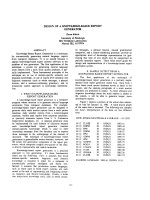
The PEDV binding protein was detected in porcine
enterocytes and ST cells. Interestingly, PEDV bound to a
150 kDa protein in porcine enterocyte. However, PEDV
binding to a 66 kDa band was more dominant rather than
that to a 150 kDa band in ST cells. No PEDV binding
proteins were detected in Vero cells (Fig. 1).
$VDSRVLWLYHFRQWUROLQWKH923%$DUHFHSWRURI7*(9
ZDVGHWHFWHG7KHN'D7*(9VSHFLILFELQGLQJSURWHLQ
ZDVLGHQWLILHGLQSRUFLQHHQWHURF\WHVDQG67FHOOVE\XVLQJ
7*(9PRQRFORQDODQWLERG\
$ VHULHV RI YLUXVELQGLQJ VWXGLHV ZDV SHUIRUPHG WR
FKDUDFWHUL]HWKHUHFHSWRUOLJDQGLQWHUDFWLRQVRI.3('9
7KHDPRXQWRIS$31ERXQGYLUXVZDVGHWHUPLQHGGLUHFWO\
E\ PHDVXULQJ WKH RSWLFDO GHQVLW\ 'DWD IURP DEVRUEDQFH
VKRZHGWKDWWKHELQGLQJRI.3('9WRS$31ZDVGRVH
GHSHQGHQWDQGLQFUHDVHGWR
7&,' PODW QJRI
S$31 FRQFHQWUDWLRQ )LJ +RZHYHU WKH VDWXUDWLRQ RI
3('9 ELQGLQJ ZDV QRW UHDFKHG XQGHU WKH FRQGLWLRQ
HPSOR\HG0RFNLQIHFWHGPHGLDZDVQRWERXQGWRS$31
7KHELQGLQJRI3('9WRS$31FRXOGEHEORFNHGE\UDEELW
DQWLS$31SRO\FORQDODQWLERG\XSWRGLOXWLRQVLQ
(/,6$
$IWHUWUHDWPHQWRIVROXEOHS$31DQGWU\SVLQHDFKYLUXV
ZDV KDUYHVWHG ZKHQ WKH &3(ZDVREVHUYHG3('9
ZDV SUROLIHUDWHG VLJQLILFDQWO\ E\ DGGLWLRQ RI VROXEOH
S$31
In the experiment I, infectious titers of PEDV grown in
A-VM ranged from 10
5.1
TCID
50
/0.1 ml at 2.4~0.024 pg/ml
of pAPN concentrations. The maximum PEDV titer was
10
5.3
TCID
50
/0.1 ml in the A-VM at 2.4 pg/ml of pAPN
concentration. As controls, the titers of PEDV were 10
4.1
TCID
50
/0.1 ml in T-VM, and 10
1.0
TCID
50
/0.1 ml in VM
without trypsin and pAPN (Fig. 3).
,Q WKH H[SHULPHQW ,, WKH KDUYHVWHG 3('9 JURZQ LQ
S$31 SUHWUHDWHG 9HUR FHOOV UDQJHG
7&,' PO
DFFRUGLQJWRWKHFRQFHQWUDWLRQRIS$31+RZHYHU3('9
JURZQ LQ 790 ZDV
7&,' PO 7KH WLWHU RI
3('9 FXOWXUHG LQ 90 ZLWKRXW WU\SVLQ ZDV
7&,'
POLQ9HURFHOOVZKLFKZDVQRWSUHWUHDWHGZLWKS$31
F
ig. 1.
Virus overlay protein binding assay. (a) TGEV using monoclonal antibody. Lane 1,2 porcine enterocytes, Lane 3-ST cells, La
ne
4
-Vero cells, Lane 5-negative control. (b) PEDV using monoclonal antibody. Lane 1,2-porcine enterocytes, Lane 3-Vero cells, Lane
4-
S
T cells, Lane 5, 6-negative control. (c) PEDV using polyclonal antibody. Lane 1-negative control, Lane 2-Vero cells, Lane 3,4-S
T
c
ells, Lane 5,6-porcine enterocyte.
272 Jin Sik Oh
et al.
7KHPD[LPXP3('9WLWHUZDVREWDLQHGZLWK 7&,'
PO DW µJPO FRQFHQWUDWLRQ RI S$31 RQ 9HUR FHOO
EHIRUH 3('9 LQRFXODWLRQ 7KH EHVW WLPH RI S$31
WUHDWPHQWZDVGHWHUPLQHGZLWKKDWWKHFRQFHQWUDWLRQRI
S$31HPSOR\HG)LJ
7KH YLUXV JURZWK SDWWHUQV ZHUH YHU\ VLPLODU LQ ERWK
H[SHULPHQWV ,Q WKH H[SHULPHQW ,,, 3('9 ZDV FXOWXUHG
ZLWK S$31 SJPO VLPXOWDQHRXVO\ LQ 90 7KH
UHSOLFDWLRQNLQHWLFVLVLOOXVWUDWHGLQ)LJ,QWUDFHOOXODU,&
3('9 JURZWK SDWWHUQV ZHUH YHU\ VLPLODU WR WKRVH RI $
90DQG790)URPVL[KDIWHUDGVRUSWLRQWKHDPRXQWRI
,&3('9EHJDQWRLQFUHDVHDQGUHDFKHGWKHSHDNEHWZHHQ
DQGK7KHH[WUDFHOOXDU(&3('9ZDVUHOHDVHG
LQWRPHGLDIURPKDIWHUDGVRUSWLRQDQGSHDNHGDW KLQ
$90 ,Q 790 KRZHYHU WKH (& 3('9 ZDV UHOHDVHG
IURPKDQGSHDNHGDWK7KHWLWHUWUHQGVRI(&3('9
ZHUHGLIIHUHQWGXULQJDOOLQFXEDWLRQWLPHV7KHYLUXV\LHOG
RI(&LQ$90
7&,' POZDVKLJKHUDVPXFK
DV
IROGVWKDQWKDWLQ790 7&,' PODW K
DIWHUDGVRUSWLRQ
,Q WKH H[SHULPHQW ,9 9HUR FHOOV ZHUH SUHWUHDWHG ZLWK
S$31 µJPO IRU K EHIRUH 3('9 DGVRUSWLRQ 7KH
YLUXVJURZWKNLQHWLFVLVLOOXVWUDWHGLQ)LJ)URPKDIWHU
DGVRUSWLRQWKHDPRXQWRI,&3('9EHJDQWRLQFUHDVHDQG
UHDFKHGWKHSHDNDW K7KH(&3('9ZDVUHOHDVHGLQWR
PHGLD IURP K DIWHU DGVRUSWLRQ DQG SHDNHG DW K LQ
F
ig. 3.
PEDV infectivity in Vero cell cultured with pAP
N
s
imultaneously (Experiment I). The viral titers of PEDV we
re
d
escribed in Mean ±S.D.
F
ig. 4.
PEDV infectivity in Vero cell pretreated with pAPN before inoculation (Experiment II). PEDV was cultured in virus replicati
on
m
edium without trypsin in pAPN pretreatment group.
F
ig. 2.
PEDV binding activity to pAPN in ELISA. The micr
o-
E
LISA plate was coated at 0.5 ng of pAPN concentration p
er
w
ell. (a) Binding activities between PEDV and pAPN. The tit
er
o
f PEDV-infected cell lysate was 10
5.5
TCID
50
/0.1 ml. (
b)
B
locking of PEDV binding to pAPN by an anti-pAPN antibody
.
A cellular receptor of PEDV 273
S$31 WUHDWPHQW,Q790KRZHYHUWKH(&3('9ZDV
UHOHDVHGIURP KDQGSHDNHGDW K 7KHWLWHUWUHQGVRI
(&3('9 ZHUHGLIIHUHQWGXULQJWKH DOOLQFXEDWLRQWLPHV
7KHYLUXV\LHOGRI(&LQS$31WUHDWPHQW
7&,'
POZDVKLJKHUDVPXFKDV
IROGVWKDQWKDWLQ790
7&,' PO DW K DIWHU DGVRUSWLRQ 0RUHRYHU
WKH WLWHU RI (& 3('9 FXOWXUHG LQ 90 ZDV KLJKHU
FRPSDUHGWRWKDWRI(&3('9FXOWXUHGLQ790
7KH3('9UHSOLFDWHVLQHQWHURF\WHVRIVXFNOLQJSLJVDQG
FDXVHVXOWUDVWUXFWXUDOFKDQJHVPDLQO\LQWKHF\WRSODVPRI
HQWHURF\WHV>@8VLQJ923%$WKH DXWKRUV LGHQWLILHGD
N'DELQGLQJSURWHLQ RI3('9LQSRUFLQHHQWHURF\WHV
DQGWKH3('9ELQGLQJWRS$31FRXOGEHGHPRQVWUDWHGE\
(/,6$XVLQJ3('9PRQRFORQDODQWLERG\,QDGGLWLRQWKLV
ELQGLQJFRXOGEHEORFNHGE\DQWLS$31DQWLERG\
In a similar disease, pAPN is known as receptor for
TGEV. APN is an 150 kDa ectoenzyme which is
abundantly expressed at the apical membrane of the
enterocytes. There were increasing evidences that APN is a
common receptor for coronavirus group I [6,29].
Interestingly, feline APN (fAPN) acts as a common
receptor for coronavirus in group I, whereas human and
porcine APN glycoproteins serve only for human and
porcine coronaviruses, respectively [29]. These facts lead
to the speculation that PEDV may gain entry into the
enterocytes through APN which is an 150 kDa
ectoenzyme. But because of the lack of permissiveness of
the APN-expressing porcine cell lines, it has been very
difficult to confirm the receptor of PEDV. One of the most
convincing methods of receptor identification is to
transfect a putative receptor gene into a cell line
(nonpermissive cell line) to which the virus can not bind
and demonstrate that the cell acquires the ability to bind
virus and be infected through it. Another method, such as
VOPBA, has also been used to identify receptor [2]. By
using this method, the APN was identified as the receptor
of TGEV [7]. By using VOPBA, a binding protein of
PEDV was identified in porcine enterocytes and ST cells.
In addition, APN was detected in ST cells and porcine
enterocytes (not in Vero cells) by anti-APN monoclonal
antibody (Data not shown). These results suggested that
VOPBA was a useful screening procedure for identifying a
virus receptor. A similar assay had been used successfully
to identify putative receptors for several viruses including
reovirus, Sendai virus, MHV-A59, Theiler’s murine
encephalomyelitis virus, echovirus, and cytomegalovirus
[1,2,4,13,19,24,28,30]. The proteins of cells or their
membranes were separated by SDS-PAGE, blotted, and
overlaid with virus to determine whether virus could bind
to any of the separated proteins [14].
As a positive control of VOPBA, the 150 kDa specific
binding protein to TGEV was detected in porcine
enterocytes and ST cells. Also the authors could detect the
150 kDa binding protein specific to PEDV in porcine
enterocytes and about 66 kDa binding protein in ST cell.
The distinction of specific proteins of PEDV in enterocyte
and ST cells in size was supposed to allow the difference
of permissiveness. But, inability of PEDV to replicate in
ST cells suggests that there may be other factors required
for virus replication likewise in Vero cells as well [31].
Although PEDV was replicated in Vero cell, the specific
binding proteins to PEDV were impossible to be identified.
Therefore, at present, the replication of PEDV in Vero cell
could be explained as the following reasons. First, the
trypsin, added to virus replication media when PEDV is
cultured, may change the cell membrane so that the virus
can bind to the cell membrane. As other coronaviruses like
infectious bronchitis virus (IBV) and murine coronavirus,
proteolytic cleavage of peplomeric glycoproteins may play
an important role in the function of viral glycoprotein
[20,27]. This cleavage is required for the activation of cell-
fusing or neuraminidase activity [23]. Second, the
attachment of virus to cell receptor may not be the only
essential step for a virus to infect a target cell. In fact,
neurotropic murine coronavirus has undergone cell
receptor-independent infection [12]. This may suggest that
PEDV infection in Vero cells is probably not mediated by
an interaction between the virus and a relevant receptor.
Because Vero cells are widely used to grow heterologous
viruses, it could be assumed that broad permission of virus
in Vero cells is probably due to an intrinsic property of the
cells, and not due to the presence of a receptor.
In this study, the authors showed that binding of PEDV
to pAPN was dose-dependent and blocked by anti-pAPN
antibody. However, saturation of PEDV binding was not
F
ig. 5.
One-step growth curve of PEDV cultured in Vero ce
lls
p
retreated with pAPN before inoculation (Experiment IV) a
nd
i
noculated with pAPN (Experiment III). EC: Extracellular PED
V,
I
C: Intracellular PEDV, A-VM: Virus replication medium wi
th
p
APN, T-VM: Virus replication medium with trypsin, pr
e:
p
retreated with pAPN before inoculation.
274 Jin Sik Oh
et al.
reached under the condition used because the virus titers
exceeding 10
5.5
TCID
50
/0.1 ml could not prepare in PEDV
propagation. As a similar study, porcine reproductive and
respiratory syndrome virus (PRRSV) bound specifically to
alveolar macrophage in a dose-dependent manner [25].
2QHVWHSJURZWKFXUYHIRU3('9VKRZHGWKDWWKHYLUXV
ZKLFK ZDV FXOWXUHG LQ 9HUR FHOO ZLWK VLPXOWDQHRXV
WUHDWPHQW RU SUHWUHDWPHQW ZLWK $31 \LHOGV KLJKHU WLWHUV
WKDQ WKDW LQ 790 (VSHFLDOO\ LQ S$31SUHWUHDWHG 9HUR
FHOO(&3('9VKRZHGKLJKHUWLWHUVFRPSDUHGWRWKDWLQ7
90 +RZHYHU WKHUH ZDV QR V\QHUJLVP ZLWK WU\SVLQ DQG
S$31 %HFDXVH VROXEOH IRUP RI WKH +,9 UHFHSWRU &'
FRXOG HQKDQFH WKH LQIHFWLRQ RI FHOOV E\ &'LQGXFHG
IXVRJHQLF FRQIRUPDWLRQDO FKDQJHV RI WKH HQYHORSH
JO\FRSURWHLQV >@ RXU GDWD LQGLFDWHG WKDW 3('9 PLJKW
ELQG E\ PHDQV RI S$31 DQG LQGXFHG IXVRJHQLF
FRQIRUPDWLRQ IRU SURPRWLQJ LQIHFWLRQ LQ 9HUR FHOOV RI
3('9 $QRWKHU H[SODQDWLRQ RI LQFUHDVHG 3('9 WLWHU LQ
9HUR FHOOV WUHDWHG ZLWK S$31 LV WKDW S$31 FRXOG SOD\ D
UROHDVDFRIDFWRUIRU WKH UHSOLFDWLRQRI3('9 ,QKXPDQ
LPPXQRGHILFLHQF\ YLUXV LQIHFWLRQ ELQGLQJ RI WKH JS
HQYHORSH JO\FRSURWHLQ WR WKH &' UHFHSWRU ZDV QRW
VXIILFLHQW LQ LWVHOI WR DOORZ YLUXV HQWU\ DQG DGGLWLRQDO
FRPSRQHQWV LQ WKH PHPEUDQH ZHUH UHTXLUHG IRU FHOO
LQIHFWLRQ DV D FRIDFWRU VHULQH SURWHDVH QDPHG WU\SWDVH
7/LQWKHPHPEUDQHRI&'O\PSKRF\WHV>@
&RQFOXVLYHO\ WKH DXWKRUV GHPRQVWUDWHG WKDW 3('9
ERXQG N'DSURWHLQLQHQWHURF\WHVXVLQJ923%$7KH
3('9 ELQGLQJ WR S$31 ZDV EORFNHG E\ DQWLS$31
DQWLERG\ ,W VXSSRUWV WKDW VROXEOH IRUP RI S$31 FRXOG
LQFUHDVHWKHYLUXV\LHOGLQFHOOFXOWXUH7KHVHUHVXOWVPLJKW
VXJJHVWWKDWS$31SOD\VDQLPSRUWDQWUROHLQLQIHFWLRQDQG
UHSOLFDWLRQRI3('9LQHQWHURF\WHV
Acknowledgment
This work was supported by the 2000 University-
Industry Cooperative Activities Program of Korea Science
and Engineering Foundations (Grant#2000-22200-001-1),
the Brain Korea 21 Project, and the Research Institute for
Veterinary Science, Seoul National University.
References
1. Borrow, P. and Olastone, M. B. A. Characterization of
lymphocytic choriomeningitis virus-binding protein(s). A
candidate cellular receptor for the virus. J. Virol. 1992, 66,
7270 - 7281.
2. Boyle, J. F., Weismiller, D. G. and Holmes, K. V. Genetic
resistance to mouse hepatitis virus correlates with absence of
virus-binding activity on target tissues. J. Virol. 1987, 61, 185
-189.
3. Bridgen, A., Kocherhans, R., Tobler, K., Carvajal, A. and
Ackermann, M. Further analysis of the genome of porcine
epidemic diarrhea virus. Adv. Exp. Med. Biol. 1998, 440,
781-786.
4. Dalziel, R. G., Hopkins, J., Watt, N. J., Dutia, B. M.,
Clarke, H. A. K. and McConnell, I. Identification of a
putative cellular receptor for the lentivirus visna virus. J. Gen.
Virol. 1991, 72, 1905-1911.
5. de Bouck, P., Pensaert, M. and Coussement, W. The
pathogenesis of an enteric infection in pigs, experimentally
induced by the coronavirus-like agent, CV777. Vet.
Microbiol. 1981, 6, 157-165.
6. Delmas, B., Gelfi, J., L'Haridon, R. and Sjostrom, H.
Further chracterization of aminopeptidase N as a receptor for
coronavirus. Adv. Exp. Med. Biol. 1994, 342, 293-298.
7. Delmas, B., Gelfi, J., L'Haridon, R., Vogel, L.K.,
Sjostrom, H., Noren, O. and Laude, H. Aminopeptidase N
is a major receptor for the enteropathogenic coronavirus
TGEV. Nature 1992, 357, 417-420.
8. Duarte, M. and Laude, H. Sequence of the spike protein of
the porcine epidemic diarrhea virus. J. Gen. Virol. 1994, 75,
1195-1200.
9. Duarte, M., Tobler, K., Bridgen, A., Rasschaert, D.,
Ackermann, M. and Laude, H. Sequence analysis of the
porcine epidemic diarrhea virus genome between the
nucleocapsid and spike protein genes reveals a polymorphic
ORF. Virology 1994, 198, 466-476.
10. Ducatelle, R., Coussement, W., Pensaert, M., de Bouck, P.
and Hoorens, J.
In vivo
morphogenesis of a new porcine
enteric coronavirus, CV777. Arch. Virol. 1981, 68, 35-44.
11. Enserink, M. Calling all coronavirologists. Science 2003,
300, 413-414.
12. Gallagher, T. M., Buchmeier, M. J. and Perlman, S. Cell
receptor-independent infection by a neurotropic murine
coronavirus. Virology 1992, 191, 517-522.
13. Gershoni, J. M., Lapidot, M., Zakai, N. and Loyter, A.
Protein blot analysis of viral receptors: Identification and
characterization of the Sendai virus receptor. Biochim.
Biophys. Acta. 1986, 856, 19-26.
14. Haywood, A. M. Virus receptors: binding, adhesion
strengthening, and changes in viral structure. J. Virol. 1994,
68, 1-5.
15. Hoffman, M. and Wyler, R. Propagation of the virus of
porcine epidemic diarrhea in cell culture. J. Clin. Microbiol.
1988, 26, 2235-2239.
16. Horvath, I. and Moscari, E. Ultrastructural changes in the
small intestinal epithelium of suckling pigs affected with
transmissible gastroenteritis (TGE)-like disease. Arch. Virol.
1981, 68, 103-113.
17. Kessler, M., Acuto, O., Storelli, C., Murer, H., Muller, M.
and Semenza, G. A modified procedure for the rapid
preparation of efficiently transporting vesicles from small
intestinal brush border membrane. Biochim. Biophys. Acta.
1978, 506, 136-154.
18. Kido, H., Niwa, Y., Beppu, Y. and Towatari, T. Cellular
proteases involved in the pathogenicity of enveloped animal
viruses, human immunodeficiency virus, influenza virus A
and Sendai virus. Adv. Enzyme Regul. 1996, 36, 325-347.
19. Kilpatrick, D. R. and Lipton, H. L. Predominant binding of
Theiler's viruses to a 34-kilodalton receptor protein on
susceptible cell lines. J. Virol. 1991, 65, 5244-5249.
A cellular receptor of PEDV 275
20.
Klenk, H. D. and Rott, R.
Cotranslational and
posttranslational processing of viral glycoproteins. Curr. Top.
Microbiol. Immunol. 1980,
90
, 19-48.
21.
Kusanagi, K., Kuwahara, H., Katoh, T., Nunoya, T.,
Ishikawa, Y., Samejima, T. and Tajima, M.
Isolation and
serial propagation of porcine epidemic diarrhea virus
infection in cell cultures and partial characterization of the
isolate. J. Vet. Med. Sci. 1992,
54
, 303-318.
22.
Kweon, C. H., Kwon, B. J., Lee, J. G., Kwon, G. O. and
Kang, Y. B.
Derivation of attenuated porcine epidemic
diarrhea virus (PEDV) as vaccine candidate. Vaccine 1999,
17
, 2546-2553.
23.
Lazarowitz, S. G. and Choppin, P. W.
Enhancement of the
infectivity of influenza A and B viruses by proteolytic
cleavage of hemagglutinin polypeptide. Virology 1975,
68
,
440-454.
24.
Mbida, A. D., Pozzetto, B., Gaudin, O. G., Grattard, F.,
Bihan, J-C. L., Akono, Y. and Ros, A.
A 44,000
glycoprotein is involved in the attachment of echovirus-11
onto susceptible cells. Virology 1992,
189
, 350-353.
25.
Nauwynck, H. J., Duan, X., Favoreel, H. W., van
Oostveldt, P. and Penasert, M. B.
Entry of porcine
reproductive and respiratory syndrom virus into porcine
alveolar macrophages via receptor-mediated endocytosis. J.
Gen. Virol. 1999,
80
, 297-305.
26.
Schenten, D., Marcon, L., Karlsson, G. B., Parolin, C.,
Kodama, T., Gerard, N. and Sodroski, J.
1999. Effect of
soluble CD4 on simian immunodeficiency virus infection of
CD4-positive and CD4-negative cells. J. Virol. 1999,
73
,
5373-5380.
27.
Sturman, L. S., Ricard, C. S. and Holmes, K. V.
Proteolytic cleavage of the E2 glycoprotein of murine
coronavirus: activation of cell-fusing activity of virions by
trypsin and separation of two different 90K cleavage
fragments. J. Virol. 1985,
56
, 904-911.
28.
Taylor, H. P. and Cooper, N. R.
The human cytomegalovirus
receptor on fibroblasts is a 30-kilodalton membrane protein.
J. Virol. 1990,
64
, 2484-2490.
29.
Tresnan, D. B. and Holmes, K. V.
Feline aminopeptidase N
is a receptor for all group I coronaviruses. Adv. Exp. Med.
Biol. 1998,
440
, 69-75.
30.
Verdin, E. M., King, G. L. and Maratos-flier, E.
Characterization of a common high affinity receptor for
reovirus serotypes 1 and 3 on the endothelial cells. J. Virol.
1989,
63
, 1318-1325.
31.
Xue, W. and Minocha, H.C.
1996. Identification of bovine
viral diarrhea virus receptor in different cell types. Vet.
Microbiol. 1996,
49
, 67-79.