Báo cáo khoa học: "An immunohistochemical study of the gastrointestinal endocrine cells in the ddY mice" doc
Bạn đang xem bản rút gọn của tài liệu. Xem và tải ngay bản đầy đủ của tài liệu tại đây (7.42 MB, 9 trang )
-2851$/ 2)
9H W H U L Q D U \
6FLHQFH
J. Vet. Sci.
(2004),
/
5
(2), 87–95
An immunohistochemical study of the gastrointestinal endocrine cells
in the ddY mice
Sae-kwang Ku, Hyeung-sik Lee
1,
*, Jae-hyun Lee
2
Pharmacology & Toxicology Lab., Central Research Laboratories, Dong-Wha Pharm. Ind. Co., Anyang 430-017, Korea
1
Department of Herbal Biotechnology, Daegu Haany University, Daegu 712-715, Korea
2
Department of Histology, College of Veterinary Medicine, Kyungpook National University, Daegu 702-701, Korea
The distributions and frequencies of some endocrine cells
in the gastrointestinal (GI) tract of ddY mice were studied
with immunohistochemical method using 7 types of
antisera against bovine chromogranin (BCG), serotonin,
gastrin, cholecystokinin (CCK)-8, somatostatin, glucagon
and human pancreatic polypeptide (HPP). All of 7 types of
immunoreactive (IR) cells were identified. Most of IR cells
in the intestinal portion were generally spherical or spindle
in shape (open typed cell) while cells showing round in
shape (close typed cell) were found in the intestinal gland
and stomach regions occasionally. Their relative
frequencies were varied according to each portion of GI
tract. BCG-IR cells were demonstrated throughout whole
GI tract except for the cecum and they were most
predominant in the fundus and pylorus. Serotonin-IR cells
were detected throughout whole GI tract and they were
most predominant cell types in this species of mice. Gastrin-
IR cells were restricted to the pylorus and CCK-8-IR cells
were demonstrated in the pylorus, duodenum and jejunum
with numerous frequencies in the pylorus. Somatostatin-IR
cells were detected throughout whole GI tract except for the
cecum and rectum and they showed more numerous
frequencies in the stomach regions. In addition, glucagon-
IR cells were restricted to the fundus, duodenum and
jejunum with rare frequencies, and HPP-IR cells were
restricted to the rectum only with rare frequency. In
conclusion, some strain-dependent unique distributional
patterns of gastrointestinal endocrine cells were found in
GI tract of ddY mice.
Key words:
Gastrointestinal tract, endocrine cell, ddY mouse,
immunohistochemistry, PAP method
Introduction
The ddY mouse is an closed colony albino mouse. From
non-inbred dd of Institute of Infectious Diseases, University
of Tokyo, 1953 [39], most suitable one among 6 strains was
tested for the culture of
Clonorchis sinensis
[16]. Now it is
widely distributed and one of the most widely used inbred
mouse strains in Japan and other countries. This strain is
particularly well known for the induction of osteoporosis by
ovariectomy [42] and sciatic neurectomy [30].
Gastrointestinal endocrine cells dispersed in the epithelia
and gastric glands of the digestive tract synthesized various
kinds of gastrointestinal hormones and played an important
role in the physiological functions of the alimentary tract [2].
Until now, the investigation of gastrointestinal endocrine
cells is considered to be an important part of a phylogenic
study [5]. In addition, the regional distributions and relative
frequencies of these endocrine cells were varied with animal
species and feeding habits [34]. Many studies have
elucidated the regional distribution and relative frequency of
different endocrine cells in the gastrointestinal (GI) tract of
the various vertebrates including various species of rodents,
and also the researches or data processing about
gastrointestinal endocrine cells in the mouse strains have
been widely executed. In the Rodentia, the localization of
endocrine cells in the GI tract of the Manchurian chipmunk
[23] and gerbil [26] was demonstrated, and the distribution
of the endocrine cells in the intestinal tract was also detected
in the Korean tree squirrel [22,24]. And also endocrine cells
in the GI tract of homozygous obese mouse were
investigated [36,37] and Pinto
et al
. [27] showed that the
gastrointestinal endocrine cells in genetically diabetic (db/
db) mouse had quite different distributional patterns
compared to those of nondiabetic control (db/+) mouse. In
addition, the changes of regional distribution and relative
frequency of some gastrointestinal endocrine cells in mice
with ageing were also reported [31-33] and the regional
distribution and relative frequency of gastrointestinal
endocrine cells in hairless [21] and C57BL/6 [20] were also
*Corresponding author
Tel: 82-53-819-1436; Fax: 82-53-819-1574
E-mail:
88 Sae-kwang Ku
et al.
demonstrated. Although many studies have elucidated the
regional distribution and relative frequency of different
endocrine cells in the GI tract of the various vertebrates
including various species and strains of rodents, the reports
dealing with the endocrine cells in GI tract of ddY mice
were seldom in spite of their biological, physiological and
anatomical differences from the other rodents and
usefulness in many research fields.
The objective of this study was to clarify the regional
distribution and relative frequency of the endocrine cells in
the GI tract of ddY mice by specific immunohistochemistry
using 7 types of antisera against bovine chromogranin
(BCG), serotonin, gastrin, cholecystokinin (CCK)-8,
somatostatin, glucagon and human pancreatic polypeptide
(HPP).
Materials and Methods
Five adult female ddY mice (6-wk old, 24-26 g body
weight upon receipt) were acquired from Japan SLC
(Shizuoka, Japan) and used in this study after
acclimatization for one week. Animals were allocated 5 per
polycarbonate cage in a temperature (20-25
o
C) and humidity
(30-35%) controlled room during acclimatization periods.
Light: dark cycle was 12hr: 12hr and feed (Samyang, Korea)
and water were supplied
ad libitum
.
After anesthetizing, the GI tract of mouse was divided into
8 portions according to general classification of mammalian
GI tract [29]. For inducing gastric and/or intestinal empty,
animals were fasted about 24 hours. After phlebotomization,
samples from the fundus, pylorus, duodenum, jejunum,
ileum, cecum, colon and rectum were fixed in Bouin's
solution. After paraffin embedding, 3-4
µ
m serial sections
were prepared. Representative sections of each tissue were
stained with hematoxylin and eosin for light microscopic
examination of the normal gastrointestinal architecture.
The each representative section was deparaffinized,
rehydrated and immunostained with the peroxidase-anti
peroxidase (PAP) method
[38]. Blocking of nonspecific
reaction was performed with normal goat serum prior to
incubation with the specific antisera (Table 1). After rinsing
in phosphate buffered saline (PBS; 0.01 M, pH 7.4), the
sections were incubated in secondary antiserum. They were
then washed in PBS buffer and finally the PAP complex was
prepared. The peroxidase reaction was carried out in a
solution 3,3'-diaminobenzidine tetrahydrochloride containing
0.01% H
2
O
2
in Tris-HCl buffer (0.05 M, pH 7.6). After
immunostaining, the sections were lightly counterstained
with Mayer's hematoxylin and the immunoreactive (IR)
cells were observed under light microscope.
The specificity of each immunohistochemical reaction
was determined as recommended by Sternberger [38],
including the replacement of specific antiserum by the same
antiserum, which had been preincubated with its
corresponding antigen. The relative frequency of IR cell was
placed into one of five categories, not detected (
−
), rare (±;
mean values were below 2/one field), a few (+; mean values
were below 5/one field); moderate (++; mean values were
below 10/one field) and numerous (+++; mean values were
up to 20/one field), according to their observed mean
numbers as seen under one filed of light microscope (×200)
and the observation of each regions of GI tract was
conducted as triplet by 3 histologists.
Results
In this study, all of seven kinds of the IR endocrine cells
were detected with the antisera against BCG, serotonin,
gastrin, CCK-8, somatostatin, glucagon and HPP in the GI
tract of ddY mice (Table 2). According to the location of the
GI tract, different regional distributions and relative
frequencies of these IR cells were observed and these
differences are shown in Table 2. The regional distribution
and relative frequency of gastrointestinal endocrine cells
were varied with the GI tract, and some peculiar
distributional patterns were found in ddY mice. Most of
these IR cells in the intestinal portions were generally
spherical or spindle in shape (open typed cell), while
occasionally round in shape (close typed cell) cells were
also found in the intestinal gland and stomach regions.
BCG-IR cells
BCG-IR cells were observed throughout the whole GI
tract except for the cecum and they showed highest
Table 1. Antisera used in this study
Antisera raised
1
Code Source Diluton
BCG
2
805398 Dia Sorin, Stillwater, Minnesota, USA 1 : 1,000
Serotonin BO68082C BioGenex Lab., San Ramon, CA, USA 1 : 20
Gastrin PUO190796 BioGenex Lab., San Ramon, CA, USA 1 : 20
CCK-8
2
750257 Dia Sorin, Stillwater, Minnesota, USA 1 : 500
Somatostatin PUO421295 BioGenex Lab., San Ramon, CA, USA 1 : 20
Glucagon 927604 Dia Sorin, Stillwater, Minnesota, USA 1 : 2,000
HPP
2
A610 DAKO Corp., Carpinteria, CA, USA 1 : 600
1
All antisera were raised in rabbits;
2
BCG: bovine SP-1/chromogranin, CCK-8: cholecystokinin-8, HPP: human pancreatic polypeptide
Gastrointestinal endocrine cells of ddY mouse 89
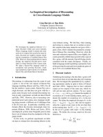
frequencies in the fundus and pylorus (Table 2). Close typed
cells with rare frequencied open typed cells were dispersed
in the basal portions of the mucosa, between chief and
parietal cells, of the fundus with numerous frequency (Fig.
1a, b). In the pylorus, close typed or occasionally open typed
BCG-IR cells were situated in the basal portions of the
gastric mucosa especially in the pyloric gland regions with
numerous frequency but no cells were observed in the upper
part of the pyloric mucosa and epithelial lining (Fig. 1c, d).
In the duodenum, close typed cells were located in the
intestinal glands, which were located in the basal portion of
duodenal mucosa and open typed cells were located in the
inter-epithelial cell regions with moderate frequency (Fig.
1e). In the jejunum and ileum, open typed BCG-IR cells
having long cytoplasmic process which was extended to the
lumen, were mainly located in the inter-epithelial cell
regions with moderate or a few frequencies, respectively and
close typed cells were restricted to the intestinal gland
Table 2.
Regional distributions and relative frequencies of the gastrointestinal endocrine cells in the gastrointestinal tract of the ddY
mice
Fundus Pylorus Duo
1
Jej
1
Ileum Cecum Colon Rectum
BCG
2
+++
3
+++ ++ ++ + - + ±
Serotonin + +++ +++ +++ ++ ++ +++ +++
Gastrin-+++
CCK-8
2
-++++±
Som
2
+++++++-±-
Glucagon±-±±
HPP
2
±
1
Duo: duodenum, Jej: jejunum;
2
BCG: bovine SP-1/chromogranin, Som: somatostatin, HPP: human pancreatic polypeptide, CCK-8: cholecystokinin-8;
3
Relative frequencies; +++: numerous, ++: moderate, +: a few,
±
: rare, -: not detected.
F
ig. 1.
BCG-IR cells in the in the GI tract of ddY mice. They were demonstrated in the fundus (a, b), pylorus (c, d), duodenum (
e),
j
ejunum (f), ileum (g), colon (h) and rectum (i) but no cells were detected in the cecum. Scale bars = 40
µ
m. PAP method.
90 Sae-kwang Ku
et al.
regions (Fig. 1f, g). In the colon, open typed BCG-IR cells
were demonstrated in the inter-epithelial cell regions with a
few frequency (Fig. 1h) and close typed cells were
demonstrated in the intestinal gland regions of the rectum
with rare frequency (Fig. 1i). In the cecum, no BCG-IR cells
were detected in this study.
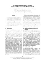
Serotonin-IR cells
Serotonin-IR cells were observed throughout the whole
GI tract with variable relative frequencies in each portion of
the GI tract and they were predominant cell type in this
strain of mice (Table 2). In the fundus, open and close typed
cells were dispersed in the whole gastric mucosa, between
chief and parietal cells, with a few frequency (Fig. 2a).
Although some open typed cells were also observed in the
relatively upper parts of pyloric gastric mucosa, most of
cells were mainly situated in the basal portions with
numerous frequency and they were poly-morphic but close
typed appearances (Fig 2b~d). In the small intestine,
serotonin-IR cells were demonstrated in the inter-epithelial
cells or intestinal glands, which were located in the basal
portion of mucosal layer with moderate to numerous
frequencies, respectively. Open typed cells were restricted to
the inter-epithelial cell regions while most of close typed
cells were found in the intestinal gland regions (Fig. 2eg).
Similar to that of the small intestine, open and close typed
serotonin-IR cells were widely dispersed in the mucosa of
the large intestine (Fig. 2h~j) with moderate to numerous
frequencies, respectively.
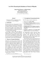
Gastrin- and CCK-8-IR cells
Gastrin-IR cells were restricted to the pylorus with
numerous frequency (Table 2). Close typed gastrin-IR cells
were exclusively located in the basal portion of pyloric gastric
mucosa but occasionally open typed cells were situated in that
regions mixed with close type cells (Fig. 3a, b).
CCK-8-IR cells were detected in the pylorus, duodenum
and jejunum with numerous, a few and rare frequencies,
respectively (Table 2). In the pylorus, CCK-8-IR cells were
located in the basal portion of pyloric gastric mucosa with
numerous frequency and most of these cells were close
typed cells but occasionally open typed cells were situated in
that regions mixed with close-typed cells similar to that of
gastrin-IR cells (Fig. 3c, d). Open and close typed cells were
F
ig. 2.
Serotonin-IR cells in the in the GI tract of ddY mice. These IR cells were located in the epithelium and gastric or intestinal gla
nd
r
egions of the fundus (a), pylorus (b~d), duodenum (e), jejunum (f), ileum (g), cecum (h), colon (i) and rectum (j). Scale bars = 40
µ
m
.
P
AP method.
Gastrointestinal endocrine cells of ddY mouse 91
demonstrated in the inter-epithelial cell and intestinal gland
regions of the duodenum (Fig. 3e) and open typed CCK-8-
IR cells were restricted to the inter-epithelial cell regions of
the jejunum (Fig. 3f). However, no gastrin- and CCK-8-IR
cells were observed in the remaining portions of the GI tract
of this strain of mice.
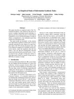
Somatostatin-IR cells
Somatostatin-IR cells were demonstrated throughout the
whole GI tract except for the cecum and rectum, and they
showed highest frequencies in the fundus and pylorus (Table
2). They were dispersed in the whole gastric mucosa,
between chief and parietal cells (Fig. 4a). However, close
typed cells were exclusively located in the basal portion of
pyloric gastric mucosa but occasionally open typed cells
were situated in that regions mixed with close type cells
(Fig. 4b). In the duodenum (Fig. 4c, d), jejunum (Fig. 4e, f)
and ileum (Fig. 4g), open typed cells having long
cytoplasmic processes were located in the inter-epithelial
cell regions with a few frequencies, respectively. However,
no close typed cells were demonstrated in that portion of the
GI tract. In the colon, open typed somatostatin-IR cells were
detected in the inter-epithelial cell regions with rare
frequency (Fig. 1h).
Glucagon-IR cells
Close typed glucagon-IR cells were restricted to the
fundus, duodenum and jejunum with rare frequencies (Table
2). In the fundus, they were located in the gastric mucosa
between chief and parietal cells (Fig. 5a). In the duodenum
(Fig. 5b) and jejunum (Fig. 5c), close typed glucagon-IR
cells were demonstrated in the basal regions of the epithelial
lining.
HPP-IR cells
Open typed HPP-IR cells were restricted to the inter-
epithelial cell regions of the rectum (Fig. 6a, b) with rare
frequency (Table 2).
Discussion
It is generally accepted that the endocrine cells in the
alimentary tracts appeared remarkably different depending
on the regional distribution, relative frequency, cell types
with animal species and each regional part of the GI tract. In
addition, many studies have elucidated the regional
distribution and relative frequency of different endocrine
cells in the GI tract of the various vertebrates including
various species of rodents. Also the researches or data
processing about gastrointestinal endocrine cells in the mice
strains have been widely executed [20,21,27,37]. The
gastrointestinal endocrine cells were generally divided into
two types, one was round to spherical shaped close typed
cells which were located in the stomach regions, and the
other was spherical to spindle shaped open typed cells which
were situated in the intestinal regions. In this study, close
typed cells were mainly located in the gastric or intestinal
gland regions whereas most of open typed cells were found
in the epithelial regions of ddY mice.
Chromogranin (CG) belongs to a family of large anionic
proteins (CG A, B and secretogranin), the members of
which are known to be present in the secretory granules of a
broad spectrum of amine and peptide-producing cells of
adrenal medulla and gastrointestinal endocrine system, as
well as in some neurons of the peptidergic and
catecholaminergic nervous system of several mammals [12,
28]. CGs have been found to occur in large variety of
endocrine organs and cells outside the adrenal medulla, and
they have been claimed as common “markers” of all
neuroendocrine cells [4,11]. Although, the distributional
patterns of these CG-IR cells in the GI tract of Rodentia
F
ig. 3.
Gastrin- and CCK-8-IR cells in the GI tract of ddY mic
e.
G
astrin-IR cells were restricted to the pylorus (a, b), and CCK-
8-
I
R cells were demonstrated in the pylorus (c, d), duodenum (
e)
a
nd jejunum (f). Scale bars = 40
µ
m. PAP method.
92 Sae-kwang Ku
et al.
were seldom, Hawkins
et al.
[14] reported that CGA-IR
cells were demonstrated throughout the whole GI tract of 7
species of laboratory animals including mouse. In the
present study, BCG-IR cells were detected throughout the
whole GI tract of ddY mice except for the cecum. These
results were, well corresponded to those of previous studies
[12,14,28]. However, it is considered that single use of BCG
is not suitable as a marker of endocrine cells because the
relative frequencies of BCG-IR cells were not detected or
lower than those of serotonin- and other IR cells in case of
some regions. If mixed or concomitantly immunostained
with other types of CGs, it is considered that CGs are
suitable as marker of other endocrine cells in this strain of
mouse similar to that of C57BL/6 mouse [20].
Serotonin, consisted of monoamines, was widely
distributed in nervous system and in gastric epithelial
endocrine cells [9]. The main functions of serotonin were
inhibition of gastric acid secretion and contraction of
smooth muscle in the GI tract [13]. El-Salhy
et al
. [9]
reported that serotonin-IR cells were detected throughout
the GI tract of all species and established in the GI tract at
the early stage of vertebrate evolution. In addition, these IR
cells were detected in the whole alimentary tract including
esophagus of low vertebrates [19]. Serotonin-IR cells were
detected in the whole GI tract of the gerbil [26], common
tree shrew [41],
Philippine carabao
[1], Manchurian
chipmunk [25], rat [15] and several strains of mice [20,21,
33]. In the present study, serotonin-IR cells were detected
throughout the whole GI tract and they were most
predominant endocrine cell types in ddY mice. These results
were considered as similar to most of other mammals [1,15,
20,21,25,26,31,41]. However, it is considered that serotonin-
IR cells in this strain of mouse showed somewhat higher
relative frequency than those of other mammals especially
other rodents [20,21].
It is generally accepted that gastrin and CCK-8 originated
from the same ancestor and in the human duodenum a large
fraction of these cells, besides reacting with non-C terminal
CCK antibodies and C-terminal gastrin/CCK antibodies,
also show immunoreactivity with C-terminal gastrin-34
antibodies, colocalised with CCK in a variable portion of
secretory granules [35]. Gastrin secretion by intestinal G cell
promotes gastric acid secretion, and CCK secretion by
intestinal I cell stimulates the secretion of pancreatic enzyme
secretion. In present study, gastrin-IR cells were restricted to
the pylorus and CCK-8-IR cells were demonstrated in the
pylorus, duodenum and jejunum of ddY mice and these
results were well corresponded to those of hairless mice [21]
and C57BL/6 mice [20]. Generally, it is well known that
gastrin- and CCK-8-IR cells were located in the gastric
mucosa and whole small intestinal tract in mammals
[18,41]. However, Lee
et al
., [22] reported that gastrin/CCK-
IR cells were abundant in the pyloric gland region but scarce
in the duodenum and no cells were found in the other
gastrointestinal regions of the Korean tree squirrel. In the GI
tract of gerbil [26], gastrin-IR cells were restricted to the
pylorus and CCK-8-IR cells were located in the pylorus and
duodenum similar to that of Korean tree squirrel. Although
somewhat different aspects were also demonstrated, these
results were well corresponded to those of present study.
However, in the GI tract of Manchurian chipmunk, gastrin-
IR cells were demonstrated from the fundus to ileum and
CCK-8-IR cells were detected from the duodenum to ileum
[23]. These differences were considered that it might be due
F
ig. 4.
Somatostatin-IR cells in the GI tract of ddY mice. Somatostatin-IR cells were demonstrated in the fundus (a), pylorus (b), duodenu
m
(
c, d), jejunum (e, f), ileum (g) and colon (h) but no cells were detected in the cecum and rectum. Scale bars = 40
µ
m. PAP method.
Gastrointestinal endocrine cells of ddY mouse 93
to the differences of the antisera tested or the methods and/or
species differences used in the each study [6,7,40].
Somatostatin, consisting of 14 amino acids, was first
isolated from hypothalamus of sheep and can be divided into
straight form and cyclic form [3]. This substance inhibits the
secretion of the other neuroendocrine hormones [17]. It is
known that somatostatin-IR cells show the widest
distribution in the whole GI tract except for the large
intestine of all vertebrate species investigated, including the
primitive agnathans with serotonin-IR cells [10]. However,
somewhat species-dependent variations on the distributional
pattern of these IR cells have been reported. In the GI tract
of Manchurian chipmunk, they were detected throughout
the whole GI tract and showed the highest frequencies in the
pylorus [25] but they were restricted to the pylorus of the
gerbil [26]. In mice strains, somatostatin-IR cells decrease in
the duodenum of NMRI mice with age [31] and in the
antrum of diabetic mouse regardless of their obesity [8,37].
In hairless [21] and C57BL/6 mice [20], somatostatin-IR
cells were demonstrated from fundus to ileum and showed
highest frequency in the fundus. In the present study,
somatostatin-IR cells were detected throughout the whole
GI tract except for cecum and rectum. These results were
somewhat similar to those of other mouse strains [8,20,21,
37] and the Manchurian chipmunk [25] but quite different
from those of the gerbil [26].
Glucagon is synthesized in the A cells of the pancreas and
regulates serum glucose levels. These IR cells have been
demonstrated in various mammals. They were demonstrated
in the whole GI tract of the common tree shrew [41] and
musk shrew [17] but Baltazar
et al
. [1]
persisted that these
IR cells were only detected in the intestinal tract of the
Philippine carabao
and Lee
et al
. [22] reported that they
were restricted to the cardia and fundus of the Korean tree
squirrel. In addition, glucagon-IR cells were detected in the
stomach and small intestine of Manchurian chipmunk [23]
and they were restricted to the fundus with relatively low
frequencies in hairless mice [21]. In addition, they were
restricted to the fundus, ileum and colon [20]. Collectively it
is considered that the distributional patterns of glucagon-IR
cells in the GI tract of the mammals show species-dependent
variation. Especially, appearances of these IR cells in large
intestine were also reported in mouse [31,32]. However, no
glucagon-IR cells were demonstrated in the GI tract of the
gerbil [26]. In the present study, glucagon-IR cells were
restricted to the fundus, duodenum and jejunum with rare
F
ig. 5.
Glucagon-IR cells in the GI tract of ddY mice. Close typ
ed
g
lucagon-IR cells were restricted to the fundus (a), duodenum (
b)
a
nd jejunum (c). Scale bars = 40
µ
m. PAP method.
F
ig. 6.
HPP-IR cells in the GI tract of ddY mice. Open typ
ed
H
PP-IR cells were restricted to the rectum (a, b). Scale bars
=
4
0
µ
m. PAP method.
94 Sae-kwang Ku
et al.
frequencies, respectively. These findings were somewhat
different from those of previous studies [18,20-23,26,31,32,
41] and these differences were considered as species and/or
strain-dependent variations.
Since PP was isolated from insulin extraction of pancreas
at 1961, the regional distribution of PP-IR cells in the
mammalian species was relatively well known but species-
dependant differences existed among the mammals [1,22,
25,26,41]. These IR cells were demonstrated from the
fundus to the jejunum of the Manchurian chipmunk [25] and
restricted to the fundus of C57BL/6 mice [20] but no cells
were detected in the GI tract of gerbil [26] and hairless
mouse [21]. In the present study, somewhat differed from
that of other rodents, HPP-IR cells were restricted to the
rectum with rare frequency.
In conclusion, the regional distribution and relative
frequency of the GI tract endocrine cells in the ddY mice is
similar to that of other rodents. However, strain-dependent
unique distributional patterns of gastrointestinal endocrine
cells were also found in ddY mice especially to the CCK-8-,
glucagons- and PP-IR endocrine cells.
References
1. Baltazar ET, Kitamura N, Hondo E, Yamada J, Maala
CP, Simborio LT. Immunohistochemical study of endocrine
cells in the gastrointestinal tract of the Philippine carabao
(
Bubalus bubalis
). Anat Histol Embryol 1998, 27, 407-411.
2. Bell FR. The relevance of the new knowledge of
gastrointestinal hormones to veterinary science. Vet Sci
Commun 2, 1979, 305-314.
3.
Brazeau P, Vale W, Burgurs R, Ling N, Butcher M, Rivier
J, Guillemin R.
Hypothalamic polypeptide that inhibits the
secretion of immunoreactive pituitary growth hormone.
Science 1973,
179
, 77-79.
4. Cohn DV, Elting JJ, Frick M, Elde R. Selective localization
of the parathyroid secretory protein I/adrenal medulla
chromogranin A protein familiy in a wide variety of
endocrine cells of the rat. Endocrinology 1984, 144, 1963-
1974.
5. DEste L, Buffa R, Pelagi M, Siccardi AG, Renda T.
Immunohistochemical localization of chromogranin A and B
in the endocrine cells of the alimentary tract of the green
frog,
Rana esculenta
. Cell Tissue Res 1994, 277, 341-349.
6. Dockray GJ. Molecular evolution of gut hormones.
Application of comparative studies on the regulation of
digestion. Gasteroenterology 1977, 72, 344-358.
7. El-Salhy M, Grimelius L. The endocrine cells of the
gastrointestinal mucosa of a squamata reptile, the grass lizard
(
Mabuya quinquetaeniata
). A histological and
immunohistological study. Biomedical Res 1981, 2, 639-658.
8. El-Salhy M, Spangeus A. Antral endocrine cells in
nonobese diabetic mice. Dig Dis Sci 1998, 43, 1031-1037.
9. El-Salhy M, Winder E, Lundqvist M. Comparative studies
of serotonin-like immunoreactive cells in the digestive tract
of vertebrates. Biomedical Res 1985, 6, 371-375.
10. Falkmer S, Van Noorden S. Ontogeny and phylogeny of
glucagon cell. In: Lefebrve P (ed.). Handbook of
Experimental Pharmacology. Vol. 66, pp. 81-119, Springer-
Verlag, Berlin, 1983.
11. Fujita T, Kanno T, Kobayashi S. The paraneuron. pp. 1-
286, Springer, New York, 1988.
12. Grube D, Yoshie S. Immunohistochemistry of chromogranin
A and B, and secretogranin II in the canine endocrine
pancreas. Arch Histol Cytol 1989, 52, 287-298.
13. Guyton AC. Secretory functions of the alimentary tract. In:
Guyton AC (ed). Textbook of Medical Physiology, pp. 801-
815, Saunders, Philadelphia, 1988.
14. Hawkins KL, Lloyd RV, Toy KA. Immunohistochemical
localization of Chromogranin A in normal tissues from
laboratory animals. Vet Pathol 1989, 26, 488-498.
15. Inokuchi H, Kawai K, Takeuchi Y, Sano Y.
Immunohistochemical demonstration of EC cells in rat
gastrointestinal tract. Histochemistry 1982, 27, 453-456.
16. Kim JI, Chung DI, Choi DW. Egg production of
Clonorchis
sinensis
in different strains of inbred mice.
Korean J
Parasitology 1992,
30, 169-175
.
17. Kitamura N, Yamada J, Calingasan YN, Yamashita T.
Immunocytochemical distribution of endocrine cells in the
gastro-intestinal tract of the horse. Equine Vet J 1984, 16,
103-107.
18. Kitamura N, Yamada J, Watanabe T, Yamashita T. An
immunohistochemical study on the distribution of endocrine
cells in the gastrointestinal tract of the musk shrew,
Suncus
murinus
. Histol Histopathol 1990, 5, 83-88.
19. Ku SK, Lee HS, Lee JH. Immunohistochemistry of
endocrine cells in the alimentary tract of the tree frog,
Hyla
arborea japonica
. Korean J Biol Sci 2000, 4, 95-100.
20. Ku SK, Lee HS, Lee JH. An immunohistochemical study of
the gastrointestinal endocrine cells in the C57BL/6 mice.
Anat Histol Embryol 2003, 32, 21-28.
21. Ku SK, Lee JH, Lee HS, Park KD. The regional distribution
and relative frequency of gastrointestinal endocrine cells in
SKH-1 hairless mice: an immunohistochemical study. Anat
Histol Embryol 2002, 31, 78-84.
22. Lee HS, Hashimoto Y, Kon Y, Sugimura M. An
immunohistochemical study of the gastro-entero-pancreatic
endocrine cells in the alimentary tract of the Korean tree
squirrel,
Sciurus vulgaris
corea. Jpn J Vet Res 1991, 39, 117-
131.
23. Lee HS, Ku SK, Lee JH. Distributional patterns of
immunoreactivities for gastrin and secretin families in the
gastrointestinal tract of the Manchurian Chipmunk,
Tami as
sibiricus barberi
. Korean J Lab Anim Sci 1997, 13, 167-171.
24. Lee HS, Ku SK, Lee JH. An immunohistochemical study on
the endocrine cells in the intestines of the Korean tree
squirrel,
Sciurus vulgalis corea
. Korean J Lab Anim Sci
1997, 13, 173-178.
25. Lee HS, Ku SK, Lee JH. Localization of endocrine cells in
the gastrointestinal tract of the Manchurian chipmunk,
Tamias sibiricus barberi
. Korean J Biol Sci
1998, 2, 395-401.
26. Lee JH, Lee HS, Ku SK, Park KD, Kim KS.
Immunohistochemical study of the gastrointestinal endocrine
cells in the Mongolian gerbils,
Meriones unguiculatus
.
Gastrointestinal endocrine cells of ddY mouse 95
Korean J Vet Res 2000,
40
, 653-660.
27.
Pinto HC, Portela-Gomes GM, Grimelius L, Kohnert
KD, de Sousa JC, Albuquerque MA.
The distribution of
endocrine cell types of the gastrointestinal mucosa in
genetically diabetic (db/db) mice. Gastroenterology 1995,
108
, 967-974.
28.
Reinecke M, Höög A, Östenson CG, Efendic S, Grimelius
L, Falkmer S.
Phylogenetic aspects of pancreastatin- and
chromogranin-like immunoreactive cells in the gastro-entero-
pancreatic neuroendocrine system of vertebrates.
Gen Comp
Endocrinol 1991,
83
, 167-182.
29.
Ross MH, Romrell LJ, Kaye GI.
Histology, A text and
atlas. pp. 440-495, Williams & Wilkins, Baltimore, 1995.
30.
Sakai A, Nakamura T, Tsurukami H, Okazaki R, Nishida
S, Tanaka Y, Norimura T, Suzuki K.
Bone marrow
capacity for bone cells and trabecular bone turnover in
immobilized tibia after sciatic neurectomy in mice. Bone
1996,
18
, 479-486.
31.
Sandstrom O, El-Salhy M.
Duodenal endocrine cells in
mice with particular regard to age-induced changes. Histol
Histopathol 2000,
15
, 347-353.
32.
Sandstrom O, Mahdavi J, El-Salhy M.
Effect of ageing on
colonic endocrine cell population in mouse. Gerontology
1998,
44
, 324-330.
33.
Sandstrom O, Mahdavi J, El-Salhy M.
Age-related
changes in antral endocrine cells in mice. Histol Histopathol
1999,
14
, 31-36.
34.
Solcia E, Capella C, Vassallo G, Buffa R.
Endocrine cells
of the gastric mucosa. Int Rev Cytol 1975,
42
, 223-286.
35.
Solcia E, Usellini L, Buffa R, Rindi G, Villani L, Aguzzi
A, Silini E.
Endocrine cells producing regulatory peptides.
In: Polak JM (ed). Regulatory Peptides. pp. 220-246,
Birkhäuser, Basel, 1989.
36.
Spangeus A, El-Salhy M.
Large intestinal endocrine cells in
non-obese diabetic mice. J Diabetes Complications 1998,
12
,
321-327.
37.
Spangeus A, Kand M, El-Salhy M.
Gastrointestinal
endocrine cells in an animal model for human type 2
diabetes.
Dig Dis Sci 1999,
44
, 979-985.
38.
Sternberger LA.
The unlabeled antibody peroxidase-
antiperoxidase (PAP) method. In: Sternberger LA (ed).
Immunocytochemistry, pp. 104-169, John Wiley & Sons,
New York, 1979.
39.
Tajima Y.
Standardized nomenclature for inbred strains of
mice. Experimental animals in cancer research. Japanese
Cancer Association Gann Monograph 1968,
5
, 123-128.
40.
Walsh JH.
Gastrointestinal hormones. In: Johnson LR (ed).
Physiology of the Gastrointestinal Tract. pp. 181-253, Raven
Press, New York, 1987.
41.
Yamada J, Tauchi M, Rerkamnuaychoke W, Endo H,
Chungsamarnyart N, Kimura J, Kurohmaru M, Hondo
E, Kitamura N, Nishida T, Hayashi Y.
Immunohistochemical
survey of the gut endocrine cells in the common tree shrew
(
Tupaia belangeri
). J Vet Med Sci 1999,
61
, 761-767.
42.
Yamaguchi K, Tada M, Tsuji T, Kuramoto M, Uemura D.
Suppressive effect of norzoanthamine hydrochloride on
experimental osteoporosis in ovariectomized mice. Biol
Pharm Bull 1999,
22
, 920-924.