Báo cáo khoa học: "Comparative antibody response of five recombinant antigens in related to bacterial shedding levels and development of serological diagnosis based on 35 kDa antigen for Mycobacterium avium subsp. Paratuberculosis" pot
Bạn đang xem bản rút gọn của tài liệu. Xem và tải ngay bản đầy đủ của tài liệu tại đây (517.8 KB, 7 trang )
-2851$/ 2)
9H W H U L Q D U \
6FLHQFH
J. Vet. Sci.
(2004),
/
5
(2), 111–117
Comparative antibody response of five recombinant antigens in related to
bacterial shedding levels and development of serological diagnosis based on
35 kDa antigen for
Mycobacterium
avium
subsp.
paratuberculosis
Sung Jae Shin
1,2
, Han Sang Yoo
2
, Sean P. McDonough
1
, Yung-Fu Chang
1,
*
1
College of Veterinary Medicine, Cornell University, Ithaca, NY 14853, USA
2
Department of Infectious Diseases, College of Veterinary Medicine and School of Agricultural Biotechnology, Seoul National
University, Seoul 151-742, Korea
Eighty-five complex (85A, 85B and 85C), 35-kDa and
superoxide dismutase (SOD) were cloned, expressed and
purified as antigens in an enzyme-linked immunosorbent
assay (ELISA) to compare the serological reactivity of cows
with different shedding levels of
Mycobacterium avium
subsp.
paratuberculosis
(MPT). Antibody responses to all recombinant
antigens positively increased depending on shedding levels.
In particular, antibody responses to the 35 kDa were higher
than those to the others in all shedder groups. Also, the mean
of O. D. values among Ag 85 complex, 85B showed slightly
higher response than others with high sensitivity and
specificity in all shedder groups. In receiver operating
characteristic (ROC) curve analysis, the result of 35 kDa
ELISA yielded an area under the curve value of 0.945 (95%
confidence interval = 0.895
−
0.996), which indicated that this
35 kDa is more accurate indicator of MPT infection than
other antigens. At the cut-off point recommended by the
ROC curve analysis, the sensitivity and specificity of 35 kDa
ELISA were higher than those of other antigens with 93.3%
and 86.4%, respectively. Finally, a commercially available
ELISA kit was used to clarify 200 positive and 200 negative
sera. We then re-tested these serum samples with our ELISA
test using the 35-kDa antigens. 35 kDa ELISA and
commercial kit showed almost similar results in ROC curve
analysis even though two of positive sera in commercial kit
were negative in 35 kDa ELISA. The sera, which showed
difference in the comparison with commercial ELISA kit,
they also did not react with 35 kDa in Western blot. These
results suggest that a 35-kDa based ELISA can be useful for
detecting MPT infection.
Key words:
Mycobacteium avium
subsp.
paratuberculosis,
85 A, 85 B, 85 C, 35-kDa, Superoxide dismutase, Sensitivity,
Specificity, ELISA, ROC curve analysis
Introduction
Johne’s disease is a chronic granulomatous enteritis of
ruminants caused by
Mycobacterium avium
subsp
.
paratuberculosis
(MPT). Disease occurs worldwide and
affects cattle, sheep, goats, deer and members of the camelid
family [11,15]. Johne’s disease is of tremendous economic
importance to the worldwide dairy industry, causing major
losses due to reduced production and early culling of
animals with estimates of 20% of U.S. dairy herds affected
and costs of $220 million per year to dairy industry [17,23].
In addition to direct economic losses, premature culling of
infected animals reduces the herd manager’s ability to cull
for other reasons such as low productivity or other health
problems and can result in the loss of valuable genetic
potential [20]. This organism has been suggested to cause
Crohn’s disease in people [6,11]. However, this issue is
highly controversial and others report that this bacterium is
not present in specimens obtained from Crohn’s patients [4].
Further studies are needed to resolve the question of the
zoonotic potential of MPT [22].
At present, no specific therapy or vaccination program
effectively prevents Johne’s disease [11]. Although good
management practices can lead to a reduced incidence,
eradication is dependent on early detection and culling of
infected animals [5,15]. Unfortunately, eradication and
control programs to limit the impact of this disease are
hampered by the lack of simple and specific diagnostic tests
that can detect the disease in subclinically infected (infected
but symptom-free) animals [5].
Isolation of MPT by fecal culture is the definitive test for
diagnosis of Johne’s disease. Fecal culture techniques are
currently the most sensitive and specific ante-mortem test
for MPT infection in cattle [25]. However, culture techniques
using solid media require 6 to 12 weeks to produce a result
and performance varies because of lack of standardization of
culture procedures [13,19].
Recently, gene probes and PCR assays for the detection of
*Corresponding author
Phone: 607-253-3675; Fax: 607-253-3943
E-mail:
112 Sung Jae Shin
et al.
MPT in feces have been developed. However, these nucleic
acid-based techniques require specialized equipment, are
expensive, and are less sensitive than conventional fecal
culture, especially in low shedding animals [8]. In order to
overcome these limitations, research has been directed
towards the development of new serologic tests with
improved sensitivity for the identification of paratuberculosis
[12,16,18,19]. Currently available serological tests for
Johne’s disease are the complement fixation test (CFT), agar
gel immunodiffusion (AGID) and various forms of enzyme-
linked immunosorbent assays (ELISA) [12,14,19,26].
However, since seroconversion occurs relatively late during
the course of the disease, the utility of these tests is limited
[12,18,19]. The specificity of ELISA tests for sheep and
cattle is improved by removing cross-reacting antibodies by
absorbing sera with
Mycobacterium phlei
. This increases the
specificity of serological testing to 98.8% and 99.8% in
sheep and cattle respectively. However, the sensitivity for
detection of fecal shedders under field conditions is
reportedly only 57%, depending on the disease status of the
animals tested [3,19,26].
Although current serological tests are useful in detecting
cattle with clinical paratuberculosis, the application of this
procedure in identifying cattle in early stages of infection or
in subclinical stages has proven to be of limited value. Also,
the low sensitivity of the commercially available ELISAs
might, at least in part, be due to the heterologous nature of
the antigen they are apparently based on (derived from MTB
strain 18, which was recognized to be
M. avium
subsp.
avium
) [13]. Development of sensitive serologic tests for the
rapid identification of infected animals requires identification
of protein antigens or epitopes specific for MPT [16].
Several specific antigens of MPT have been reported.
Among these antigens, the 85 A, B and C complex, 35-kDa
(p35) and superoxide dismutase (SOD) elicit strong T- and/
or B- cell immune responses in MPT infection [2,9,15].
In this study, these 5 recombinant proteins were purified
and used as ELISA antigens to evaluate the sensitivity and
specificity of the test with sera from cows with different
levels of bacterial shedding using receiver operating
characteristic (ROC) curve analysis (Analyse-it Software,
www. analyse-it.com) and, then ELISA based on 35 kDa
antigen was compared with a commercial kit.
Material and Methods
Antigen preparation
Bacterial strains and plasmid
E. coli
Top10 was used for Zero-Blunt and TA vector
cloning (Invitrogen, USA).
E. coli
DH5
α
was used as a host
for plasmids pBSK (Bluescript k+) (Stratagene, USA) and
pET22B (Novagen, USA).
E. coli
BL21 (DE3) pLysS strain
and
E. coli
BL21 (DE3) ((Novagen, USA) served as hosts
for the expression of the 85 complex, 35kDa and
sod
genes
as previously described [9]. All
E. coli
strains were cultured
in LB medium with appropriate antibiotics. MPT field
isolate (A198638) was cultured at 37
o
C in Middle brook
7H9 medium supplemented with OADC, Mycobactin J and
Tween 80 as previously described [9,25]. This strain (IS
900
positive and mycobactin dependent) was isolated from a
cow with Johne’s disease [9].
Cloning of 85A, 85B, 85C, 35-kDa protein and Sod genes
Genomic DNA of MPT strain A198638 was isolated and
used as a template in PCR as previously described [9,24].
The primers specific for 85A, 85B, 85C, 35-kDa and sod
genes were prepared as previously described and reported to
Genbank as AF280067, AF219121, AF280068, AF333435
and AF333434, respectively. PCR was performed as
previously described [9]. The amplified PCR products were
cloned and the presence of appropriate insert was confirmed
by restriction enzyme analysis.
Expression and purification of 85A, 85B, 85C, 35kDa and
SOD proteins
The specific primers for expression of 85 complex (85A,
85B and 85C) were prepared as previously described (10).
The specific primers for 35 kDa, forward 5'CAT ATG ACG
TCG GCT CAA AAT G3' and reverse 5'GAA TTC TCA
CTT GTA CTC ATG GAA CTG3'. The primers for SOD,
forward 5'CAT ATG GCT GAA TAC ACC CTG CCC
GA3' and reverse 5'CTC GAG TCA GCC GAA GAT CAG
GCC TT3'. PCR was performed as previously described [9].
The PCR products of each gene were cut with
Nde
I and
Eco
RI or
Xho
I and inserted into pET22B digested with the
same enzymes. The recombinant plasmids were transformed
into
E. coli
BL21 for protein overexpression. Cultures were
grown at 37
o
C until OD
600
= 0.6 and then induced with
different concentrations (0 mM to 5 mM) of isopropyl-
β
-D-
thiogalactopyranoside (IPTG). The overexpressed proteins
formed inclusion bodies that were partially purified as
described previously [9]. An aliquot of the cell pellet of
overexpressing bacteria was denatured in sample buffer and
subjected to 12% SDS-PAGE. All proteins were further
purified by preparative continuous elution using a
polyacrylamide tube gel apparatus (Bio-Rad, USA)
following the manufacturer’s instructions [9]. Purified
proteins were subjected to N-terminal amino acid sequence
to prove that these proteins were the desired gene products.
Refolding of proteins and determination of protein
concentration
Refolding of proteins was performed using a Refolding kit
(Novagen) following the manufacturers instructions. The
concentration of purified recombinant protein was
determined using a BCA protein assay kit (Bio-Rad, USA).
Antibody response to five recombinant antigens and sero-diagnosis with 35 kDa for
M.
avium
subsp.
paratuberculosis
113
Dialysis of proteins and removal of endotoxins
The recombinant proteins were dialyzed against PBS at
4
o
C for 3 days with three changes of PBS using
SPECTRAPOR membrane (Spectrum, USA). After
dialysis, the protein solution was centrifuged at 5,000 g for
10 min. The supernantant was collected and endotoxins
were removed using Detoxi-Gel Affinity Pak Columns
(Pierce, USA)
Polyacrylamide gel electrophoresis and Western blot
SDS-PAGE and Western blot analysis were performed as
previously described [9]. Monoclonal antibody specific to
85-complex antigen of
M. tuberculosis
was used (obtained
from Colorado State University, Clone# CS90). Also, serum
from MPT infected cows and non-infected cows, as
determined by IS
900
PCR and fecal culture, were used as
primary antibodies for identification of 35-kDa and SOD
proteins. Goat anti-mouse immunoglobulin G conjugated to
horseradish peroxidase (Cappel, USA) and anti-bovine
immunoglobulin G conjugated to alkaline phosphatase
(Sigma, USA) were used as secondary reagents.
Serological tests
A total of 82 sera from asymptomatic cows were divided
into four groups based on shedding level as shown in Table
1. Fecal culture and IS
900
PCR tests for MPT infection
were performed to determine positive and negative samples
[8,24].
Checkerboard titration was used to determine the
optimum concentration of protein (2.5, 5 or 10
µ
g/ml) and
serum dilution for use in an indirect ELISA. Flat-bottom 96-
well plates (Maxisorp, Denmark) were coated with 100
µ
l of
each antigen in carbonate-bicarbonate buffer (14.2 mM
Na
2
CO
3
, 34.9 mM NaHCO
3
, 3.1 mM NaN
3
, pH 9.5) at 4
o
C
overnight, followed by washing three times with PBS
containing 0.05% Tween 20 (PBST, washing buffer) using
microwell plate washer Bio-Tek ELx405 (BioTEK
Instruments, USA). Uncoated sites in the wells were
blocked with 5% skim milk in PBST at 37
o
C for 1 h. One
hundred microliter of sera (1 : 100 diluted) from each group
were added to plate and incubated at 37
o
C for 1 hr. The plate
was washed twice with PBST and 100
µ
l of optimally
diluted (1 : 25,000) HRP conjugated anti-bovine IgG
(Sigma, USA) was added to all the wells and incubated at
37
o
C for 1 h. The plates were again washed three times in
PBST. Next, 100
µ
l of 2-2'-Azino-Bis-Thiazoline-6-Sufonic
acid (Sigma, USA) was added to each well and the plates
were incubated for 30 minutes at 37
o
C in the dark. Stop
solution (1M HCl, 50
µ
l) was added and the plates were
read 3 times at 405 nm at 2-minute intervals in a Bio-Tek
312e ELISA reader (BioTEK Instruments, USA). Positive
and negative sera along with antigen and antibody controls
were included in each plate.
Comparison of an ELISA using the 35 kDa protein
antigen and a CSL commercial kit
Two hundred positive and negative bovine sera for MPT
infection, as determined by the Johne’s Absorbed EIA kit
(CSL Veterinary, Australia) were used in an ELISA assay
using the 35 kDa protein. Wells were coated with 1
µ
g of
35-kDa protein and alkaline phosphatase conjugated rabbit
anti-bovine IgG (Sigma, USA) was used as the secondary
reagent. Absorbance was read twice at 405 nm using a Bio-
Tek 312e ELISA reader (BioTEK Instruments, USA).
Evaluation of tests and statistical analysis
Wilcoxon signed-ranks test was used for analysis of the
mean of O. D. values between individual antigens in same
shedding levels using SAS version 8.0 Software.
Differences were considered to be significant if probability
values of
P
< 0.05 were obtained.
In addition, ROC curve analysis (Analyse-it Software,
www. analyse-it.com) was performed on the ELISA results
of individual antigens to determine the optimal cut-off point
(at which the serum of the sensitivity and specificity values
is maximal) [21] for distinguishing between positive and
negative result. The ROC curve (a plot o the true positive
rate (sensitivity) against the false positive rate (1-specificity)
that is obtained at each cut-off point) was constructed and
the area under the curve (AUC) value was calculated as a
measure of the accuracy of the test. Also, ELISA was
compared with fecal culture used as indicator in this study
by kappa statistic.
Results
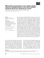
SDA-PAGE and Western blot analysis
Monoclonal antibody specific to 85-complex of
M.
tuberculosis
showed strong reaction to recombinant 85A, 85B
and 85C of MPT in Western blot as previously described (data
not shown). Also, overexpressed 35-kDa and SOD (22-kDa)
proteins in
E. coli
BL21 (DE3) pLysS cells were purified by
preparative continuous elution using a polyacrylamide tube
gel apparatus and also reacted strongly against sera from MPT
infected cattle. In contrast, no reaction was observed with sera
Table 1.
Grouping with respect to shedding level and IS
900
PCR
test
Infected level Number of cows
*Fecal culture
(CFU/g feces)
Highly infected 22 > 300
Moderately infected 15 31-300
Low infected 23 1-30
Negative control 22 0
*Based on the criteria adopted by New York State Diagnostic Laboratory
for Johne’s test, Cornell University.
114 Sung Jae Shin
et al.
from MPT-free cattle (determined by fecal culture and IS
900
PCR test) in Western blot (Fig. 1).
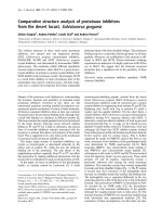
Serological evaluation of 5 recombinant antigens
Antibody responses to all recombinant antigens positively
increased depending on shedding levels. In particular, the
mean of O.D. values of 35 kDa was higher than those of
other antigens in moderate and high shedder groups
(
P
< 0.01, Wilcoxon signed-ranks test). Among Ag 85
complex, 85B showed slightly higher response than others
with high sensitivity and specificity in all shedder groups but
no statistical differences were obsereved according to
individual antigens with same shedding levels except 35
kDa (
P
> 0.05, Wilcoxon signed-ranks test) (Fig. 2).
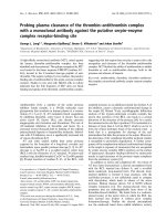
Table 2 summarized cut-off point, sensitivity and
specificity of all recombinant antigens analyzed by the ROC
curve. The result of 35 kDa ELISA yielded an area under
the curve value of 0.945 (95% confidence interval = 0.895
−
0.996) (Fig. 3), which indicated that this 35 kDa is more
accurate indicator of MPT infection than other antigens. At
the cut-off point recommended by the ROC curve analysis,
the sensitivity and specificity of 35 kDa ELISA were higher
than those of other antigens as 93.3% and 86.4%,
respectively (Table 2). Also, 35 kDa ELISA could detect
100% of MPT-infected cattle, which had shedding levels
over than moderate shedding level (Table 3).
Comparison of 35-kDa and a commercial ELISA kit
for herd screening
An ELISA test was developed using the 35-kDa and was
compared with the CLS Commercial kit that has been used
for herd screening in the field. Only 2 positive sera in CLS
were negative in the 35-kDa ELISA.
All sera tested except 2 positive sera in CLS were matched
with 35-kDa ELISA (Table 4). The sera were negative in 35-
kDa ELISA also showed non-reactivity with the antigen by
Western blot.
F
ig. 1.
Continuous purification of overexpressed SOD and 35-kDa proteins using 12% a Polyacrylamide gel with 491 Prep Cell (A a
nd
B
) and Western blot analysis of Sod and 35kDa purified proteins against serum from MPT infected cows and non-infected cow
s
d
etermined by IS
900
PCR, fecal culture and CSL ELISA test (C). (A) SDS-PAGE: M, molecular mass markers, Lane 1, Un-induc
ed
c
ell lysates, Lane 2 to 5, purified Sod protein (B) SDS-PAGE: M, molecular mass makers, Lane 2 to 5, purified 35-kDa protein fracti
on
s
, Lane 6, Induced cell lysates (C) Lane 1: Sod with negative serum, Lane 2: Sod with positive serum, Lane 3: 35-kDa with negati
ve
s
erum and Lane 4: 35-kDa with positive serum.
F
ig. 2.
IgG seroreactivity to recombinant proteins in sera for cattle infected with MPT. The data representd mean of O. D. at 405 n
m
w
ith S.D. *indicated the siginificant differences between individual antigens in the same shedding levels (
P
< 0.05, Wilcoxon signe
d-
r
anks test).
Antibody response to five recombinant antigens and sero-diagnosis with 35 kDa for
M.
avium
subsp.
paratuberculosis
115
Discussion
Control of paratuberculosis in diary herds requires
preventing transmission of MPT to calves by culling cows
that are shedding organisms in their feces (MPT shedders).
However, there are no simple and accurate diagnostic
methods for herd screening to detect MPT shedders. [5,11].
Thus, research has focused on improvedse rological
methods to demonstrate antibodies against MPT [10,12,16,
18,26]. Compared to fecal culture and PCR tests, serologic
tests are inexpensive, rapid, and easy to perform. Currently,
complement fixation test (CFT) and an absorbed enzyme-
linked immunosorbent assay (ELISA) are the serologic tests
used most frequently. Western blot analysis has also been
used for the identification of MPT infection. However, it is
too labor extensive to perform western blot analysis
especially if there are too many serum samples [5,11].
The specificity of Johne’s ELISA testing has been
increased into the range of 95% to 100% by the introduction
of absorbed ELISAs, which reduce cross-reactivity and use
species-specific recombinant proteins [3,18,26]. However,
sensitivity is still low and limited by variations in the stage
of infection. In the attempt to increase sensitivity, several
modifications have been tried including using a combination
of antigens or a single recombinant antigen [10,16].
Initial screening in this study was performed using crude
antigens such as purified protein derivate (PPD) but animals
with different levels of shedding could not be distinguished.
However, OD values were statistically significantly higher
for serum samples from cattle shedding high numbers of
MPT when the 35kDa protein was used as compared to the
other recombinant antigens tested. The ELISA data of all
recombinant antigens were subject to ROC curve analysis
[21], which estimates the sensitivity and specificity of a test
at every possible cut-off point and provides a measure of test
accuracy.
The results obtained in our study with sera in related to
shedding level confirmed by fecal culture and IS
900
PCR
Table 2. Comparison of cut-off point, sensitivity and specificity of individual recombinant antigen in ELISA assay
Antigens Cut-off point of O.D. Sensitivity (%) Specificity (%)
85A 0.170 66.7 81.8
85B 0.174 71.7 86.4
85C 0.152 68.3 81.8
35 kDa 0.210 93.3 86.4
SOD 0.160 80.8 90.9
Table 3. Comparison of identical ratio between fecal culture and ELISA based on 35 kDa antigen
Subjects
35 kDa ELISA
Infected
a
Non-infected
b
35 kDa
No. of tested 22 (H) 15 (M) 23 (L) 22 (N)
No. of positive in
ELISA (%)
22
(100%)
15
(100%)
19
(82.3%)
3
(13.6%)
a
Infected animals based on the combined results of fecal culture and IS
900
PCR
b
Non-infected cows based on fecal culture and IS
900
form healthy flock
(H = High shedder, M = moderate shedder, L = Low shedder, and N = Non-infected cattle)
Table 4. Comparison of between 35-kDa and commercial kit
CSL kit
positive negative
35-kDa
positive 198 0
negative 2* 200
*These two sera did not show any specific reactivity with 35 kDa antigen
in Western blot.
F
ig. 3. The representative of ROC curve obtained from the analys
is
o
f ELISA based on 35 kDa and fecal culture that were obtained f
or
t
he sera from 60 cattle infected with MPT and 22 of MPT-fr
ee
c
attle. The false positive rate (1-specificity (x-axis)) is plott
ed
a
gainst the true positive rate (sensitivity (y-axis)) for each cut-o
ff
p
oint applied. An optimal cut-off point (0.21) is indicated (arrow
).
A
UC = 0.945, 95% confidence interval = 0.895
−
0.996).
116 Sung Jae Shin
et al.
test that 35 kDa protein showed most sensitive against MPT
infections compared to the other recombinant antigens in the
ROC curve analysis. The sensitivity and specificity was
93.3% and 86.4%, respectively. Also, O.D. values of 35 kDa
in moderate shedders were as high those of other antigens in
high shedder and 35 kDa was much better to detect positive
sera even in low shedders.
Importantly, O.D. values to 35-kDa in moderate shedders
were as high as those in high shedder to the other antigens.
Thus, 35-kDa was superior to the other antigens testes for
detecting positive sera even in low shedders.
In a previous study, 35-kDa was recognized by sera from
all 16-reference animals with advanced Johne’s disease
(clinical stage) and 15 of 20 (75%) reference cattle with
early infection (subclinical stage) [10]. In addition, 35-kDa
did not react with sera from 15 MPT-free control cows. Use
of recombinant 35-kDa antigen and/or a monoclonal
antibody against an epitope on 35-kDa have recently been
described for detection of MPT
infection [2,10].
In this study, 85B and 85A had slightly higher seroreactivity
than 85C, consistent with a previous study performed in
tuberculosis patients. However there was no significant
difference between 85A and 85B. Thus, the low sensitivity
of these recombinant antigens were not effective for
detection of MPT infection [9]. Previous serological studies
showed the presence of antibodies to SOD in all tuberculosis
patients tested and in 84% of leprosy patients [15]. Even
though we found in this study that SOD was more sensitive
than 85 complex, it is not recommended for field use to
screen herds because the sensitivity and negative predictive
values are low compared to 35-kDa
We tried to combine the 35-kDa and SOD antigens to
detect MPT infection. The results were unsatisfactory
compare to a single antigen used due to an increased signal
to noise ratio (data not shown). An ELISA based on the
35 kDa compared favorably with the CLS commercial kit,
as only two positive CSL sera were negative in our ELISA
assay. The cut-off between positive and negative samples in
commercially available kits is extremely narrow which
makes these kits prone to subjective error [1]. In contrast,
O.D. values between positive and negative sera exceeded
0.25 in our 35-kDa-based ELISA. Also, in kappa statistic
compared with fecal culture, 35 kDa ELISA (0.51) were
relatively higher than a commercial kit (0.276) [7].
These results suggested that ELISA based on 35 kDa
antigen could be used as a sensitive tool for the
serodiagnosis of MPT infection. The technical simplicity,
speed and low cost of this serological assay, makes it very
attractive for in conjunction with a test that measures the
CMI response, such as the lymphocyte stimulation test, the
gamma interferon test or the skin test, or, possibly even as a
stand-alone screening test.
Although the results are promising, other proteins, which
have not been studied so far in the antibody-based assays in
MPT
infection and more serum samples, are necessary to be
further studied.
Acknowledgments
This work was supported by the New York State Science
and Technology Foundation. Sung Jae Shin was supported
by overseas visiting fellowship provided by the Brain Korea
21 project.
References
1. Adaska JM, Munoz-Zanzi CA, Hietala SK. Evaluation of
result variability with a commercial Johne’s disease enzyme-
linked immunosorbent assay kit and repeat testing of
samples. J Vet Diagn Invest 2002, 14, 423-426.
2. Banasure KD, Basagoudanavar SH, Chaudhury P, Tiwari
V, Parihar NS, Goswami PP. Identification and characterization
of a gene encoding a 35-kDa protein from
Mycobacterium
avium
subspecies
paratuberculosis
. FEMS Microbiol Lett
2001, 196, 195-199.
3. Bech-Nielsen S, Jorgensen JB, Ahrens P, Feld NC.
Diagnostic accuracy of a
Mycobacterium phlei
-absorbed
serum enzyme-linked immunosorbent assay for diagnosis of
bovine paratuberculosis in dairy cows. J Clin Microbiol
1992, 30, 613-618.
4. Clarkston WK, Presti ME, Petersen PF, Zachary PE, Jr.,
Fan WX, Leonardi CL, Vernava AM, 3rd, Longo WE,
Kreeger JM. Role of
Mycobacterium paratuberculosis
in
Crohn’s disease: a prospective, controlled study using
polymerase chain reaction. Dis Colon Rectum 1998, 41, 195-
199.
5. Collins MT. Clinical approach to control of bovine
paratuberculosis. J Am Vet Med Assoc 1994, 204, 208-210.
6. Collins MT, Lisby G, Moser C, Chicks D, Christensen S,
Reichelderfer M, Hoiby N, Harms BA, Thomsen OO,
Skibsted U, Binder V. Results of multiple diagnostic tests
for
Mycobacterium avium
subsp.
paratuberculosis
in patients
with inflammatory bowel disease and in controls. J Clin
Microbiol 2000, 38, 4373-4381.
7. Collins MT, Sockett DC, Ridge S, Cox JC. Evaluation of a
commercial enzyme-linked immunosorbent assay for Johne’s
disease. J Clin Microbiol 1991, 29, 272-276.
8. Cousins DV, Whittington R, Marsh I, Masters A, Evans
RJ, Kluver P. Mycobacteria distenct from
Mycobacterium
avium
subsp.
paratuberculosis
isolated from the faeces of
ruminants possess IS900-like sequences detectable IS900
polymerase chain reaction: implications for diagnosis. Mol
Cell Probes 1999, 13, 431-442.
9. Dheenadhayalan V, Shin KS, Chang CF, Chang CD,
Wang SJ, McDonough S, McDonough P, Stehman S, Shin
S, Torres A, Chang YF. Cloning and characterization of the
genes coding for antigen 85A, 85B and 85C of
Mycobacterium avium
subsp.
paratuberculosis
. DNA Seq
2002, 13, 287-294.
10. El-Zaatari FA, Naser SA, Graham DY. Characterization of
a specific
Mycobacterium paratuberculosis
recombinant
Antibody response to five recombinant antigens and sero-diagnosis with 35 kDa for
M.
avium
subsp.
paratuberculosis
117
clone expressing 35,000-molecular-weight antigen and
reactivity with sera from animals with clinical and subclinical
Johne’s disease. J Clin Microbiol 1997,
35
, 1794-1799.
11.
Harris NB, Barletta RG.
Mycobacterium avium
subsp.
paratuberculosis
in Veterinary Medicine. Clin Microbiol Rev
2001,
14
, 489-512.
12.
Jark U, Ringena I, Franz B, Gerlach GF, Beyerbach M.
Development of an ELISA technique for serodiagnosis of
bovine paratuberculosis. Vet Microbiol 1997,
57
, 189-198.
13.
Kalis CH, Barkema HW, Hesselink JW, van Maanen C,
Collins MT.
Evaluation of two absorbed enzyme-linked
immunosorbent assays and a complement fixation test as
replacements for fecal culture in the detection of cows
shedding
Mycobacterium avium
subspecies
paratuberculosis.
J Vet Diagn Invest 2002,
14
, 219-224.
14.
Kalis CH, Hesselink JW, Russchen EW, Barkema HW,
Collins MT, Visser IJ.
Factors influencing the isolation of
Mycobacterium avium
subsp.
paratuberculosis
from bovine
fecal samples. J Vet Diagn Invest 1999,
11
, 345-351.
15.
Mullerad J, Hovav AH, Fishman Y, Barletta RG,
Bercovier H.
Antigenicity of
Mycobacterium paratuberculosis
superoxide dismutase in mice. FEMS Immunol Med
Microbiol 2002,
34
, 81-88.
16.
Mutharia LM, Moreno W, Raymond M.
Analysis of
culture filtrate and cell wall-associated antigens of
Mycobacterium paratuberculosis
with monoclonal
antibodies. Infect Immun 1997,
65
, 387-394.
17.
Ott SL, Wells SJ, Wagner BA.
Herd-level economic losses
associated with Johne’s disease on US dairy operations. Prev
Vet Med 1999,
40
, 179-192.
18.
Rajukumar K, Tripathi BN, Kurade NP, Parihar NS.
An
enzyme-linked immunosorbent assay using immonoaffinity-
purified antigen in the diagnosis of caprine paratuberculosis
and its comparison with conventional ELISAs. Vet Res
Commun 2001,
25
, 539-553.
19.
Reichel MP, Kittelberger R, Penrose ME, Meynell RM,
Cousins D, Ellis T, Mutharia LM, Sugden EA, Johns AH,
de Lisle GW.
Comparison of serological tests and faecal
culture for the detection of
Mycobacterium avium
subsp.
paratuberculosis
infection in cattle and analysis of the
antigens involved. Vet Microbiol 1999,
66
, 135-150.
20.
Stabel JR.
Johne’s disease: a hidden threat. J Dairy Sci 1998,
81
, 283-288.
21.
Surujballi OP, Romanowska A, Sugden EA, Turcotte C,
Jolley ME.
A fluorescence polarization assay for the
detection of antibodies to
Mycobacterium bovis
in cattle sera.
Vet Microbiol 2002,
87
, 149-157.
22.
Van Kruiningen HJ.
Lack of support for a common etiology
in Johne’s disease of animals and Crohn's disease in humans.
Inflamm Bowel Dis 1999,
5
, 183-191.
23.
Wells SJ, Wagner BA.
Herd-level risk factors for infection
with
Mycobacterium paratuberculosis
in US dairies and
association between familiarity of the herd manager with the
disease or prior diagnosis of the disease in that herd and use
of preventive measures. J Am Vet Med Assoc 2000,
216,
1450-1457.
24.
Whipple DL, Callihan DR, Jarnagin JL.
Cultivation of
Mycobacterium paratuberculosis
from bovine fecal
specimens and a suggested standardized procedure. J Vet
Diagn Invest 1991,
3
, 368-373.
25.
Whitlock RH, Buergelt C.
Preclinical and clinical
manifestations of paratuberculosis (including pathology). Vet
Clin North Am Food Anim Pract 1996,
12
, 345-356.
26.
Whitlock RH, Wells SJ, Sweeney RW, Van Tiem J.
ELISA
and fecal culture for paratuberculosis (Johne’s disease):
sensitivity and specificity of each method. Vet Microbiol
2000,
77,
387-398.