Báo cáo khoa học: "Quantification of mitral regurgitation using proximal isovelocity surface area method in dogs" docx
Bạn đang xem bản rút gọn của tài liệu. Xem và tải ngay bản đầy đủ của tài liệu tại đây (346.55 KB, 9 trang )
-2851$/ 2)
9H W H U L Q D U \
6FLHQFH
J. Vet. Sci.
(2004),
/
5
(2), 163–171
Quantification of mitral regurgitation using proximal isovelocity surface
area method in dogs
Hojung Choi, Kichang Lee, Heechun Lee
1
, Youngwon Lee
2
, Dongwoo Chang
3
, Kidong Eom
4
,
Hwayoung Youn, Mincheol Choi, Junghee Yoon*
Department of Radiology, College of Veterinary Medicine, Seoul National University, Seoul 151-742, Korea
1
Department of Radiology, College of Veterinary Medicine, Gyeongsang National University, Jinju 660-701, Korea
2
Department of Radiology, College of Veterinary Medicine, Chungnam National University, Daejeon 305-764, Korea
3
Department of Radiology, College of Veterinary Medicine, Chungbuk National University, Cheongju 361-763, Korea
4
Department of Radiology, College of Veterinary Medicine, Kyungpook National University, Daegu 702-701, Korea
The present study was performed to determine the
accuracy and reproducibility of calculating the mitral
regurgitant orifice area with the proximal isovelocity
surface area (PISA) method in dogs with experimental
mitral regurgitation and in canine patients with chronic
mitral insufficiency and to evaluate the effect of general
anesthesia on mitral regurgitation. Eight adult, Beagle
dogs for experimental mitral regurgitation and 11 small
breed dogs with spontaneous mitral regurgitation were
used. In 8 Beagle dogs, mild mitral regurgitation was
created by disrupting mitral chordae or leaflets. Effective
regurgitant orifice (ERO) area was measured by the PISA
method and compared with the measurements simultaneously
obtained by quantitative Doppler echocardiography 4
weeks after creation of mitral regurgitation. The same
procedure was performed in 11 patients with isolated
mitral regurgitation and in 8 Beagle dogs under two
different protocols of general anesthesia. ERO and
regurgitant stroke volume (RSV) by the PISA method
correlated well with values by the quantitative Doppler
technique with a small error in experimental dogs
(r = 0.914 and r = 0.839) and 11 patients (r = 0.990 and
r = 0.996). The isoflurane anesthetic echocardiography
demonstrated a significant decrease of RSV, and there was
no significant change in fractional shortening (FS), ERO
area, LV end-diastolic and LV end-systolic volume. ERO
area showed increasing tendency after ketamine-xylazine
administration, but not statistically significant. RSV, LV
end-systolic and LV end-diastolic volume increased
significantly (p < 0.01), whereas FS significantly decreased
(p < 0.01). The PISA method is accurate and reproducible
in experimental mitral regurgitation model and in a
clinical setting. ERO area is considered and preferred as a
hemodynamic-nondependent factor than other traditional
measurements.
Key words:
dog, mitral regurgitation, PISA method, color
Doppler imaging
Introduction
One of the major goals of clinical cardiology is more
accurate quantification of valvular regurgitation, which has
proven to be a difficult task with both invasive and
noninvasive methods in human medicine and veterinary
practice. Color Doppler mapping, the length or area of the
mitral regurgitant jet has been used as an index of severity
[10,16,17,23]. However, it could be influenced not only by
the severity of mitral regurgitation, but also by hemodynamics,
the size of the regurgitant orifice, and the setting of
instruments [3,13,22]. To overcome these limitations, a new
method for analyzing the proximal isovelocity surface area
(PISA) was proposed as an alternative quantitative
approach. The validity of this PISA method has been
reported in in vitro experiments [3,13,27] and in clinical
human patients [8,26]. However, a few studies were carried
out on PISA method in experimental dogs [20] and in canine
patients [6,11]. The purpose of the present study was the
evaluation of the feasibility and reproducibility of “PISA”
method in dogs with experimentally induced mitral
regurgitation, and spontaneous mitral insufficiency
diagnosed by color flow Doppler echocardiography. To
prove the usefulness, this method was prospectively
compared with simultaneously performed quantitative
Doppler and echocardiography examinations.
Mitral regurgitation may be dynamic, and regurgitant
volume may be affected by variations in loading conditions.
General anesthesia is known to result in alterations in
*Corresponding author
Phone: +82-2-880-1265; Fax: +82-2-880-8662
E-mail:
164 Hojung Choi
et al.
patients heart rate, blood pressure, and systemic vascular
resistance [2]. As the effects of echocardiographic alterations
of mitral regurgitation accompanying general anesthesia are
unknown in dogs, this study was also undertaken to evaluate
the effect of general anesthesia on mitral regurgitation using
color Doppler imaging in dogs with experimentally induced
mitral regurgitation.
Materials and Methods
Animals
Eight adult, conditioned Beagle dogs were used. Body
weights ranged from 7.7 to 13 kg with a mean of 9 kg.
Preliminary data included complete physical examination
with emphasis on the cardiovascular system. All dogs were
examined for circulating microfilaria and had thoracic
radiographs and echocardiograms. The experimental protocol
was approved by the Animal Care and Use Committee at
Seoul National University.
Creation of mitral regurgitation
Dogs were sedated with 0.03 mg/kg of acepromazine
(Sedaject, Samu chem Co, Seoul, Korea) and 15 mg/kg of
thiopental sodium (Thionyl, Daihan Pharm, Korea). After
anesthesia was induced, it was maintained at least with 2%
of isoflurane (Isoflurane, Rhodia, UK). During the procedure,
the pulmonary arterial temperature was maintained at
38.0
±
0.5
o
C using a circulating warm water pad. An
anterior cervical site and a femoral site were shaved and
aseptically prepared. With sterile technique, 1- inch cutdown
were performed, and the carotid artery, external jugular vein,
and femoral artery were isolated. A Swan-Ganz catheter
(Cook, USA) was passed into the pulmonary artery via the
external jugular vein. The measurements of pulmonary
capillary wedge pressure and cardiac output were performed
with the anesthetic patient monitoring system (S-3
anesthesia monitor, Datex-Ohmeda, Finland). A 14-cm, 8-
Fr sheath was inserted into the carotid artery and passed into
the left ventricle. A 6-Fr pigtail angiographic catheter (Pig-
tail catheter, Cook, USA) was placed into the left ventricle
via the femoral artery. A 5-Fr, 120-cm long, 4 prong
grasping forceps (4-prong grasping forceps, ESS Inc, USA)
were guided into the left ventricle via the placed sheath in
carotid artery. The forceps were manipulated to engage the
mitral valve chordae or mitral valve leaflets. The disruption
of chordae or mitral valve leaflet was performed until there
was 100% increase in pulmonary capillary wedge pressure,
a grade II to VI or greater left apical holosystolic murmur,
and/or a reduction in cardiac output. All manipulations of
catheters and forceps were performed with fluoroscopic
guidance (DXG-525RF, Dong-A X-ray, Korea). The
catheters were then removed, and vascular incisions were
repaired. Echocardiographic examination was performed at
1 month after creation of mitral regurgitation.
Echocardiographic imaging and analysis
Echocardiography was performed with a multifrequency
sector probe (Logiq 400 pro, General Electric, USA)
imaging at 6 MHz and recording Doppler at 4 MHz. The
data were recorded on thermal printing paper (UP-895
MDW, Sony, Japan).
Measurement of proximal isovelocity surface area
The theoretic basis for calculating the effective regurgitant
orifice (ERO) area has been described previously. The
calculation was based on following formulas [9,27,28].
Flow = Area
×
Velocity
Regurgitant flow = ERO area
×
Regurgitant velocity
ERO area = Regurgitant flow/Regurgitant velocity
Integrated over the cardiac cycle,
ERO area = Regurgitant volume/Regurgitant time velocity
integral
The frame rate of color Doppler imaging was 30/s and the
sector arc was 30
ο
. First aliasing velocity was set to 20-50
cm/s for all examinations. Imaging was obtained from an
apical four-chamber view, and color gain was adjusted to
eliminate random color in areas without flow. The
regurgitant orifice was imaged in the center of the echo
beam and adjusted to best visualize the flow convergence
region on the left ventricular side of the mitral valve. Color
M-mode interrogation was set to pass through the center of
the PISA, all measurements were obtained from all three
beats and then averaged. The PISA radius was measured as
the distance from the first alias to the leading edge of the
mitral valve tracing using ultrasonographic unit internal
caliper.
The regurgitant flow rate was determined by the following
equation where PISA is assumed to be a hemisphere:
FR = 2
π×
r
2
×
V
Where FR is the regurgitant flow rate (ml/s), r is the radius
of the PISA (cm), and V is the aliasing velocity (cm/s).
Regurgitant stroke volume (ml) using PISA method was
calculated by multiplying the mean regurgitant flow rate by
the regurgitant time.
Quantitative Doppler echocardiography
Quantitative Doppler study was performed as previously
described [9]. The diameters of the aortic annulus in systole
and the mitral annulus in diastole were measured at the point
of inner edge of the leaflets. The apical 4 chamber view was
used to record and digitize the pulsed wave Doppler signal
at the mitral and aortic annuli, and the time-velocity
integrals were computed. At least three measurements of
each variable were averaged. Continuous wave Doppler
echocardiography was recorded with an apical or para-
apical window to obtain the maximal velocities of the
regurgitant jet. Once full envelope was obtained, the outline
Quantification of mitral regurgitation using PISA method in dogs 165
was digitized, and the time-velocity integral of the
regurgitant jet was computed. The cross-sectional areas of
the mitral and aortic annuli were calculated
π
R
2
formula,
assuming a circular shape. The mitral and aortic stroke
volumes were obtained by multiplying the cross-sectional
area by the respective time-velocity integral determined by
pulsed wave Doppler imaging at each specific location.
Regurgitant volume = Mitral stroke volume
−
Aortic
stroke volume
The regurgitant fraction = Regurgitant volume/Mitral or
aortic stroke volume.
Anesthetics
To assess the effects of general anesthesia and loading
conditions on mitral valve function, dogs with mitral
regurgitation were initially sedated with 0.03 mg/kg of
acepromazine and 15 mg/kg of thiopental sodium. After
tracheal intubation, anesthesia was maintained with 2% of
isoflurane in oxygen at flow rate 100 ml/kg/hr. A period of 7
days was allowed to elapse following isoflurane anesthesia.
Then, dogs were premedicated with 0.03 mg/kg acepromazine
and 0.04 mg/kg of atropine (Daihan Pharm, Korea)
following intravenous injection of 10 mg/kg of ketamine
(Ketalar, Yuhan, Korea) with 2.2 mg/kg of xylazine
(Rompun, Bayer Korea, Korea). Under anesthesia, M-mode
and quantitative Doppler measurements were performed
prior to PISA method. The latter values were measured as
previously described. Preanesthetic and postanesthetic heart
rates were monitored.
Clinical applications
Clinical characteristics of patients with chronic mitral
insufficiency were summarized in Table 1. PISA method
was utilized on 11 small breed dogs semiquantitatively
assessed as having moderate to severe mitral regurgitation
with physical examination, thoracic radiography, and routine
echocardiography. Their ages ranged from 6 to 12 years
(mean: 8.2 years) and their body weights from 2.1 to 5.8 kg
(mean: 3.5 kg). Enlargement of the left atrium and left
ventricle was confirmed in every animal by radiography, and
vertebral heart size ranged from 10.2 to 12.5 (mean: 11.3 v).
Clinical observations revealed that all dogs had a normal
appetite and normal vigor. Most of the dogs had mild to
severe cough, and dogs with severe cough had intolerance to
exercise. Evaluations were performed by the same method
on induced-mitral regurgitant group.
Statistical analysis
Measurements are expressed as the mean value
±
SD.
Using linear regression, the ERO area and regurgitant stroke
volume determined by the PISA method were compared with
that values obtained by the quantitative Doppler method.
Since a wide range of values may yield a high correlation
coefficient even when data are in poor agreement, the
differences between pairs of measurements were additionally
determined according to Bland-Altman method. To test the
reproducibility of PISA calculation, measurements of the
proximal accelerating flow variables were examined by the
same observer after an interval of 1 week. To determine the
interobserver variability, all measurements were repeated by a
second independent observer on the separate day. The
interobserver variability was measured by the Bland-Altman
method. These were also expressed as the coefficient of
variation of the repeated measurements (COVr). The COVr
was calculated from the following formula: COVr = (SD of
the mean differences/mean)
×
100 %.
The paired samples t-test was used to assess the statistical
significance of preanesthetic and postanesthetic changes in
hemodynamic and Doppler echocardiographic parameters.
Results
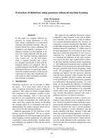
Comparison of the PISA method with the quantitative
Doppler technique
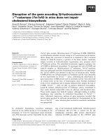
ERO area by the PISA method correlated well with values
by the quantitative Doppler technique (y = 0.641x + 3.023, r
= 0.914) with a small error (mean difference = 2.73
±
2.11;
Table 1.
Clinical characteristics of the patients with chronic mitral regurgitation
Breed BW (kg) Clinical sign VHS (v) Pulmonary edema
1 Pomeranian 3.5 Cough 10.5 None
2 Maltese 2.5 Cough 11.3 None
3 Maltese 3.9 Syncope, cyanosis 12.5 Mild
4 Miniature Pinscher 4.2 Syncope 10.2 None
5 Pomeranian 2.1 Dyspnea, depression 12.1 Mild
6 Maltese 5.8 Cough, dyspnea, panting 12.0 Moderate
7 Maltese 2.5 Cyanosis, panting 10.6 Mild
8 Chihuahua 3.6 Dyspnea, hemoptysis 10.2 Moderate
9 Yorkshire Terrier 3.0 Exercise intolerance, cough 11.5 Mild
10 Maltese 3.0 Cough 12.0 Mild
11 Maltese 4.0 Cough, syncope 11.8 Mild
166 Hojung Choi
et al.
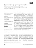
Fig. 1). A good correlation was also found between
regurgitant stroke volume (RSV) by PISA and the
quantitative Doppler technique (y = 0.724x + 6.589, r =
0.839) with a small error (mean difference =
−
2.62
±
1.80
ml; Fig 2).
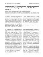
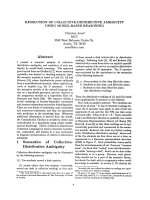
Reproducibility
The intraobserver variability was 0.101
±
3.030 mm
2
(mean difference
±
SD) with COVr = 10.63 % for ERO area
and 0.631
±
4.848 ml with 21.67% for regurgitant stroke
volume. The interobserver variability was 0.58
±
2.34 mm
2
with 12.52 %, and 1.81
±
3.85 ml with 18.42%, respectively
(Fig. 3 and 4).
The effect of anesthetics on echocardiographic parameters
There was no significant change in fractional shortening,
ERO area, and LV (left ventricle) end-diastolic and LV end-
systolic volume under isoflurane anesthesia (Table 2). The
echocardiography under isoflurane anesthesia demonstrated
a significant decrease of RSV (16.97
±
3.33 vs 11.54
±
4.17
ml, p < 0.05). ERO area showed the tendency of increase
after administration of ketamine-xylazine combination, but
not statistically significant (13.78
±
3.43 vs 17.34
±
6.69
mm
2
, p = NS). RSV increased significantly from 16.97
±
3.34 to 26.37
±
7.19 ml (p < 0.01), and end-diastolic volume
also increased significantly from 35.95
±
7.72 to 53.38
±
8.80 ml (p = 0.01), whereas fractional shortening significantly
decreased from 37.13
±
3.57 to 26.42
±
3.61% (p < 0.01,
Table 3).
Clinical applications
ERO by the PISA method correlated well with values by
the quantitative Doppler technique (y = 0.920x + 0.230, r =
0.99) with a small error (mean difference = 1.886
±
5.176;
Fig. 5). A highly significant correlation was also found
between RSV by PISA and the quantitative Doppler
Fig. 1.
Results in dogs with experimental mitral regurgitation. Correlation between the effective regurgitant orifice (ERO) area obtain
ed
by the proximal isovelocity surface area (PISA) method and by quantitative Doppler echocardiography (A). The difference between
the
proximal isovelocity surface area (PISA) and the Doppler values is plotted against the average of the same data. The mean differ
ence
(mean diff.) is indicated by the dashed line; the limits of agreement (continuous lines) are indicated by
±
2SDs (B).
Fig. 2.
Results in dogs with experimental mitral regurgitation. Correlation between the regurgitant stroke volume (RSV) obtained by the
proximal isovelocity surface area (PISA) method and by quantitative Doppler echocardiography (A). The difference between the
proximal isovelocity surface area (PISA) and the Doppler values is plotted against the average of the same data. The mean differ
ence
(mean diff.) is indicated by the dashed line; the limits of agreement (continuous lines) are indicated by
±
2SDs (B).
Quantification of mitral regurgitation using PISA method in dogs 167
technique (y = 0.960x + 5.445, r = 0.996) with a small error
(mean difference =
−
4.505
±
5.253 ml; Fig. 6) in spontaneous
chronic mitral regurgitant patients.
Discussion
Mitral regurgitation (MR) was induced by handling the
grasping forceps in left ventricle via carotid artery, in this
study. An advantage of this MR model over those previously
reported surgical models [5] is that it does not require a
thoracotomy and thus is less invasive. Also, surgically
produced models of MR may not be analogous to the
volume overload seen in spontaneous MR because of the
potential restrictive effects of a postoperatively thickened
pericardium on the volume overloaded heart [12].
This study was investigated in a clinical setting and an
experimentally induced MR the potential of the proximal
flow convergence method to assess the quantitative severity
of mitral regurgitation in comparison with the quantitative
Doppler echocardiographic method as an established and
validated standard. As shown in Fig. 1 and 2, regurgitant
stroke volume (RSV) as calculated by the proximal
isovelocity surface area (PISA) method showed good
overall agreement with the values that were calculated by
the quantitative Doppler echocardiographic method (r =
0.839, mean difference =
−
2.62
±
1.80 ml). Similar
correlations were obtained for the calculated effective
regurgitant orifice (ERO) area (r = 0.914, mean difference =
2.73
±
2.11 mm
2
). In clinical trials, RSV and ERO as
calculated by the PISA method showed highly agreement
with the values that were calculated by the quantitative
Doppler method (r = 0.96, mean difference =
−
4.505
±
5.253, and r = 0.99, mean difference = 1.886
±
5.176).
These results were similar to those of several human studies
[8,19]. Although there is a good correlation and agreement
between the two methods, the tendency of underestimation
was shown in ERO, while overestimation in RSV. The
possible causes of these small errors include the existing
intraventricular flow, which is theoretically destined to pass
the left ventricular out flow tract, could superimpose the
F
ig. 3. Results in dogs with experimental mitral regurgitation. Scatterplots of the differences between the two measurements on the
y-
a
xis and the mean values obtained by the intraobservers on the x-axis for effective regurgitant orifice area (A) and regurgitant stro
ke
v
olume (B) by the PISA method.
F
ig. 4. Results in dogs with experimental mitral regurgitation. Scatterplots of the differences between the two observers on the y-ax
is
a
nd the mean values obtained by the two observers on the x-axis for effective regurgitant orifice area (A) and regurgitant stroke volum
e
(
B) by the PISA method.
168 Hojung Choi
et al.
proximal accelerating flow through the mitral regurgitant
orifice, especially when the regurgitant orifice is near the left
ventricular outflow tract. The regurgitant orifice, which is
close to the left ventricular wall may distort hemispheric
shape of the proximal flow convergent isovelocity layers
[4,15]. This may be especially true for a small left
ventricular cavity during systole in small animals. All of
these possibilities require further investigations. Also,
higher correlation in clinical series was considered that
PISA method was more accurate in chronic severe MR than
mild MR estimated by semiquantitative method. Also, it
seems that thick and irregular valvular margin doesn’t
significantly affect on measurements of PISA radius in
chronic MR patients compared to experimental dogs with
thin and smooth valve. Thus, ERO calculation by PISA
method may be useful in dogs with chronic mitral
insufficiency.
High reproducibility is important for the echocardiographic
parameters, and should be evaluated. In the present study, high
reproducibility was demonstrated in ERO and RSV by two
observers and two measurements. These close agreement is
similar to those reported in several human studies [8,19].
The authors need to discuss about some technical points
used in the present study concerning accuracy of
measurements of regurgitant flow rate or volume using
Doppler color flow mapping of the proximal accelerating
flow region. Axial and lateral resolutions of two-
dimensional Doppler color flow mapping are dependent on
the size and depth of the imaging area and the frequency of
the transducer chosen. Whenever possible, the narrowest
imaging angle, shallowest depth, highest imaging frequency
and lowest pulse repetition frequency should be chosen to
increase the resolutions of Doppler color mapping. The
proximal accelerating field should be magnified as large as
possible to minimize measurement error. The prerequisite
for accurate measurement of the proximal accelerating area
using two-dimensional scanning was through both standard
and nonstandard imaging planes with a rotating, shifting and
angulating transducer. Aotsuka et al. [1] reported that color
M-mode was useful in children with small heart size
because it provides color Doppler information and
positional information regarding the mitral surface more
clearly than B-mode color flow mapping due to its higher
signal to noise ratio. The color M-mode is also thought to be
useful to measure the flow convergence region in dogs,
because the radius of the PISA is small and heart rate is high
for color flow rate like children. The M-mode beam should
be aligned center to the accelerating region and perpendicular
to the regurgitant orifice plane.
One of the basic assumptions of the present study is that
the shape of the PISA is a hemisphere, and calculations are
based on unidirectional measurement of the PISA radius.
Several experimental studies on the relationships between
the shape of the PISA and machine setting or hemodynamic
factors have been reported [4,18,21,24]. It was found that
the contours of the PISA changed variously because of
pressure gradients between the left ventricle and left atrium,
the Nyquist limit, and orifice size [4,18,21,24,25]. If the
orifice size and the pressure gradients between left ventricle
and left atrium (almost 100 mmHg) are constant values, the
Nyquist limit is an important and controllable factor that
have influenced on the shape of PISA. For precise
Table 2.
The effect of isoflurane anesthesia on the echocardiographic parameters
Preanesthetic Postanesthetic p value
LV end-diastolic volume (ml) 35.95 ± 7.72 036.43 ± 11.35 0.879
LV end-systolic volume (ml) 11.79 ± 3.77 13.17 ± 4.76 0.358
Fractional shortening (%) 37.13 ± 3.57 33.81 ± 3.97 0.052
ERO (mm
2
) 13.79 ± 7.72 11.05 ± 3.19 0.051
RSV (ml) 16.97 ± 3.33 11.54 ± 4.17 0.013
LV: left ventricle
ERO: effective regurgitant orifice area
RSV: regurgitant stroke volume
Table 3.
The effect of ketamine and xylazine combination anesthesia on the echocardiographic parameters
Preanesthetic Postanesthetic p value
LV end-diastolic volume (ml) 35.95 ± 7.72 53.38 ± 8.80 0.001
LV end-systolic volume (ml) 11.79 ± 3.77 025.6 ± 7.36 0.001
Fractional shortening (%) 37.13 ± 3.57 26.42 ± 3.61 0.001
ERO (mm
2
) 13.79 ± 7.72 17.34 ± 6.69 0.097
RSV (ml) 16.97 ± 3.33 26.37 ± 7.19 <0.001
LV: left ventricle
ERO: effective regurgitant orifice area
RSV: regurgitant stroke volume
Quantification of mitral regurgitation using PISA method in dogs 169
estimation of the regurgitant flow or RSV, the radius should
be measured at the machine setting for most appropriate
hemispheric assumption. Shandas
et al
. [21] reported that if
the Nyquist limit is 30-55 cm/s and the pressure gradients is
between 60-100 mmHg, a hemispheric model provides the
best agreement between the calculated and actual flow rate.
In another report, Deng
et al
. [4] indicated the optimal
Nyquist limit between 30-35 cm/s is appropriate for a
hemispheric assumption in most children. To minimize the
error when measuring the PISA radius, it is better to set the
Nyquist limit as low as possible because it tends to
maximize the PISA radius; but to distinguish low
intraventricular flow from true proximal accelerating flow, it
should not be set the velocity too low. It was thought that
setting of the Nyquist limit velocity at about 20-40 cm/s is
suitable when applying the PISA method in this study. It is
not strictly necessary to use the first alias to calculate flow
rate since any isovelocity hemisphere should theoretically
provide the same result. However, the first alias is the most
apparent and reproducible region of the flow stream and is
therefore most suitable for velocity estimation and
measurement of radial distance.
In the present study, the dogs with induced MR were
anesthetized to alter ventricular loading conditions, because
general anesthesia may be a common situation that
hemodynamic alteration can be occurred in old small
animals such as scaling and surgery associated geriatric
disease. Also, general anesthesia has profound effects on
loading conditions with resulting effects on mitral valve
function and regurgitant volume. In this anesthetic study,
isoflurane anesthesia resulted in a non-significant change in
echocardiographic parameters except regurgitant volume.
Bach
et al.
[2] demonstrated general anesthesia with
isoflurane altered blood pressure and LV cavity dimensions
Fig. 6.
Correlation between the regurgitant stroke volume (RSV) obtained by the proximal isovelocity surface area (PISA) method and
by quantitative Doppler echocardiography in patients with chronic mitral regurgitation (A). The difference of regurgitant stroke
volume
(RSV) between the PISA and Doppler methods is plotted against the average of the same data in patients with chronic mitral
regurgitation. The mean difference (mean diff.) is indicated by the dashed line; the limits of agreement (continuous lines) are
indicated
by
±
2SDs (B).
Fig. 5.
Correlation between the effective regurgitant orifice (ERO) area obtained by the proximal isovelocity surface area (PISA)
method and by quantitative Doppler echocardiography in patients with chronic mitral regurgitation (A). The difference of effecti
ve
regurgitant orifice (ERO) area by between the PISA and Doppler methods is plotted against the average of the same data in patien
ts with
chronic mitral regurgitation. The mean difference (mean diff.) is indicated by the dashed line; the limits of agreement (continu
ous lines)
are indicated by
±
2SDs (B).
170 Hojung Choi
et al.
reflecting altered loading condition. These discrepancies of
the results may be due to the differences between anesthetic
protocols.
All echocardiographic parameters were markedly
changed except ERO area and regurgitant time in ketamine-
xylazine combination anesthesia. Xylazine has
cardiodepressant and arrhythmogenic effects, and induces
bradycardia and a brief period of hypertension, followed by
a longer-lasting decrease in cardiac output and blood
pressure [25]. Xylazine-induced decreases in heart rate and
cardiac output are moderated by ketamine’s sympathomimetic
action, while blood pressure and systemic vascular
resistance are increased [14]. The increase of blood pressure
and systemic vascular resistance may cause marked increase
of end-systolic volume, and decrease of aortic output, thus
RSV increased, while fractional shortening decreased in this
study.
The change in regurgitant volume was not related to
differences in heart rate, blood pressure, or technical factors
in imaging, but may be related to lower systemic vascular
resistance under isoflurane anesthesia and increase systemic
vascular resistance under ketamine-xylazine combination.
Thus, the possibility of underestimation of mitral regurgitant
severity must be considered under isoflurane anesthesia,
such as transesophageal echocardiography or surgery of
cardiovascular system. ERO area was not changed under
both isoflurane and ketamine-xylazine anesthesia that shows
ERO may be hemodynamically independent factor and
should be preferred as a factor reflecting the severity of
mitral regurgitation.
There are several limitations in this study. First, the
quantitative Doppler method was not “gold standard” to
estimate the accuracy of PISA method. Direct measurement
of the effective regurgitant orifice area should be performed,
but such a method does not exist because of inaccuracies of
measurements of flow by invasive methods [14]. Also
consistent use of quantitative Doppler echocardiography has
proved to be a very reliable method. Incompleteness of the
PISA method has been described for measuring regurgitant
flow and effective regurgitant orifice area in the previous
studies. The PISA method assumes that this orifice area is
roughly constant in systole, but its not true [19]. Thus we
just measured instantaneous maximal PISA radius. To go
beyond this limitation, total regurgitant volume might be
calculated by integrating the instantaneous flow rate over
time, or 3-dimensional reconstruction of the hemicircle into
a hemisphere. However, these methods are not available in
clinical veterinary practice. Although the theoretic problems
exist, high-resolution imaging, experienced technique, and
appropriate ascertainment of flow convergence allow
accurate quantitation of mitral regurgitation.
Enriquez-Sarano
et al.
[7] studied the progression of MR
in large clinical series. Their study suggested that regular
follow-up echocardiographic examinations should be
performed in patients with MR. They recommended the
optimal delay for follow-up examinations. It is thought that
these standards to estimate progression of MR should also
be performed in veterinary clinical fields through a large
clinical outcome using quantitative method. In conclusion,
the feasibility of the PISA method is excellent after the
initial learning phase in dogs. Flow calculations that are
based on the assumption of simple hemispheric symmetry
of the proximal flow field showed excellent correlation with
flow values that were obtained by the more cumbersome and
time-consuming Doppler two-dimensional echocardiographic
method. Veterinary practitioners do not have sufficient time
for gathering high quality recordings, because dogs with left
heart failure may be intolerant of protracted
echocardiographic examination due to severe dyspnea and
cough. Thus, PISA method is especially useful in small
animal practice considering its simplicity. Although
refinements to the proximal convergence method are to be
expected in the future, it appears to be suitable for routine
echocardiographic practice in dogs.
Acknowledgment
This work was supported by the Korean Research
Foundation Grant (KRF-2001-GN0017).
References
1. Aotsuka H, Tobita K, Hmada H, Uchishiba M, Tateno S,
Matsuo K, Fujiwara T. Validation of the proximal
isovelocity surface area method for assessing mitral
regurgitation in children. Pediatr Cardiol 1996, 17, 351-359.
2. Bach DS, Deeb M, Bolling SF. Accuracy of intraoperative
transesophageal echocardiography for estimating the severity
of functional mitral regurgitation. Am J Cardiol 1995, 76,
508-512.
3. Bolger AF, Eigler NL, Pfaff JM, Resser KJ, Maurer G.
Computer analysis of Doppler color flow mapping images
for quantitative assessment of in vitro fluid jets. J Am Coll
Cardiol 1988, 12, 450-457.
4. Deng YB, Shiota T, Shandas R, Zhang J, Shan J.
Determination of the most appropriate velocity threshold for
applying hemispheric flow convergence equations to
calculate flow rate : selected according to the transorifice
pressure gradient. Circulation 1993, 88, 1699-1708.
5. Dent JM, Jayaweera AR, Glasheen WP, Nolan SP,
Spotnits WD, Villanueva FS, Kaul S. A mathematical
model for the quantification of mitral regurgitation;
Experimental validation in the canine model using contrast
echocardiography. Circulation 1992, 86, 553-562.
6. Doiguchi O, Takahashi T. Examination of quantitative
analysis and measurement of the regurgitation rate in mitral
valve regurgitation by the “Proximal isovelocity surface area”
method. J Vet Med Sci 2000, 62, 109-112.
7. Enriquez-Sarano M, Basmadjian AJ, Rossi A, Bailey KR,
Seward JB, Tajik AJ. Progression of mitral regurgitation. J
Quantification of mitral regurgitation using PISA method in dogs 171
Am Coll Cardiol 1999,
34
, 1137-1144.
8.
Enriquez-Sarano M, Miller FA, Hayes SN, Bailey KR,
Tajik AJ, Seward JB.
Effective mitrl regurgitant orifice
area : Clinical use and pitfalls of the proximal isovelocity
surface area method. J Am Coll Cardiol 1995,
25
, 703-709.
9.
Enriquez-Sarano M, Seward JB, Bailey KR, Tajik AJ.
Effective regurgitant orifice area: A noninvasive Doppler
development of an old hemodynamic concept. J Am Coll
Cardiol 1994,
23
, 443-451.
10.
Helmeke F, Nanda NC, Hsiung MC, Hsiung MC, Sato B,
Adey CK, Goyal RG.
Color Doppler assessment of mitral
regurgitation with orthogonal planes. Circulation 1987,
75
,
175-183.
11.
Kittleson MD, Brown WA.
Regurgitant fraction measured
by using the proximal isovelocity surface area method in
dogs with chronic myxomatous mitral valve disease. J Vet
Intern Med 2003,
17
, 84-88.
12.
Kleaveland JP, Kussmaul WG, Vinciguerra T, Diters R,
Canabello BA.
Volume overload hypertrophy in a closed-
chest model of mitral regurgitation. Am J physiol 1988,
254
,
1034-1041.
13.
Krabil KA, Sung HW, Tamura T, Chung KJ, Yoganathan
AP, Sahn DJ.
Factors influencing the structure and shape of
stenotic and regurgitant jets: An in vitro investigation using
Doppler color flow mapping and optical flow visualization. J
Am Coll Cardiol 1989,
13
, 1672-1681.
14.
Lopez JF, Hanson S, Orchard RC, Tan L.
Quantification
of mitral valvular incompetence. Cathet Cardiovasc Diagn
1985,
11
, 139-152.
15.
Min PU, Vandervoort PM, Greenberg NL, Powell KA,
Griffin BP, Thomas JD.
Impact of wall constraint on
velocity distribution in proximal flow convergence zone. J
Am Coll Cardiol 1996,
27
, 706-713.
16.
Miyatake K, Izumi S, Okamoto M, kinoshita N, Asonuma
H, Nakagawa H.
Semiquantitative grading of severity of
mitral regurgitation by real-time two-dimensional Doppler
flow imaging technique. J Am Coll Cardiol 1986,
7
, 82-88.
17.
Omoto R, Yokote Y, Takamoto S, Kyo S, Ueda K, Asano
H.
The development of real-time two-dimensional Doppler
echocardiography and its clinical significance in aquired
valvular diseases with special references to the evaluation of
valvular regurgitation. Jpn Heart J 1984,
25
, 325-340.
18.
Rodriquez L, Anconina J, Flachskamp FA, Weyman AE,
Levine RA, Thomas JD.
Impact of finite orifice size on
proximal flow convergence: Implications for Doppler
quantification of valvular regurgitation. Circ Res 1992,
70
,
923-930.
19.
Schwammenthal E, Chen C, Benning F, Block M,
Breithardt G, Levine RA.
Dynamics of mitral regurgitant
flow and orifice area; Physiologic application of the proximal
flow convergence method: Clinical data and experimental
testing. Circulation 1994,
90
, 307-322.
20.
Schwammenthal E, Chen C, Giesler M, Sagie A,
Guerrero JL, Vazquez de prada JA, Hombach V,
Weymen AE, Levine RA.
New method for accurate
calculation of regurgitant flow rate based on analysis of
Doppler color flow maps of the proximal flow field.
Validation in a canine model of mitral regurgitation with
initial application in patients. J Am Coll Cardiol 1996,
27
,
161-172.
21.
Shandas R, Gharib M, Liepmann D, Shiota T, Sahn DJ.
Experimental studies to define the geometry of the flow
convergence region: laser Doppler particle tracking and color
Doppler imaging. Echocardiography 1992,
9
, 43-50.
22.
Simpson IA, Valdez-Cruz LM, Sahn DJ, Murillo A,
Tamura T, Chung KJ.
Doppler color flow mapping of
simulated in vitro regurgitation jets: Evaluation of the effects
of orifice size and hemodynamic variables. J Am Coll
Cardiol 1989,
13
, 1195-1207.
23.
Spain MG, Smith MD, Grayburn PA, Harlamert EA,
DeMaria AN.
Quantitative assessment of mitral
regurgitation by Doppler color flow imaging: Angiographic
and hemodynamic correlations. J Am Coll Cardiol 1989,
13
,
585-590.
24.
Switzer DF, Yoganathan AP, Nanda NC, Woo Y-R,
Ridgway AJ.
Calibration of color Doppler flow mapping
during extreme hemodynamic conditions in vitro: A
foundation for a reliable quantitative grading system for
aortic incompetence. Circulation 1987,
75
, 837-846.
25.
Thurmon JC, Tranguilli WJ, Benson GJ
(eds.)
.
Veterinary
Anesthesia, 3rd ed. pp. 183-209, pp. 241-296. Lippincott
Williams Wilkins, Philadelphia, 1996.
26.
Vandervoort PM, Rivera M, Mele D.
Application of color
Doppler flow mapping to calculate effective regurgitant
orifice area: an in vitro study and initial clinical observations.
Circulation 1993,
88
, 1150-1156.
27.
Vandervoort PM, Thoreau DH, Rivera JM, Levine RA,
Weyman AE, Thomas JD.
Automated flow rate calculations
based on digital analysis of flow convergence proximal to
regurgitant orifices. J Am Coll Cardiol 1993,
22
, 535-541.