Báo cáo khoa học: "Gastrointestinal impaction by Parascaris equorum in a Thoroughbred foal in Jeju, Korea" potx
Bạn đang xem bản rút gọn của tài liệu. Xem và tải ngay bản đầy đủ của tài liệu tại đây (921.71 KB, 2 trang )
-2851$/ 2)
9H W H U L Q D U \
6FLHQFH
J. Vet. Sci.
(2004),
/
5
(2), 181–182
Gastrointestinal impaction by
Parascaris equorum
in a Thoroughbred foal
in Jeju, Korea
Seung-ho Ryu, Jong-duck Jang, Ung-bok Bak, Chang-woo Lee
1,
*, Hee-jeong Youn
2
and Yonghoon Lyon Lee
3
Equine Hospital, Korea Racing Association, Bukjejugun, Jeju 695-900, Korea
1
Department of Clinical Pathology, College of Veterinary Medicine, Seoul National University, Seoul 151-742, Korea
2
Department of Parasitology, College of Veterinary Medicine, Seoul National University, Seoul 151-742, Korea
3
Department of Anesthesia, Pain Management and Perioperative Medicine, Boren Veterinary Medical Teaching Hospital and
College of Veterinary Medicine, Oklahoma State University, Stillwater, OK 74074, USA
A weanling Thoroughbred foal was admitted to Equine
Hospital, Korea Racing Association with signs of colic. On
admission the foal was sweating profusely, appeared
anxious and exhibiting signs suggestive of abdominal pain.
Clinical examination revealed: tachycardia (90 beats/min),
tachypnea (50 breaths/min) and congested and slightly
cyanotic mucous membranes. No intestinal sounds were
auscultated in all 4 abdominal quadrants. Rectal palpation
identified concurrent cecum and large colon impactions.
Treatment consisted of intravenous administration of a
balanced electrolyte solution, nasogastric siphonage and
administration of analgesics. Nasogastric reflux contained
ascarids. This treatment failed to alleviate the signs of colic.
The foal died 3 hours later following discharge because the
owner didn’t want laparatomy because of economic
constraints. Prior to admission this foal had not received
any prophylactic anthelmintic treatment. In necropsy, there
were masses of ascarids accumulation in the stomach, small
intestine and large intestine. The outcome of this report is
to describe the first diagnosed case of gastrointestinal
impaction by
P. eq u o rum
in a Thoroughbred foal in South
Korea and indicates the importance of regular anthelmintic
treatment.
Key words:
impaction,
Parascaris equorum,
Thoroughbred
foal
Ascariasis is classically associated with lethargy,
inappetence, coughing, nasal discharge and decreased
weight gain. The prevalence of ascariasis in foals less than 1
year old is 31-61%; however, most of these infections are
subclinical [1].
Parascaris equorum
is a common and
ubiquitous parasite that persists for many years in stables
and on pasture in spite of good hygiene and anthelmintic
control programs. Foals are usually infected early in life [5].
Young horses susceptible to ascarid infection are at risk,
particularly immediately after antihelmintic administration
[2]. Some foals die as a result of intestinal impaction or
rupture [5].
To our knowledge, there is no data reporting clinical cases
of intestinal impaction by
P. equorum
in South Korea. The
purpose of this report was to describe the first diagnosed
case of gastrointestinal impaction by
P. equorum
in a
Thoroughbred foal in Jeju, South Korea.
Clinical findings, therapy and course of condition:
A
weanling Thoroughbred foal was admitted to Equine
Hospital, Korea Racing Association with signs of colic. On
admission the foal was sweating profusely, appeared
anxious and exhibiting signs suggestive of abdominal pain.
Clinical examination revealed: tachycardia (90 beats/min),
tachypnea (50 breaths/min) and congested and slightly
cyanotic mucous membranes. No intestinal sounds were
auscultated in all 4 abdominal quadrants. Rectal palpation
identified concurrent cecum and large colon impactions.
Treatment consisted of intravenous administration of a
balanced electrolyte solution, nasogastric intubation and
siphonage and administration of analgesics. Mineral oil was
administered after gastric reflux had ceased. Nasogastric
reflux contained ascarids. This treatment failed to alleviate
the signs of colic. The foal died 3 hours later following
discharge because the owner didnt want laparatomy because
of economic constraints. Prior to admission this foal had not
received any prophylactic anthelmintic treatment.
Pathological findings:
A necropsy was performed. The
carcass showed poor body condition. Gastrointestinal
contents were bloody and there were a little ingesta. There
were masses of ascarids accumulate in the stomach, small
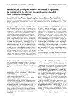
intestine and large intestine (Fig. 1, 2). Margin of the liver
was dull. The spleen was atrophic. The lung showed doughy
*Corresponding author
Phone: 82-2-880-1273; Fax: 82-2-880-8662
E-mail:
Case Report
182 Seung-ho Ryu
et al.
consistensy. The trachea was filled with foam which
extended into the smaller airways. The mucosa of the
trachea was congested and ecchymotic hemorrhagic in
appearance. Both kidneys were swollen. The remainder of
the gross necropsy was unremarkable.
Although infection with ascarids is common in horses
under 1 year of age, ascarid impactions are a relatively
uncommon cause of colic in horses. Foals are usually
infected during the first few days of life and the
gastrointestinal phase of the parasite begins 14-17 days
following infection. The prepatent period of
P. equorum
is
72-110 days; therefore, clinical signs of gastrointestinal tract
disease would be expected from approximately 3-4 months
onward, as the worms increase in number and size. Exposed
and nonexposed horses older than 6 months of age develop
an age-dependent immunity. Older horses are more likely to
develop pulmonary and hepatic signs rather than
gastrointestinal tract signs [5]. Heavy infestation of
P.
equorum
in foals, weanlings and yearlings can lead to small
intestinal impaction, particularly after the administration of a
high efficacy anthelmintic such as ivermectin, piperazine or
an organophosphate. These non-benzimidazole drugs inhibit
neuromuscular transmission and paralyze the ascarids,
thereby promoting impaction [4]. Rupture of the ascarid
cuticle following organophosphate administration reportedly
causes the release of antigenic fluid that produces
hypomotility when it is absorbed [3]. In one study, 54% of
horses were dewormed 1-5 days prior to the onset of colic
signs [6]. Controversy exists regarding the optimal
deworming regimen to prevent impaction in foals with
heavy worm burdens [6]. In this case, the new farm manager
changed the policy of anthelmintic treatment due to financial
constraints. So anthelmintics had not been administered and
shock was thought to result from a heavy worm burden
throughout the entire gastrointestinal tract.
Ascarid infections are reported to occur most commonly
in the duodenum and proximal jejunum and they may be
throughout the entire gastrointestinal tract [6] like this case.
Ascarid impactions are treated medically with intestinal
lubricants and analgesics if the obstruction is incomplete. If
a medical approach to relieve the obstruction is not
successful or if the obstruction becomes complete, surgical
intervention is necessary. In most cases surgical intervention
requiring, multiple enterotomies is necessary and the
prognosis is guarded [2]. In this case, the economic
constraints by the owner prevented from performing
laparatomy and limited therapeutic options.
Conclusively, the outcome of this report is to describe the
first diagnosed case of gastrointestinal impaction by
P.
equorum
in a Thoroughbred foal in South Korea and
indicate the importance of regular anthelmintic treatment.
References
1. Adair HS. What is your diagnosis? J Am Vet Med Assoc
1990, 196, 1023-1024.
2. Bello TR. Endoparasitism. In: Colahan PT, Mayhew IG
(Joe), Merritt AM, Moore JN (eds.). Equine Medicine and
Surgery. 5th ed. pp. 729, Mosby, St. Louis, 1999.
3. Clayton HM. Ascarids. Recent advances. Vet Clin North Am
Equine Pract 1986, 2, 313-28.
4. DiPietro JA, Boero M and Ely RW. Abdominal abscess
associated with Parascaris equorum infection in a foal. J Am
Vet Med Assoc 1983, 182, 991-992.
5. Robertson JT. Diseases of the small intestine. In: White NA
(ed.). The Equine Acute Abdomen, pp. 361-362, Lea &
Febiger, Philadelphia, 1990.
6. Southwood LL, Ragle CA, Snyder JR, Hendrickson DA.
Surgical treatment of Ascarid impactions in horses and foals.
AAEP Proceedings, 1996, 42, 258-261.
F
ig. 1. The ascarid mass in the stomach.
F
ig. 2. The ascarid mass in the small intestine.