Báo cáo khoa học: "Electroacupuncture ameliorates experimental colitis induced by acetic acid in rat" doc
Bạn đang xem bản rút gọn của tài liệu. Xem và tải ngay bản đầy đủ của tài liệu tại đây (2.82 MB, 7 trang )
-2851$/ 2)
9H W H U L Q D U \
6FLHQFH
J. Vet. Sci.
(2004),
/
5
(3), 189–195
Electroacupuncture ameliorates experimental colitis induced by
acetic acid in rat
Jeoung-Woo Kang
1
, Tae-Wan Kim
2
, Jun-Ho La
1
, Tae-Sik Sung
1
, Hyun-Ju Kim
1
, Young-Bae Kwon
3
,
Jeum-Yong Kim
3
, Il-Suk Yang
1,
*
1
Department of Physiology, College of Veterinary Medicine, Seoul National University, Seoul 151-742, Korea
2
Department of Physiology, College of Veterinary Medicine, Kyungpuk National University, Daegu 712-715, Korea
3
Institute of Bioscience and Biotechnology, Daewoong Pharm Co. LTD., Yongin 449-814, Korea
The effect of electroacupuncture (EA) on experimental
colitis was investigated in Sprague-Dawley rats. Colitis was
induced by intracolonic instillation of 4% acetic acid. EA (2
Hz, 0.05 ms, 2 V for 20 min) was applied to bilateral Hoku (LI-
4) and Zusanli (ST-36) on 12 hrs and 36 hrs after induction of
colitis. EA-treatment significantly reduced the macroscopic
damage and the myeloperoxidase activity of colonic samples at
3 days post-induction of colitis. Colitic colon showed a
decreased
in vitro
motility. However, colonic motility of EA-
treated group was not significantly different from that of
normal group. The anti-inflammatory effect of EA was not
inhibited by a glucocorticoid receptor antagonist, RU-486, but
suppressed by a
β
-adrenoceptor antagonist, propranonol.
These results suggest that EA-treatment has a beneficial effect
on colitis, and its anti-inflammatory effect is mediated by
β
-
adrenoceptor activation but not by endogenous glucocorticoid-
dependent mechanism.
Key words:
colitis, electroacupuncture, glucocorticoid,
β
-
adrenoceptor
Introduction
Inflammatory bowel disease (IBD) is a chronic
inflammatory disorder with unknown etiology and
pathogenesis. In patients with IBD, gut inflammation is
associated with intestinal muscle dysfunction [25,34]. These
observations have been confirmed in a variety of animal
models of experimental intestinal inflammation [9,18],
showing that smooth muscle dysfunction is linked to the
inflammatory reaction.
Aminosalicylic acid and corticosteroids are the drugs most
commonly used in treatment of IBD, but long-term use of
these drugs may give rise to adverse effects, such as
nephrotoxicity, pulmonary toxicity and male infertility
[5,11]. Therefore, many researchers are recently interested
in an alternative medical treatment such as acupuncture.
Acupuncture therapy has been utilized to relieve and treat
various inflammatory diseases [3,13,38]. However, few
studies have evaluated the effect of acupuncture on IBD, and
moreover, its therapeutic mechanism is still unclear.
Electroacupuncture (EA) has been reported to activate
hypothalamic-pituitary-adrenal (HPA) axis and consequently
release glucocorticoids that have potent anti-inflammatory
properties [15,16]. EA was also reported to modulate the
secretion rates of catecholamines from adrenal medulla by
influencing sympathetic activity [20,23]. Catecholamines
are known to induce anti-inflammatory responses through
β
-
adrenoceptor activation [36]. Therefore, we hypothesized
that EA has therapeutic effect on IBD and the anti-
inflammatory effect of EA is mediated by glucocorticoids
and/or catecholamines acting on
β
-adrenoceptor. The
present study was designed to examine this hypothesis using
a widely used animal model of colitis, the rat model of acetic
acid induced-colitis [6,24]. In this model, we investigated
(1) whether the EA treatment would reduce the tissue
inflammatory responses and the smooth muscle
dysfunction, and (2) whether an antagonist of either
glucocorticoids receptor or
β
-adrenoceptor could modulate
the effect of EA on colitis.
Materials and Methods
Animal preparation and experimental groups
Male Sprague-Dawley rats, weighing 250-300 g were
used. The rats were housed in stainless steel hanging cages
in colony room maintained under a 12 h light/dark cycle
with a room temperature of 22 ± 1
o
C and humidity of 65-
70%. Water and food were available
ad libitum
.
*Corresponding author
Tel: 82-2-880-1261; Fax: 82-2-885-2732
E-mail:
190 Jeoung-Woo Kang
et al.
Induction of experimental colitis
All experimental animals were fasted for 24 hrs before
induction of colitis. Each rat was lightly anesthetized with
ether, and a polyethylene cannula (PE-60) was inserted into
the lumen of the colon via the anus. The tip of the cannula
was positioned at 8 cm proximal to the anus. Either 1 ml of
acetic acid (4% vol/vol in 0.9% NaCl) or saline as the sham
control was slowly infused into the distal colon. After 30
seconds exposure, 1 ml of saline (0.9%) was instilled in
order to withdraw the previous solution from colon.
Treatment of electroacupuncture
Two acupoints, bilateral Zusanli (ST-36), located at the
lateral upper tibia, and bilateral Hoku (LI-4), located at the
junction of the first and the second metacarpal bones, were
selected for the experiments. Stimulation of these two points
is known to have therapeutic effect on gastrointestinal
diseases [7,14]. Animals were anesthetized with ketamine.
An acupuncture needle (
Φ
0.18 mm, length 15 mm) was
soldered to a flexible electrical wire, and the needle was
inserted about 3 mm deep into the muscle layer at the
acupoint. The second identical needle, as a positive pole,
was inserted into the other point approximately 5-10 mm
from the first one. An electric current of square wave pulses
(2 Hz, 0.05 ms, 2 V for 20 min) were applied from
stimulator (S88, Grass-telefactor, West Warwick, RI, USA)
through a stimulus isolation unit (SIU5B, Grass-telefactor,
West Warwick, RI, USA) on 12 hrs and 36 hrs after the
induction of experimental colitis.
Measurement of myeloperoxidase (MPO) activity
At 3 days post-induction of colitis, rats were sacrificed by
cervical dislocation. MPO activity was estimated in the
whole colonic tissue obtained from the rats with and without
colitis [2]. A segment of colon was minced finely with
scissors in 5 ml of 50 mmol/L potassium phosphate buffer,
pH 6.0 containing 14 mmol/L hexadecyl-trimethylammonium
bromide and homogenized for 3 min. The sample were
frozen in liquid nitrogen and thawed three times and
centrifuged for 20 min in cold at 20000 g using
microcentrifuge. Aliquots of supernatants (20
µ
l) were
mixed with 980
µ
l of
o
-dianisidine solution which was
made of 16.5 mg of
o
-dianisidine-HCl (Sigma, St louis, MI,
USA), 90 ml of distilled water, 10 ml of potassium
phosphate buffer, pH 6.0 and 50
µ
l of 1% H
2
O
2
(Sigma, St
louis, MI, USA). Absorbance was measured at 450 nm
every 1 min over a period of 10 min. MPO activity was
expressed as units/g of tissue. The enzyme unit was defined
as the conversion of 1
µ
mol of H
2
O
2
per min at 25
o
C.
Measurement of colonic motility
At 3 days post-induction of colitis, rats were sacrificed by
cervical dislocation, and a 2 cm distal colonic segment was
removed. The segments were suspended in a 20 mL organ
bath containing oxygenated (95% O
2
, 5% CO
2
) Krebs
solution at 37
o
C. The distal end of the colonic segment was
tied around the mouth of J-tube and this was connected via a
3-way connector to a syringe and to a pressure transducer
(RP-1500, Narco Bio-systems Inc, Houston, TX, USA). The
ligated proximal end was secured with a silk thread to an
isometric force displacement transducer (FT-03, Grass-
telefactor, West Warwich, RI, USA). The signals from both
transducers were processed through Powerlab/400 and
Chart 4.2 (AD Instruments, Castle Hill, NSW, Australia).
The motilities of the colonic segments were detected as both
longitudinal muscle contraction (isometric tension) and
intraluminal pressure, which has been reported to reflect the
contractile activity of circular muscle [4].
For calculation of spontaneous motility, we measured the
mean longitudinal contraction and mean intraluminal
pressure in steady states for 5 min. Mean longitudinal
contraction or mean intraluminal pressure were calculated
by the area under tension curve or pressure curve for 5-min
period divided by duration of periods (5
×
60 sec) and
expressed per gram wet weight of the colonic segment.
In order to determine the effects of carbachol (CCh) and
N
ω
-nitro-L-arginine methyl ester (L-NAME), we measured
mean longitudinal contraction and mean intraluminal
pressure at the end of the equilibration time and when the
new stable level reached after each drug administration. The
concentration-response curves to CCh (0.1-10 mM) were
obtained cumulatively by adding each concentration to the
bath.
Involvement of glucocorticoid and
β
-adrenoceptor in
the action of electroacupuncture
To investigate the mechanisms of EA, the corticosteroid
receptor antagonist (RU 486 in DMSO: 20 mg/kg) or
β
-
adrenoceptor antagonist (propranolol in saline: 10 mg/kg)
was intraperitoneally administrated at 2 hrs before EA
stimulation.
Solutions and drugs
The Krebs-solution contained (in mM) 118.5 NaCl, 4.75
KCl, 2.54 CaCl
2
, 1.19 MgSO
4
, 25 NaHCO
3
, 1.19 NaH
2
PO
4
,
and 11.0 dextrose. The solution was continusously gassed
with 95% O
2
and 5 % CO
2
(v/v), and the pH ranged from 7.3
to 7.4. Carbamylcholine chloride (CCh), acetic acid, RU
486, propranolol, N
ω
-nitro-L-arginine methyl ester (L-
NAME) were obtained from Sigma Chemical Co. All drugs
were added to the baths in volumes less than 1% of the total
bath volume.
Statistical analysis
Data are expressed as means ± S.E.M. with
n
, the number
of animals. The responses were statistically tested using
ANOVA followed by the Newman-Keuls multiple
comparison test, or using Student’s
t
-test. The value of
Effect of EA on experimental colitis 191
p
< 0.05 considered to be significantly different.
Results
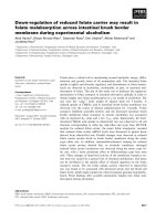
Macroscopic observation
Rats developed diarrhea 2-3 days after colitis induction.
The colitis + EA group showed less severe diarrhea than the
colitis group. However, the normal group did not develop
diarrhea (Data not shown). The colonic tissue of colitis
group showed prominent congestion and swelling, while the
macroscopic inflammatory features of colon in the
colitis+EA group were moderate (Fig. 1).
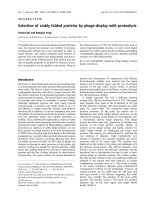
MPO activity
MPO activity in the colitis group was significantly higher
than that in the normal group (0.93 ± 0.17 Unit/g, n = 8
vs
0.15 ± 0.02 Unit/g, n = 5,
p
< 0.01). But, MPO activity of
the colitis + EA group was significantly lower than that of
the colitis group (0.37 ± 0.09 Unit/g, n = 7
vs
0.93 ± 0.17
Unit/g, n = 8,
p
< 0.01). There was no statistical difference
in MPO activity between the normal group and the colitis
+ EA group, implying that EA has an anti-inflammatory
effect on the acetic acid-induced experimental colitis (Fig. 2).
Colonic smooth muscle motility
All colonic segments from normal group exhibited a
spontaneous and highly synchronized rhythmic longitudinal
phasic contractions and intraluminal pressure waves.
However, most colonic muscles from the colitis group
showed spontaneous motility with small amplitude and
irregular pattern. But, colonic segments from the colitis +
EA group showed spontaneous and regular motility with
considerable amplitude (Fig. 3A).
The mean weight of the colonic segments from the
normal, colitis and colitis + EA group were 365 ± 37,
410 ± 35 and 378 ± 31 mg, respectively. There was no
significant difference between them (n = 11,
p
> 0.05).
The mean longitudinal contraction of colitic colonic
segments (25.6 ± 3.6 mN/g wet segment wt; n = 11) was
significantly less than that of normal colonic segments (10.5
± 3.5 mN/g wet segment wt; n = 11;
p
< 0.05). In contrast,
the mean longitudinal contraction of colonic segments in the
colitis + EA group (24.7 ± 4.8 mN/g wet segment wt; n =
11) was significantly higher than that in the colitis group
(10.5 ± 3.5 mN/g wet segment wt; n = 11;
p
< 0.05). The
mean intraluminal pressure of colonic segments from
normal, colitis, colitis + EA group were 4.2 ± 0.8, 1.4 ± 0.2
and 3.5 ± 0.9 mmHg/g wet segment wt; n = 11), respectively
(Fig. 3B).
CCh (0.1-10
µ
M), a potent cholinergic agonist, increased
both mean longitudinal contraction and mean intraluminal
pressure of all groups in a concentration-dependent manner.
The increases of mean longitudinal contraction and
intraluminal pressure by CCh in the normal and the
colitis+EA group were higher than that in colitis group
(n = 6, Fig. 4).
L-NAME (100
µ
M), a nitric oxide synthase inhibitor,
significantly increased both mean longitudinal contraction
and mean intraluminal pressure in the normal and the
colitis + EA group. However, the colonic segments of colitis
group did not respond to L-NAME (n = 5, Fig. 5).
Effects of RU486 and propranolol
To determine whether glucocorticoid, a pivotal mediator
of HPA axis, was involved in the anti-inflammatory effect of
EA, a corticosteroid receptor antagonist, RU486, was
pretreated 2 hrs before the EA treatment. RU486 did not
affect the EA induced anti-inflammatory effect (Fig. 6). But,
pretreatment with
β
-adrenoreceptor antagonist, propranolol,
significantly suppressed the effect of EA (Fig. 7).
Discussion
The present study demonstrates that EA stimulation at
F
ig. 1. The macroscopic features of colonic tissue of norm
al,
c
olitis and colitis + EA group.
F
ig. 2. MPO activity of each experimental group. **
p
<0.01
as
c
ompared with normal group, ##
p
< 0.01 as compared wi
th
c
olitis group.
192 Jeoung-Woo Kang
et al.
Zusanli (ST-36) and Hoku (LI-4) has therapeutic effect on
experimental colitis. The colitis+EA group showed milder
macroscopic lesion in colon than the colitis group, implying
that EA treatment can effectively improve the colonic
mucosal lesions. More convincingly, tissue MPO activity in
the colitis + EA group was significantly less than that of the
colitis group. The MPO activity was known to be a marker
for tissue neutrophil content and be useful to quantify the
extent of inflammation [2]. It has been reported that the
accumulation of neutrophil is a characteristic feature of such
gastrointestinal inflammatory disease as colitis [2].
Therefore, the decrease of the MPO activity in the colitis
+ EA group evidences that EA indeed reduced the
inflammation in colitic tissue.
The decreased colonic motility is generally observed in
IBD patients [12,25,34] and in the animal models of
experimental colitis [17]. In the current study, colonic
segments of colitis group also showed significantly
decreased spontaneous longitudinal and circular motilities,
compared with those of normal group. However, the
spontaneous colonic contractile activities of colitis + EA
group were significantly higher than those of colitis group.
These findings suggest that EA treatment suppresses the
inflammatory response and restores the ability of the colonic
F
ig. 3.
Typical recordings showing the spontaneous mechanical activity of colonic segments in normal, colitis and colitis + EA grou
p,
d
etected as isometric tension (upper trace) and intraluminal pressure (lower trace) (A). B and C are statistical graphs for me
an
l
ongitudinal contraction and mean intraluminal pressure, respectively. *
p
< 0.05 as compared with normal group, #
p
< 0.05 as compar
ed
w
ith colitis group.
F
ig. 4.
Effects of CCh on mean longitudinal contraction and mean intraluminal pressure of colonic segments in normal, colitis a
nd
c
olitis + EA group. *
p
< 0.05 as compared with normal group, #
p
< 0.05 as compared with colitis group.
Effect of EA on experimental colitis 193
muscle to develop spontaneous motility.
In acetic acid-induced colitis, it was reported that CCh-
induced contraction was significantly decreased, compared
with that of normal group [9]. In the present study, the CCh-
induced increases of longitudinal and circular motilities in
the colitis group were significantly less than those in the
normal and in the colitis + EA groups. These results indicate
that EA treatment improves the colitis-induced damage in
the colonic contractile function.
Because NO has been shown to act as a major nonadrenergic,
noncholinergic (NANC) inhibitory neurotransmitter in the
gut, the changes in the gastrointestinal motility have been
attributed to an impairment of NO function in the various
dysfunctional condition [21,30]. It was also reported that
nitrergic neurons were impaired in the rat model of
experimental colitis [19]. The damage of nitrergic neural
function was also observed in the present study. In the colitis
group, L-NAME, a nitric oxide synthase inhibitor, failed to
further increase the amplitude of the spontaneous motility.
On the other hand, in the normal and the colitis + EA group,
L-NAME increased the spontaneous longitudinal and
circular mechanical activilty, implying that tonic nitrergic
neural function was maintained in the colitis + EA group as
in the normal group. Taken together, these data support that
EA treatment can suppress intestinal inflammation and
reverses intestinal smooth muscle dysfunction caused by
colitis.
IBD is a chronic relapsing inflammation of the intestine
mediated by the activation of immune cells and the release
of inflammatory mediators. It is well established that
neuroendocrine and immune systems communicate
bidirectionally [28]. Increased tissue production of
interleukin (IL)-1, IL-6, IL-8, and tumor necrosis factor
(TNF)-
α
has been found during the episodes of active IBD
in patients with ulcerative colitis or Crohn’s disease [10].
Cytokines produced by immune cells during inflammation
can stimulate the HPA axis to release corticosteroids, which
are important immunoregulators. The corticosteroids are
F
ig. 5.
Effects of L-NAME on the spontaneous mechanical activity of colonic segments in normal, colitis and colitis + EA grou
p,
m
onitored as isometric tension and intraluminal pressure. *
p
< 0.05 as compared with control.
F
ig. 6.
Effect of RU486, a glucocorticoid receptor antagonist
on
t
he lowering MPO activity by EA. Vehicle: DMSO, n = anim
al
n
umber, *
p
<0.05.
F
ig. 7.
Effect of propranolol (PPN), ß-adrenoceptor antagoni
st,
o
n the lowering MPO activity by EA. Vehicle: saline, n = anim
al
n
umber, **
p
<0.01.
194 Jeoung-Woo Kang
et al.
known to effectively shut off the immune response [27,33].
In addition to the HPA axis activation, pro-inflammatory
cytokines (e.g., IL-1
β
) can also enhance the sympathetic
activity, including the release of catecholamines from
sympathetic terminals and adrenal medulla. It has been
proposed that catecholamines function as endogenous anti-
inflammatory agents [1,29].
Although the hypotheses on mechanisms of acupuncture
are various, it is often proposed that EA activates the HPA
axis [15,16,22] or sympathetic nervous system [20,23]. In
the present study, a glucocorticoid receptor antagonist,
RU486, did not alter the anti-inflammatory effect of EA on
colitis. This indicates that glucocorticoids do not participate
in the EA-induced anti-inflammation on colitis, at least in
this experimental condition. However, the possibility cannot
be excluded that the released glucocorticoids by EA was not
enough to reduce the acetic acid-induced colitis.
We found that pretreatment with a
β
-adrenoceptor
antagonist, propranolol, blocked the anti-inflammatory
effect of EA. This result implies that the anti-inflammatory
effect of EA on colitis is mediated by catecholamines acting
through ß-adrenoceptor. The mechanisms involving
β
-
adrenoceptor in the anti-inflammatory effect of EA remain
to be elucidated. It is noteworthy that immune cells can bind
different neurotransmitters [29]. For example, catecholamines
are known to act on macrophages and monocytes through
binding to the cell surface
β
-adrenergic receptors.
β
-
adrenoceptors are coupled to the GTP-binding protein of the
adenylate cyclase complex for increasing intracellular
cAMP levels and activating protein kinase A upon
stimulation [35]. In this way, catecholamines reduce the
production of pro-inflammatory cytokines such as IL-1
β
,
IL-6, and TNF-
α
, and enhance the secretion of anti-
inflammatory cytokines such as IL-10 [36]. Indeed, it was
reported that EA greatly inhibited the expression of IL-1
β
and IL-6 mRNA in the rat model of ulcerative colitis
[32,37]. Oral administration of enteric-coated recombinant
human IL-11 (rhIL-11), a potent anti-inflammatory
cytokine, suppresses intestinal inflammation and restores the
ability of the smooth muscle to develop active tension in
both jejunum and colon in HLA-B27 transgenic rats with
chronic intestinal inflammation [8].
It should be mentioned that opioid receptors are suggested
to be involved in the anti-inflammatory action of
acupuncture [3,26] and opioids have anti-inflammatory
effects on synovitis in rheumatoid arthritis [31]. Therefore, it
will be necessary to test whether endogenous opioid system
is also involved in the EA-induced anti-inflammation on
experimental colitis. Future experiments will attempt to
elucidate the relationship between opioid receptors and the
anti-inflammatory effect of EA.
In conclusion, EA therapy ameliorates intestinal
inflammation and reverses intestinal smooth muscle
dysfunction in experimental colitis induced by acetic acid in
rat. The anti-inflammatory effect of EA does not involve the
endogenous glucocorticoid-dependent mechanism but
requires the
β
-adrenoceptor activation. Further studies are
needed to elucidate the exact mechanism of EA action on
experimental colitis.
Acknowledgment
This work was supported by the Research Institute for
Veterinary Science, College of Veterinary Medicine, Seoul
National University.
References
1. Bhattacharya SK, Das N, Sarkar MK. Inhibition of
carrageenin-induced pedal oedema in rats by immobilisation
stress. Res Exp Med (Berl) 1987, 187, 303-313.
2. Bradley PP, Priebat DA, Christensen RD, Rothstein G.
Measurement of cutaneous inflammation: estimation of
neutrophil content with an enzyme marker. J Invest Dermatol
1982, 78, 206-209.
3. Ceccherelli F, Gagliardi G, Visentin R, Sandona F, Casale
R, Giron G. The effects of parachlorophenylalanine and
naloxone on acupuncture and electroacupuncture modulation
of capsaicin-induced neurogenic edema in the rat hind paw.
A controlled blind study. Clin Exp Rheumatol 1999, 17, 655-
662.
4. Coupar IM, Liu L. A simple method for measuring the
effects of drugs on intestinal longitudinal and circular
muscle. J Pharmacol Toxicol Methods 1996, 36, 147-154.
5. Di Paolo MC, Paoluzi OA, Pica R, Iacopini F, Crispino P,
Rivera M, Spera G, Paoluzi P. Sulphasalazine and 5-
aminosalicylic acid in long-term treatment of ulcerative
colitis: report on tolerance and side-effects. Dig Liver Dis
2001, 33, 563-569.
6. Elson CO, Sartor RB, Tennyson GS, Riddell RH.
Experimental models of inflammatory bowel disease.
Gastroenterology 1995, 109, 1344-1367.
7. Fireman Z, Segal A, Kopelman Y, Sternberg A, Carasso
R. Acupuncture treatment for irritable bowel syndrome. A
double-blind controlled study. Digestion 2001, 64, 100-103.
8. Greenwood-Van Meerveld B, Venkova K, Keith JC Jr.
Recombinant human interleukin-11 restores smooth muscle
function in the jejunum and colon of human leukocyte
antigen-B27 rats with intestinal inflammation. J Pharmacol
Exp Ther 2001, 299, 58-66.
9. Grossi L, McHugh K, Collins SM. On the specificity of
altered muscle function in experimental colitis in rats.
Gastroenterology 1993, 104, 1049-1056.
10. Isaacs KL, Sartor RB, Haskill S. Cytokine messenger RNA
profiles in inflammatory bowel disease mucosa detected by
polymerase chain reaction amplification. Gastroenterology
1992, 103, 1587-1595.
11. Jankauskiene A, Druskis V, Laurinavicius A. Cyclosporine
nephrotoxicity: associated allograft dysfunction at low trough
concentration. Clin Nephrol 2001, 56, S27-29.
12. Koch TR, Carney JA, Go VL, Szurszewski JH.
Effect of EA on experimental colitis 195
Spontaneous contractions and some electrophysiologic
properties of circular muscle from normal sigmoid colon and
ulcerative colitis. Gastroenterology 1988,
95
, 77-84.
13.
Kumar AM, Wen XL.
Acupuncture treatment for
osteoarthritic pain and inflammation of the knee. Altern Ther
Health Med 2002,
8
, 128
14.
Li Y, Tougas G, Chiverton SG, Hunt RH.
The effect of
acupuncture on gastrointestinal function and disorders. Am J
Gastroenterol 1992,
87
, 1372-1381.
15.
Liao YY, Seto K, Saito H, Fujita M, Kawakami M.
Effect
of acupuncture on adrenocortical hormone production: I.
Variation in the ability for adrenocortical hormone
production in relation to the duration of acupuncture
stimulation. Am J Chin Med 1979,
7
, 362-371.
16.
Liao YY, Seto K, Saitoh H, Kawakami M.
Effect of
acupuncture on adrenocortical hormone production in rabbits
with a central lesion. Am J Chin Med 1981,
9
, 61-73.
17.
Lu G, Qian X, Berezin I, Telford GL, Huizinga JD, Sarna
SK.
Inflammation modulates in vitro colonic myoelectric
and contractile activity and interstitial cells of Cajal. Am J
Physiol 1997,
273
(6 Pt 1), G1233-45.
18.
Martinolle JP, Garcia-Villar R, Fioramonti J, Bueno L.
Altered contractility of circular and longitudinal muscle in
TNBS-inflamed guinea pig ileum. Am J Physiol 1997,
272
(5
Pt 1), G1258-67.
19.
Mizuta Y, Isomoto H, Takahashi T.
Impaired nitrergic
innervation in rat colitis induced by dextran sulfate sodium.
Gastroenterology 2000,
118
, 714-723.
20.
Mori H, Uchida S, Ohsawa H, Noguchi E, Kimura T,
Nishijo K.
Electro-acupuncture stimulation to a hindpaw and
a hind leg produces different reflex responses in
sympathoadrenal medullary function in anesthetized rats. J
Auton Nerv Syst 2000,
79
, 93-98.
21.
Mule F, Serio R.
Spontaneous mechanical activity and
evoked responses in isolated gastric preparations from
normal and dystrophic (
mdx
) mice. Neurogastroenterol Mot
2002,
14
, 667-675.
22.
Pan B, Castro-Lopes JM, Coimbra A.
Activation of
anterior lobe corticotrophs by electroacupuncture or noxious
stimulation in the anaesthetized rat, as shown by
colocalization of Fos protein with ACTH and beta-endorphin
and increased hormone release. Brain Res Bull 1996,
40
,
175-182.
23.
Sato A, Sato Y, Suzuki A, Uchida S.
Reflex modulation of
catecholamine secretion and adrenal sympathetic nerve
activity by acupuncture-like stimulation in anesthetized rat.
Jpn J Physiol 1996,
46
, 411-421.
24.
Singh VP, Patil CS, Jain NK, Singh A, Kulkarni SK.
Effect of nimesulide on acetic acid- and leukotriene-induced
inflammatory bowel disease in rats. Prostaglandins Other
Lipid Mediat 2003,
71
, 163-175.
25.
Snape WJ Jr, Williams R, Hyman PE.
Defect in colonic
smooth muscle contraction in patients with ulcerative colitis.
Am J Physiol 1991,
261
(6 Pt 1), G987-G991.
26.
Son YS, Park HJ, Kwon OB, Jung SC, Shin HC, Lim S.
Antipyretic effects of acupuncture on the lipopolysaccharide-
induced fever and expression of interleukin-6 and
interleukin-beta mRNAs in the hypothalamus of rats.
Neurosci Lett 2002,
319
, 45-48.
27.
Sternberg EM.
Neuroendocrine factors in susceptibility to
inflammatory disease: focus on the hypothalamic-pituitary-
adrenal axis. Horm Res 1995,
43
, 159-161.
28.
Straub RH, Herfarth H, Falk W, Andus T, Scholmerich J.
Uncoupling of the sympathetic nervous system and the
hypothalamic-pituitary-adrenal axis in inflammatory bowel
disease? J Neuroimmunol 2002,
126
, 116-125.
29.
Straub RH, Westermann J, Scholmerich J, Falk W.
Dialogue between the CNS and the immune system in
lymphoid organs. Immunol Today 1998,
19
, 409-413.
30.
Takahashi T.
Pathophysiological significance of neuronal
nitric oxide synthase in the gastrointestinal tract. J
Gastroenterol 2003,
38
, 421-430.
31.
Takeba Y, Suzuki N, Kaneko A, Asai T, Sakane T.
Endorphin and enkephalin ameliorate excessive synovial cell
functions in patients with rheumatoid arthritis. J Rheumatol
2001,
28
, 2176-2183.
32.
Tian L, Huang YX, Tian M, Gao W, Chang Q.
Downregulation of electroacupuncture at ST36 on TNF-
alpha in rats with ulcerative colitis. World J Gastroenterol
2003,
9
, 1028-1033.
33.
Turnbull AV, Rivier CL.
Regulation of the hypothalamic-
pituitary-adrenal axis by cytokines: actions and mechanisms
of action. Physiol Rev 1999,
79
, 1-71.
34.
Vermillion DL, Huizinga JD, Riddell RH, Collins SM.
Altered small intestinal smooth muscle function in Crohn’s
disease. Gastroenterology 1993,
104
, 1692-1699.
35.
Woiciechowsky C, Asadullah K, Nestler D, Eberhardt B,
Platzer C, Schoning B, Glockner F, Lanksch WR, Volk
HD, Docke WD.
Sympathetic activation triggers systemic
interleukin-10 release in immunodepression induced by brain
injury. Nat Med 1998,
4
, 808-813.
36.
Woiciechowsky C, Schoning B, Lanksch WR, Volk HD,
Docke WD.
Mechanisms of brain-mediated systemic anti-
inflammatory syndrome causing immunodepression. J Mol
Med 1999,
77
, 769-780.
37.
Wu HG, Zhou LB, Pan YY, Huang C, Chen HP, Shi Z,
Hua XG.
Study of the mechanisms of acupuncture and
moxibustion treatment for ulcerative colitis rats in view of the
gene expression of cytokines. World J Gastroenterol 1999,
5
,
515-517.
38.
Zijlstra FJ, van den BeLI, Huygen FJ, Klein J.
Anti-
inflammatory actions of acupuncture. Mediators Inflamm
2003,
12
, 59-69.