Báo cáo khoa học: "Development of a novel antigen capture-ELISA using IgY against porcine interleukin-6 and its application" potx
Bạn đang xem bản rút gọn của tài liệu. Xem và tải ngay bản đầy đủ của tài liệu tại đây (290.55 KB, 7 trang )
-2851$/ 2)
9H W H U L Q D U \
6FLHQFH
J. Vet. Sci.
(2004),
/
5
(4), 337–343
Development of a novel antigen capture-ELISA using IgY against porcine
interleukin-6 and its application
Deog Yong Lee, Young Wook Cho, Sang Gyun Kang, Sung Jae Shin, Han Sang Yoo*
Department of Infectious Diseases, College of Veterinary Medicine and School of Agricultural Biotechnology,
Seoul National University, Seoul, 151-742, Korea
Interleukin-6 (IL-6) is introduced as a marker of
disease. At present, a variety of method may be used to
quantify expression of this protein. Antigen capture-
ELISA is a sensitive and accurate quantification method
previously used with ovine, rat, and human IL-6 proteins.
However, it has never been reported to quantify porcine
IL-6 protein using capture ELISA. In this study, we
generated and characterized a set of IgY and mono-
specific polyclonal antibodies to recombinant porcine IL-6
(rpIL-6), and combining these with a sensitive and specific
capture-ELISA for a diagnostic purpose. cDNA encoding
the mature protein coding region of porcine IL-6 was
cloned and expressed with pQE-30UA expression vector.
rpIL-6 was then expressed and purified by using Ni-NTA
resin. Protein mass of 24 kDa was found with SDS-PAGE
and the identity of the protein was confirmed by Western-
blot. Production of polyclonal antibodies against rpIL-6
was performed using the purified rpIL-6 in mice and
hens. An antigen capture-ELISA was developed with the
antibodies after their extraction. To compare the IL-6
level in the different sanitary state of farms, pig sera were
randomly collected and concentration of IL-6 in the sera
was measured with the antigen capture-ELISA. The
capture-ELISA with the optimal concentration of antibodies,
in this study, was able to detect about 10 ng/ml of rpIL-6.
IL-6 levels determined with the capture-ELISA in pig sera
showed positive correlation with the sanitary states of the
farms. These results suggested that the developed antigen
capture-ELISA could be a good tool for the screening of
microbial infection in pig farms.
Key words:
Antigen capture-ELISA, porcine interleukin-6,
IgY, protein expression
Introduction
Interleukin-6 (IL-6) is a 21 to 28 kDa glycoprotein [23,
30] and a multifocal cytokine, produced by both lymphoid
and non-lymphoid cells [18]. IL-6 plays an important role in
immune response, hematopoiesis, and acute-phase reaction.
IL-6 induces B cell proliferation and differentiation [12,13,
27], antibody production [11], and T-cell activation and
differentiation [35]. In addition, IL-6 stimulates hematopoietic
stem cells and macrophage differentiation in several human
and murine cell lines. Also, a variety of acute-phase
proteins, such as fibrinogen,
α
1
-antichymotrypsin,
α
1
-acid
glycoprotein, and haptoglobin, are induced by IL-6 [10].
This study introduces the use of IL-6 as a marker of
disease in swine. The appearance of IL-6 positive pigs
coincided with the onset of clinical signs of disease and
increased body temperature associated with acute bacterial
infection [3]. In challenge studies of SIV-vaccinated pigs,
levels of IL-6 with IFN-
α
and TNF-
α
were correlated with
both clinical and viral protection [17]. Used as a marker of
disease, measurement of IL-6 concentration in serum
predicts the disease status of pigs or farms.
Antigen capture-ELISA is a sensitive and accurate
quantification method [24] which usually uses monoclonal
antibodies to increase sensitivity. However, monoclonal
antibody preparation for capture-ELISA requires great skill
and laborious job. Therefore, for cheap and easy preparation,
we used IgY as a capture-antibody instead.
IgY is the typical low-molecular-weight (LMW) egg yolk
serum antibody of birds, reptiles, amphibians and lungfish,
whereas IgG occurs in mammals [8]. Because of evolutionary
difference, chicken IgY reacts with more epitopes on a
mammalian antigen, producing an amplification of the signal.
IgY also has the advantage in that it avoids the interference
in immunological assays caused by the complement system,
rheumatoid factors, anti-mouse IgG antibodies or human
and bacterial Fc receptors [2].
Quantification of IL-6 protein using capture ELISA has
been done using ovine, rat, and human IL-6 [9,24,28].
Detection of porcine cytokines using capture-ELISA has
*Corresponding author
Tel:+82-2-880-1263; Fax: +82-2-874-2738
E-mail:
338 Deog Yong Lee
et al.
only been performed with IFN-gamma, IL-8 and IL-18 [21,
26,33]. To our knowledge this is the first study to use the
ELISA capture method to quantify concentrations of
porcine IL-6, which were then used as a marker for disease.
This study generated and characterized a set of IgY and
polyclonal antibodies to recombinant porcine IL-6 (rpIL-6),
and then combined these antibodies to develop a sensitive
and specific capture-ELISA for the diagnosis of a farm’s
sanitary state.
Materials and Methods
Production of recombinant pig IL-6
Cloning of cDNA encoding mature protein:
Total RNA
was extracted from PBMCs using Trizol reagent (Gibco,
USA) and chloroform after stimulation with 20
µ
g/ml of
phytohemagglutinin (PHA, Invitrogen, Carlsbad, USA) for
4 hr. Single-stranded cDNA was synthesized using the
Superscript preamplification system for First strand cDNA
synthesis kit (Gibco, USA). PCR primers were designed to
amplify the mature protein-coding region of IL-6, without a
signal peptide sequence (F, 5'-GAACGC CTGGAAGAAG
ATGCC-3'; R, 5'-CTACATTATCCGAATGGCCCTC-3').
Purified PCR products were cloned into the pQE30-UA
expression vector (Qiagen, Germany). Sequence identity of
the cloned cDNA encoding the pIL-6 gene was confirmed
using an automated DNA sequence (ABI PRISM 377xL,
Perkin Elmer, USA).
Screening of clones producing porcine IL-6:
A single
colony was inoculated into 1.5 ml of LB broth containing
100
µ
g/ml of ampicillin and 25
µ
g/ml of kanamycin and
was then grown at 37
o
C. Five hundred
µ
l of this culture was
used to inoculate a 10 ml of pre-warmed medium with the
antibiotics listed above and cultured at 37
o
C for 100 min at
300 rpm until the OD
600
reached 0.6~0.7. After 5 hours
culture in the presence of 1 mM of IPTG, the cells were
harvested by centrifugation at 15,000
×
g for 10 minutes.
Identification of porcine IL-6 producing clones was
performed by SDS-PAGE analysis of uninduced and
induced cell lysates followed by Western-blot using an
antibody against rpIL-6 (Endogen, USA).
Western-blot was performed with purified anti-pig IL-6
antibody (Endogen, USA). Briefly, 5
µ
l of lysates were
loaded into a 12% SDS-PAGE and run under reducing
condition. The separated lysates were then electro-blotted
onto a nitrocellulose membrane and blocked with 3%
gelatin in phosphate buffered saline (PBS; pH7.4) also
containing 0.05% Tween 20. After treatment with anti-pig
IL-6 antibody, the blot was incubated for 1hr at room
temperature with anti-mouse-IgG-HRP (BioRad) then
washed and visualized using HRP substrate reagent
(BioRad, USA).
Protein purification:
The cells producing rpIL-6 were
cultured in 500 ml of media and harvested by centrifugation.
The cells were resuspended and then lysed with lysis buffer
(100 mM NaH
2
PO
4
, 10 mM Tris-Cl, 6 M GuHCl, pH 8.0).
The cell suspension was additionally lysed by sonication
and then incubated with 4-volumes of Ni-NTA resin for 1 hr.
This lysate-resin mixture was loaded into a column and
washed with washing buffer (100 mM NaH
2
PO
4
, 10 mM
Tris-Cl, 8 M Urea, pH 6.3). Protein elution was done using
elution buffer with serial pH from 8.0 to 4.5 (100 mM
NaH
2
PO
4
, 10 mM Tris-Cl, 8 M Urea). Fractions from each
elution were analyzed by SDS-PAGE and Western-blot
assay to show purity and specificity, respectively. Concentration
of the purified protein was measured using protein assay kit
(Bio-Rad, USA) with bovine serum albumin (Bio-Rad,
USA) being used as a standard protein.
Production of polyclonal antibodies against rpIL-6
Immunization of mice and hens with rpIL-6:
Four
week-old female mice (ICR) were immunized by injection
of 500
µ
g/ml of rpIL-6. At first, 200
µ
l of rpIL-6 were
injected subcutaneous with the same volume of Freund's
complete adjuvant (Sigma, USA). The second and the third
boosting were done 10 days after each immunization.
Freund’s incomplete adjuvant was used for second and third
immunization.
Twenty-four week old white egg laying hens were used to
produce IgY antibody. Hens were injected intramuscularly
with 500
µ
g/ml of rpIL-6 emulsified with Freund’s complete
adjuvant. The second and the third were carried out at 10
day after each immunization with Freund's incomplete
adjuvant. Eggs were collected 7 days after the third
immunization, to extract IgY antibodies.
Antibody extraction:
Mouse whole blood was obtained
from abdominal vein and incubated at 4
o
C overnight. Mouse
sera was then collected by centrifugation and stored at
−
20
o
C
until use.
Egg yolk antibody was extracted from eggs collected
weekly after immunization [32]. Egg yolk was separated
from the egg white and homogenized with an equal volume
of PBS (pH 7.2). Homogenized egg yolk was mixed with an
equal volume of chloroform and incubated at room
temperature for 2 hr. The supernatant was separated by
centrifugation at 5,500 rpm for 10 min and collected.
Finally, extracted IgY was filtered using a membrane filter
with 0.45
µ
m pore size and stored at
−
20
o
C until use.
Specificity of the antibodies were confirmed by ELISA with
different porcine cytokines such as IFN-
γ
, GM-CSF.
Titration of antibodies to rpIL-6
Optimization of the antibody titer was conducted using a
check board titration of ELISA. In each microplate well,
Development of a novel antigen capture-ELISA using IgY against porcine interleukin-6 339
one-hundred
µ
l of purified rpIL-6, ranging from 580
µ
g to
1.09 ng, was coated by overnight incubation at 4
o
C. After
unbound antigens were removed by washing, the wells were
each blocked with 100
µ
l of 1% bovine serum albumin
(BSA) in PBST per well. One-hundred
µ
l of mouse serum
and egg yolk were 4-fold serial diluted with PBST and
incubated for 1 hr at 37
o
C. Plates were then washed three
times with PBST. Horse radish peroxidase- conjugated goat-
anti mouse IgG (Bio-Rad, USA) or horse radish peroxidase-
conjugate rabbit IgG fraction to chicken (Cappel, USA)
were added to the microplates with 1 : 2,000 dilution and
incubated for 1 hr at 37
o
C. The plates were then washed as
above. 2,2'-azino-bis-3-ethylbenz-thiazoline-6-sulfonic acid
(ABTS) substrate was added and the optical density value
was determined at 405 nm using a microplate reader after
30 min.
Antigen capture-ELISA
The egg yolk was 1 : 1,000 diluted in coating buffer and
coated by incubation at 4
o
C overnight. Plates were blocked
and washed as described above. Purified rpIL-6 was 10-fold
serial diluted in PBST and 100
µ
l of diluted sample was
added to each well and incubated for 1 hr at 37
o
C. Mouse
anti-rpIL-6 serum was used to detect captured rpIL-6
(diluted 500:1 in PBST containing 1% gelatin). Horseradish
peroxidase-conjugated goat-anti mouse IgG (BioRad, USA)
was used as the secondary antibody and developed with
ABTS. Microplates were read at 405 nm by the optical
density reader. Detection limit of the ELISA was determined
at the lowest concentration of rpIL-6 shown P/N
≥
2.
Pig’s sera
Serum samples were collected randomly from the middle
region of Korea from 5 farms showing different sanitary
states between July to October 2003. The sera were stored at
−
20
o
C prior to use. The ages of these pigs varied and there
was no association between collection of the samples and
the presence of a known recent disease. However, grades of
the sanitary states were evaluated based on our guide- lines
(Table 1). Concentrations of IL-6 in the sera were measured
with the developed antigen capture-ELISA after being 100-
fold diluted with PBST.
Results
Expression and purification of rpIL-6
Transformants harboring inserted cDNA, (the 552 base
pair encoding the mature protein region of IL-6), were
selected by colony PCR and restriction enzyme digestion
(data not shown). A twenty-four kDa component, the
expressed recombinant porcine IL-6 (rpIL-6) was identified
by 12% SDS-PAGE and Western-blot (Data not shown).
rpIL-6 expressed in
E. coli
M15 by IPTG induction was
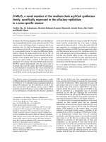
purified using Ni-NTA resin. The molecular mass of this
protein was 24 kDa in SDS-PAGE and the identity of the
protein was confirmed by Western-blot (Fig. 1).
Titration of antibodies to rpIL-6
Extracted antibodies had a specificity to rpIL-6 but not
rpIFN-
γ
and rpGM-CSF which were expressed and purified
by the same method of rpIL-6. Indirect ELISA was used to
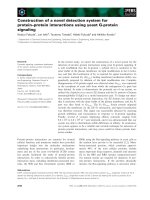
titrate mouse IgG and IgY antibodies to rpIL-6. Sixteen-fold
diluted IgY responded up to 1 ng/ml of rpIL-6 and
1 : 64~1 : 4,096 dilution of IgY was up to 30~250 ng/ml.
Based on the results, optimal IgY concentration was about
1 : 1,000 dilution (Fig. 2). Four-hundred fold diluted mouse
Table 1.
Sanitation check lists of pig farms
Title Check Points Score
a
Biosecurity Isolation Is it distant from the nearest swine herd?
Is it distant from the road to the nearest swine building above 100m?
Entrance Is there a separate change area for staff or visitors?
Is quarantine area always used for incoming stock?
Building Are cats or dogs allowed into building?
Are rodents, other wild life, or birds present in buildings?
Management Feeding Is there a chance of cross-contamination at feed delivery
Ventilation Is there a ventilation system for air condition?
Shipping Is own truck/trailer used for shipping?
Dead stock Is disposal by burial, composting or dead stock service?
Manure Removal Is there a risk of yard contamination by out side hauler?
Health issue Outbreak Is there an experience of outbreak?
Vaccine Is there a good vaccine program?
Clinics Is there a clinical symptom related disease?
Sum
a
Grades from 5 to 0. Total score was 70.
340 Deog Yong Lee
et al.
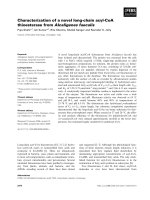
serum responded up to 1 ng/ml of rpIL-6 and
1 : 1,600~100,000 dilution of IgG was effective to
30~760 ng/ml. Optimal IgG concentration was about 1 : 400
dilution (Fig. 3).
Antigen capture-ELISA
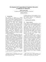
Condition of capture-ELISA was optimized with mouse IgG
and IgY antibodies on the basis of the titration. To organize
antibody titers, the optimal condition of antigen capture-ELISA
was followed: chicken IgY, 400~1,000 dilution, 25~50
µ
g/ml;
mouse IgG, 1 : 400 dilution; horse radish peroxidase-
conjugated goat-anti mouse IgG, 1 : 2,000. With those
optimized conditions, the optimal antigen capture-ELISA could
reliably detect at about 10 ng/ml of rpIL-6 (Fig. 4).
Measurement of pIL-6 in swine sera with the ELISA
Antigen capture-ELISA was applied to detect pIL-6 in
porcine sera. In one-hundred samples of total serum, most
samples were below the 10 ng/ml of pIL-6 (67%). However,
a number of samples were in the 1,000 to 10,000 ng/ml
range (16%). 27
µ
g/ml was the maximum concentration of
pIL-6 detected in the sera. A- and E- farms were lower
sanitary states on our guidelines, which were 20-30 scores.
C- and D- farms were higher states, which were 50-60
scores and B-farm was middle states, relatively. However,
the sanitary states of B- farm closed to C- and D- farms,
which was about 40 scores. IL-6 levels in pigs from farms A
and E were higher than those from other farms (Fig. 5). This
distribution positively correlated to the sanitary states of the
farms that provided the porcine sera.
Discussion
This paper describes the purification of rpIL-6, the
production of antibodies and the development of a sensitive
antigen capture-ELISA for porcine IL-6 for clinical
diagnostic purpose.
F
ig. 1.
Analysis of purified IL-6 after expression in
E. coli
by
S
DS-PAGE (A) and Western-blot (B). M, standard molecul
ar
w
eight marker; Lanes 1-8: eluted fractions from 1-8. Protein si
ze
w
as approximately 24 kDa as shown by SDS-PAGE. IL
-6
I
dentity of IL-6 was confirmed by Western-blot using
a
p
olyclonal antibody against IL-6.
F
ig. 2.
Antibody titer of chicken IgY against rpIL-6. Concentrati
on
o
f IL-6 protein ranged from 0 to 250 ng/ml and IgY was 4-tim
es
s
erial diluted. IgY was up to 1 : 1,024 dilutions for reaction wi
th
I
L-6 protein.
F
ig. 3.
Antibody titer of mouse IgG against rpIL-6. IL-6 and Ig
G
w
ere diluted and IgG was up to 1 : 10,240 dilution ratios f
or
r
eaction with IL-6 protein.
F
ig. 4.
Antigen capture-ELISA for detection of rpIL-6. Concentrati
on
o
f rpIL-6 ranged from 0 to 30 ngml/ and was normalized by PB
S.
D
eveloped capture-ELISA detected about 10 ng/ml of rpIL
-6
c
oncentration.
Development of a novel antigen capture-ELISA using IgY against porcine interleukin-6 341
To express the porcine IL-6 in
E. coli
, cDNA encoding
mature protein was amplified and transformed into
E. coli
.
of the transformants, positive clones were screened by
growth on LB plates containing appropriate antibiotics and
colony PCR. To confirm the identity of the cloned gene,
restriction enzyme analysis and sequencing of the plasmid
DNA were performed because of appearance of false
positive in colony PCR. Purified rpIL-6 was identical in size
with a previous report [35]. The identity of the protein was
also confirmed by Western-blot with a polyclonal antibody
after expression in
E. coli
by IPTG induction.
The expressed protein, rpIL-6, in
E. coli
was purified
using Ni-NTA resin and elution buffer with pH gradients.
The concentration of purified rpIL-6 was 580
µ
g/ml. To
improve the purification efficacy, washing with appropriate
pH and imidazole concentration were the most important
factors [8,29]
Five to ten ml of egg yolk was harvested from each egg
with the approximate concentration of IgY reaching 20 mg/
ml. One to two hundred
µ
l of serum was obtained per
mouse, with approximately 10% constituting the specific
antibody. This data shows that 100~200 mg/ml of IgY per
yolk was produced and this value is similar to another report
[25]. Furthermore, pIL-6 specific IgY production might be
estimated at 2 to 20 mg per yolk, because the constitution of
specific IgY is between 2-to10 % [34].
Antibody titer against rpIL-6 was evaluated by direct-
ELISA. Mouse serum responded effectively on coated rpIL-
6 at 1 : 400 ratio and chicken IgY at 1 : 1,000 ratios. In the
optimization of this capture-ELISA, there were differences
in mouse IgG titer but no difference of tendency for using
first antibody in capture-ELISA. However, there was a
difference of tendency in IgY titer for using coated protein
in a microtiter plate. Optimal dilution for IgY coating was
1 : 400~1 : 1,000 ratio. Low dilution ratio of IgY was less
detective than optimal concentration as well as high ratio. In
capture-ELISA, monoclonal antibody was used generally
with 300 ng/ml of concentration [22]. Because the specific
antibody portion is less than 5% in produced polyclonal
antibody [5], optimal concentration of IgY coated was 25~
50
µ
g/ml. This result revealed that the concentration of
specific antibody is 12.5
µ
g/ml on coated IgY.
High concentration of urea may interfere with the ELISA
cross-reaction [1]. In this study, the concentration of urea
was diluted well below the minimum level (0.1 M) by
dilution of the purified rpIL-6. Therefore, the effect of urea
should not be seen.
Most versions of capture-ELISA have used a monoclonal
antibody for the capture-antibody and a biotinylated
antibody to increase detection limit [6,9,26,33]. However,
pico-gram levels in any case of using monoclonal antibody
without biotinylation for detection antibody have not been
reached [21]. That reports suggest that modification of the
detection antibody plays an important role in the sensitivity
of capture-ELISA. However, there were some exceptions
[22,24]. The sensitivity could be increased by other detection
antibody modification, such as immunopurification [20] and
by using an IL-6 dependent cell line [16]. Although IgY was
used instead of a monoclonal antibody for the capturing
antibody, the sensitivity could still reach the nano-gram
level. Monoclonal antibody usefulness stems from three
characteristics: specificity of binding, homogeneity, and
capacity for unlimited production. In practice, however,
producing the right monoclonal antibody is often a difficult
and laborious job [4]. IgY has the amount of specific
extractable antibody in egg yolk than in rabbit in the same
period, besides the advantage of a non-invasive antibody
sampling [31]. An egg contains 100~150 mg of IgY per
yolk [25]. Furthermore, the detection of capture-ELISA
using IgY coating reached the same level as using a
monoclonal antibody without biotinylation has the detection
antibody [21]. Our results correctively matched with
previous reports as described above.
Detection level of pIL-6 was increased via dilution ratio
rather than non-diluted porcine sera and reached the critical
point at 100-fold dilution. Interference with some serum
components to perform the antigen capture-ELISA is
possible [36]. However, high level of pIL-6 was detected in
many samples (33%) by developed capture-ELISA and it
revealed that it is detectable IL-6 secretion in pig.
Pig sera were collected from 5-farms showing different
sanitary states. These farms were different in their
management and control of disease. Distribution of pIL-6
concentration was positively correlated with the sanitary
F
ig. 5.
Distribution of IL-6 in pig sera. Sera were collected fro
m
t
he different farms. (A-, B-, C-, D- and E-farms) Each farm h
as
d
ifferent management system and methods of disease control.
It
w
as different distribution to detect IL-6 concentration in the ea
ch
f
arm.
342 Deog Yong Lee
et al.
status of farms. A- and E-farms have the similar conditions
of old-fashioned equipment and have several problems with
chronic infectious diseases such as respiratory diseases and
old-traditional management. They are small-scale farms
with 1,200 pigs in A-farm and 1,500-2,000 pigs in E-farm.
However, the farmers have difficulty because of their old age
and deficient education. pIL-6 were relatively high
distributed from 2,500 to 11,000 ng/ml in mean values.
Although they have similar conditions and symptoms, E-
farm detected a higher concentration. This result indicates
that A- and E-farms have on-going infection and that in
addition, E-farm might have a new infection recently.
C- and D-farms are similar to each other in their
conditions and management, with newly introduced 3,000-
3,500 pigs and good equipments. Also, these farms have a
larger scale, more modernized than the A- and E-farms.
Here samples distributed lower than the 10 ng/ml of pIL-6
indicating that the control of disease was well performed.
Finally, B-farm has a good management system and has
about 3,000 head of pigs. However, there are sometimes
outbreaks of pluropneumoniae and diarrhea. Some samples
distributed relatively high between 100 to 10,000 ng/ml of
pIL-6 which may indicate an infection is just beginning.
These results demonstrate the clinical diagnostic use of
antigen capture-ELISA for pIL-6.
It is necessary to study protein characteristics for improving
immunogenicity by using native protein rather than denatured
[15] and IL-6, like other cytokines are critically regulated
secretion and inactivation by lymphocytes [19].
Acknowledgments
This research was supported by BioGreen 21, BrainKorea
21 and the Research Institute for Veterinary Science (RIVS),
Seoul National University, Korea.
References
1.
Bouvet JP, Stahl D, Rose S, Quan CP, Kazatchkine MD,
Kaveri SV.
Induction of natural autoantibody activity
following treatment of human immunoglobulin with
dissociating agents. J Autoimmun 2001,
16
, 163-172.
2.
Carlander D, Stalberg J, Larsson A.
Chicken antibodies: a
clinical chemistry perspective. Ups J Med Sci 1999,
104
,
179-189.
3.
Caroline F, Eva W, Lisbeth F, Klaus TJ, Per W.
Evaluation
of various cytokine (IL-6, IFN-
α
, IFN-
γ
, TNF-
α
) as markers
for acute bacterial infection in swine-a possible role for
serum interleukin-6. Vet Immunol Immunopathol 1998,
64
,
161-172.
4.
Ed H, David L.
Antibodies a laboratory manual, p. 142-143.
Cold Spring Harbor Laboratory Press, Cold Spring Harbor.
New York, 1988.
5.
Ed H, David L.
Using antibodies: A laboratory Manual. p. 3-
21. Cold Spring Harbor Laboratory Press, Cold Spring
Harbor. New York, 1999.
6.
Granger J, Remick D, Call D, Ebong S, Taur A, Williams
B, Nauss M, Millican J, OReilly M.
A sandwich enzyme-
linked immunosorbent assay for measurement of picogram
quantities of murine granulocyte colony-stimulating factor. J
Immunol Methods 1999,
225
, 145-156.
7.
Gregory WW, Katharine EM, David AH.
IgY: clues to the
origins of modern antibodies. Immunology Today. 1995,
16
,
392-398.
8.
Hardin C, Pinczes J, Riell A, Presutti D, Miller W,
Robertson D.
Cloning, gene expression, and protein
purification, p. 196-384. Oxford University Press, Oxford,
2001.
9.
Helle M, Boeije L, de Groot E, des Vos A, Aarden L.
Sensitive ELISA for Interleukin-6. Detection of IL-6 in
biological fluids: synovial fluids and sera. J Immunol
Methods 1991,
138
, 47-56.
10. Heinrich PC, Castell JV, Andus T. Interleukin-6 and the
acute phase response. Biochem J 1990, 265, 621-636.
11.
Hilbert DM, Cancro MP, Scherle PA, Nordan RP, Van
Snick J, Gerthard W, Rudikoff S.
T cell derived IL-6 is
differentially required for antigen-specific antibody secretion
by primary and secondary cells. J Immunol 1989,
143
, 4019-
4024.
12.
Hirano T, Teranishi T, Lin BH, Onoue K.
Human helper T
cell factor(s). IV. Demonstration of a human late-acting B
cell differentiation factor acting on
Staphylococcus aureus
Cowman I-stimulated B cells. J Immunol 1984,
133
, 798-
802.
13.
Hirano T, Teranishi T, Onoue K.
Human helper T cell
factor(s). III. Characterization of B cell differentiation factor-
I (BCDF- I). J Immunol 1984,
132
, 229-234.
14.
Kishimoto T, Hirano T.
Molecular regulation of B
lymphocyte response. Ann Rev Immunol 1998,
6
, 485-512.
15.
Koch C, Jensen SS, Oster A, Houen G.
A comparison of
the immunogenicity of the native and denature form of a
protein. APMIS 1996,
104
, 115-125.
16.
Krakauer T.
A sensitive, specific immunobioassay for
quantitation of human interleukin-6. J Immunoassay 1993,
14
, 267-277.
17.
Kristien VR, Steven VG, Maurice P.
In vivo
studies on
cytokine involvement during acute viral respiratory disease
of swine: troublesome but rewarding. Vet Immunol
Immunopathol 2002,
87
, 161-168.
18.
Le JM, Vilcek J.
Interleukin-6: a multifunctional cytokine
regulating immune reaction and the acute phase protein
response. Lab Invest 1989,
61
, 588-602.
19.
Lust JA, Donovan KA, Kline MP, Greipp PR, Kyle RA,
Maile NJ.
Isolation of an mRNA encoding a soluble form or
the human interleukin-6 receptor. Cytokine 1992,
4
, 96-100.
20.
Manie S, Proudfoot A, Ferrua B.
Human interleukin-6:
detection of 10 attomoles by colorimetric sandwich ELISA
using immunopurified polyclonal anti-IL-6 antibodies. Eur
Cytokine Netw 1993,
4
, 51-56.
21.
Mateu de Antonio E, Husmann RJ, Hansen R, Lunney
JK, Strom D, Martin S, Zuckermann FA.
Quantitative
detection of porcine interferon-gamma in response to
mitogen, superantigen and recall viral antigen. Vet Immunol
Development of a novel antigen capture-ELISA using IgY against porcine interleukin-6 343
Immunopathol 1998,
61
, 265-277.
22.
Mathew JA, Guo YX, Goh KP, Chan J, verburg-van
Kemenade BML, Kwang J.
Characterization of a
monoclonal antibody to carp IL-1
β
and the development of a
sensitive capture ELISA. Fish & Shellfish Immunology
2002,
13
, 85-95.
23.
May LT, Grayeb J, Santhanam U, Tatter SB, Sthoeger Z,
Helfgott DC, Chiorazzi N, Grieninger G, Sehgal PB.
Synthesis and secretion of multiple forms of
β
2
-interferon/B-
cell differentiation factor-2/hepatocyte-stimulating factor by
human fibroblasts and monocytes. J Biol Chem 1988,
263
,
7760-7766.
24.
McWaters P, Hurst L, Chaplin PJ, Collinins RA, Wood
PR, Scheerlinck JPY.
Characterization of monoclonal
antibodies to ovine interleukin-6 and the development of a
sensitive capture ELISA. Vet Immunol Immunopathol 2000,
73
, 155-165.
25.
Mine Y, Kovacs-Nolan J.
Chicken egg yolk antibodies as
therapeutics in enteric infectious diseases: a review. J Med
Food 2002,
5
, 159-169.
26.
Muneta Y, Mikami O, Shimoji Y, Nakajima Y, Yokomozo
Y, Mori Y.
Detection of porcine interleukin-18 by sandwich
ELISA and immunohistochemical staining using its
monoclonal antibodies. J Interferon Cytokine Res 2000,
20
,
331-336.
27.
Okada M, Sakaguchi N, Yoshimura N, Hara H, Shimizu
K, Yoshida H, Yoshizaki K, Kishimoto S, Yamamura Y,
Kishimoto K.
B cell growth factor (BCGF) and B cell
differentiation factor from human T hybrodomas: two
distinct kinds of BCGFs and their synergism in B cell
proliferation. J Exp Med 1983,
157
, 583-590.
28.
Rees GS, Ball C, Ward HL, Gee CK, Tarrant G, Mistry
Y, Poole S, Bristow AF.
Rat interleukin-6: Expression in
recombinant
Escherichia coli
, purification and development
of a novel ELISA. Cytokine 1999,
11
, 95-103.
29.
Sambrook J, Russel DW.
Molecular Cloning: A Laboratory
Manual, 3rd ed. pp. 18.1-18.125, Cold Spring Harbor Press,
Cold Spring Harbor, New York, 2001.
30.
Santhanam U, Ghyrayeb J, Sehgal PB, May LT.
Post-
translational modifications of human interleukin-6. Arch
Biochem Biophys 1989,
274
, 161-170.
31.
Schade R, Schniering A, Hlinak A.
Polyclonal avian
antibodies extracted from egg yolk as an alternative to the
production of antibodies in mammals a review. ALTEX
1992,
9
, 43-56.
32.
Shin NR, Choi IS, Kim JM, Hur W, Yoo HS.
Effective
methods for the production of immunoglobulin Y using
immunogen of
Bordetella bronchoseptica
,
Pasteurella
multocida
and
Actinobacillus pleuropneumoniae
. J Vet Sci
2002,
3
, 47-57.
33.
Splichal I, Muneta Y, Mori Y, Takahashi E.
Development
of a pig IL-8 ELISA detection system. J Immunoassay
Immunochem 2003,
24
, 219-232.
34.
Tini M, Jewell UR, Camenisch G, Chilov D, Gassmann
M.
Generation and application of chicken egg-yolk
antibodies. Comp Biochem Phys Part A 2002,
131
, 569-574.
35.
Van Snick J.
Interleukin-6: an overview. Annu Rev Immunol
1990,
8
, 253-278.
36.
Vilim V, Vob rka Z, Vytásek R, Senolt L, Tchetverikov I,
Kraus VB, Pavelka K.
Monoclonal antibodies to human
cartilage oligomeric matrix protein: epitope mapping and
characterization of sandwich ELISA. Clin Chim Acta 2003,
328
, 59-69.
u
·