Báo cáo khoa học: "Serum immunoglobulin fused interferon-Ձ inhibited tumor growth in athymic mice bearing colon 26 adenocarcinoma cells" ppsx
Bạn đang xem bản rút gọn của tài liệu. Xem và tải ngay bản đầy đủ của tài liệu tại đây (433.57 KB, 6 trang )
JOURNAL OF
Veterinary
Science
J. Vet. Sci. (2008), 9(1), 45
50
*Corresponding author
Tel: +82-2-880-1276; Fax: +82-2-873-1268
E-mail:
Serum immunoglobulin fused interferon-
α
inhibited tumor growth in
athymic mice bearing colon 26 adenocarcinoma cells
Jun-Sung Kim
1
, Kyeong Nam Yu
1
, Mi Suk Noh
1,2
, Min-Ah Woo
1,2
, Sung-Jin Park
1
, Jin Hong Park
1
, Jin Hua
1
,
Hyun Sun Cho
1
, Soon Kyung Hwang
1
, Eun-Sun Lee
1
, Youn-Sun Chung
1
, In-Young Choi
3
, Se-Chang Kwon
3
,
Myung-Haing Cho
1,2,
*
1
Laboratory of Toxicology, College of Veterinary Medicine and
2
Interdisciplinary Program in Nano-Science and Technology, Seoul
National University, Seoul 151
‐
742, Korea
3
Hanmi Pharmaceutical Research Center, Hwaseong 445-813, Korea
Interferon (IFN) has therapeutic potential for a wide range
of infectious and proliferative disorders. However, the
half-life of IFN is too short to have a stable therapeutic effect.
To overcome this problem, serum immunoglobulin has been
fused to IFN. In this study, the efficacy of serum immuno-
globulin fused INFs (si-IFN1 and si-IFN2) was evaluated on
athymic mice bearing colon 26 adenocarcinoma cells. Seven
days after the implantation of tumor cells, each group of
mice was injected once a week with si-IFN1 and si-IFN2 at
two different concentrations (10
×
: 30
µ
g/kg and 50
×
: 150
µ
g/kg). A slight anti-tumoral effect was observed in all 10
×
groups compared to the control. In the 50
×
groups, however,
si-IFN1 and si-IFN2 showed significant anti- tumoral effects
compared to the control. To gain more information on the
mechanisms associated with the decrease of tumor size, a
Western blot assay of apoptosis-related molecules was
performed. The protein expression of cytochrome c, caspase
9, 6, and 3 were increased by si-IFN1 and si-IFN2. These 2
IFNs also increased the expressions of p53, p21, Bax and
Bad. Interestingly, si-IFN1 and si-IFN2 decreased the
expression of VEGF-
β
. Taken together, serum immuno-
globulin fused IFNs increased therapeutic efficacy under
current experimental condition.
Keywords: adenocarcinoma cell, interferon, serum immuno-
globulin, tumor growth inhibition
Introduction
Interferon (IFN) is a cytokine produced and secreted by
eukaryotic cells in response to stimulation by viruses,
bacteria, and mitogens and mediates diverse biological
activities by binding to a specific receptor on the cell
surface [1,10]. IFNs have therapeutic potential for a wide
range of infectious and proliferative disorders due to their
pleitropic effects on multiple metabolic, immunologic and
pathologic events [3,6,9]. However, the half-life of IFN is
too short for a stable therapeutic effect. To overcome this
problem, several methods are used for improving the
stability of proteins. One of the methods is chemical
modification of a polypeptide with highly soluble macro-
molecules such as polyethylene glycol (PEG) which
prevents the polypeptides from making contact with
proteases. However, such pegylated polypeptides have the
disadvantage of lowering both the activity and production
yield of an active substance as the molecular weight of
PEG increases. Another approach for enhancing the in vivo
stability of the IFN is to conjugate the IFN with a stable
serum immunoglobulin. As an improved method for enhancing
the stability of an active polypeptide and simultaneously
maintaining the in vivo activity thereof, the two IFN
conjugate si-IFN1 and si-IFN2 comprising an IFN, PEG
and serum immunoglobulin, were interlinked to one another.
The present paper reports that IFN-α modified with
serum immunoglobulin (si-IFN1 and si-IFN2) inhibits
tumor growth in athymic mice bearing colon 26 adenocar-
cinoma cells.
Materials and Methods
Reagents and cell culture
Recombinant IFN-α, serum immunoglobulin fused IFN-
α (si-IFN1 and si-IFN2), and peginterferon α-2a were
supplied by Hanmi Pharmaceutical (Korea). The efficacy
of several IFNs was compared to efficacy-proven inter-
feron, peginterferon α-2a, which is a covalent conjugate of
recombinant α-2a interferon with a single branched
bis-monomethoxy PEG chain (Roche, USA). The si-IFN1,
46 Jun-Sung Kim et al.
si-IFN2, and PEGASYS were administered at two different
concentrations (10 × groups: 30 µg/kg and 50 × groups:
150 µg/kg). The mouse colon 26 adenocarcinoma cells
(CT-26) were purchased from the Korean Cell Line Bank
(KCLB, Korea). Colon 26 adenocarcinoma cells were
cultured with RPMI 1640 medium (Gibco, USA) supple-
mented with 10% fetal bovine serum (FBS; Gibco, USA)
in an atmosphere of 5% CO
2
in an incubator at 37
o
C.
Experimental animals
Specific pathogen-free male athymic BALB/c nude mice
were purchased from the SLC (Shizuoka Institute for
Laboratory Animals Center, Japan). Athymic nude mice
were maintained at 23 ± 2
o
C , with a relative humidity of 50
± 20% and a 12 h light/dark cycle. After 7 days of tumor
cell inoculation, the mice were grouped as follows: control,
10 × and 50 × IFN concentrations. All the procedures for
handling and caring for the animals were followed by the
guidelines given in the NIH Guide for the Care and Use of
Laboratory Animals (NIH Publication No. 85-23, 1985,
revised 1996, USA). All of the experiments were conducted
to minimize the number of animals used and the suffering
caused by the procedures used in the present study.
Tumor cell inoculation and treatment procedure
Mice were inoculated subcutaneously with 5 × 10
5
colon
26 adenocarcinoma cells in RPMI 1640 with 10% FBS.
Seven days after tumor cell inoculation, each mouse was
treated with recombinant IFN-α, si-IFN1, si-IFN2 and
peginterferon α-2a by intratumoral injection. IFN-α was
injected every day for 21 days, the others were injected on
days 7, 14 and 21 [5].
Serum and hematological analysis
Blood samples for hematology were obtained from 5 mice
in each group every week. Every week clinical biochemistry
determinations were also made on the serum harvested from
blood samples obtained from the mice. The following
parameters were assayed: total protein (TPROT), albumin
(ALB), total bilirubin (TBILI), aspartate aminotransferase
(AST), alanine aminotransferase (ALT), glucose (GLU),
blood urea nitrogen (BUN), creatinine (CREAT).
Measurement of tumor volume
Tumor volume was measured on days 7, 14 and 21 with
the aid of vernier calipers. At necropsy, the tumor volume
was estimated for the largest (a) and the smallest (b)
diameter, and the tumor volume was calculated as V =
ab
2
/2.
Western blot
Protein was extracted from tumor tissues in recombinant
IFN-α, si-IFN1, si-IFN2, and peginterferon α-2a in 50 × .
The extracts were added to the sample buffer. Samples
were boiled for 10 min, and proteins were separated on
15% SDS-PAGE for 18 h. The nitrocellulose membrane
was rinsed twice in Tween 20-TBS (T-TBS) with 5% skim
milk. Subsequently, the membranes were incubated with a
1:2,500 dilution of primary antibody (cytochrom c,
caspase 3, 6, and 9, and p53, p21, Bax, Bad, VEGF-β, and
FGF-2) in T-TBS buffer for 3 h. They were then washed
twice for 10 min in T-TBS buffer, incubated with a 1:
5,000 dilution of secondary antibody conjugated to HRP
(Santa Cruz, USA) in T-TBS buffer for 1 h. After washing,
the bands-of-interest were pictured by luminescent image
analyzer LAS-3000 (Fujifilm, Japan).
Statistical analysis
The results were expressed as mean ± SD values for
independent experiments. Statistical analysis was performed
on all groups using the t test.
Results
Clinical hematology and biochemistry
In the hematological test of the 10 × group, there was no
significant change after 1, 2 and 3 weeks (data not shown),
however, in the 50 × group, the WBC value was increased
in si-IFN2, peginterferon α-2a and IFN- α compared to the
control after 2 weeks. This pattern was also observed in the
lymphocyte value (Fig. 1A). After 3 weeks, the increase of
WBC’s and lymphocytes was observed in the si-IFN2,
peginterferon α-2a and IFN-α groups compared to the
control (Fig. 1B). Interestingly, the significant change in
the monocyte level observed at 2 weeks was not main-
tained until 3 weeks (Fig. 1). In the biochemical test of 10
× group, a decrease of BUN was observed at 2 weeks in
si-IFN2, peginterferon α-2a and IFN-α group compared to
the control and this decrease was maintained at 3 weeks
(Fig. 2). In the biochemical test of the 50 × group, a
decrease of BUN was observed only at 3 weeks in the
si-IFN2, peginterferon α-2a and IFN-α group comparing to
the control (Fig. 3). In particular, the level of ALT was
decreased in peginterferon α- 2a and IFN-α group com-
paring to the control at 3 weeks (Fig. 3).
Tumor growth was inhibited by si-IFN1 and
si-IFN2 treatment
In the 10 × group, the tumor volume decreased slightly in
the si-IFN1, si-IFN2, peginterferon α-2a and IFN-α group
compared to control (data not shown). In the 50 × group,
however, the tumor volume decreased significantly in mice
treated with si-IFN1, si-IFN2, peginterferon α-2a and IFN-
α compared to the control (Fig. 4).
Level of pro-apoptotic molecules was increased in
tumor tissue by modified IFNs
The expression levels of cytochrome c, caspase 6, and
Tumor-inhibitory effects of modified IFN-α 47
Fig. 2. Concentrations of blood urea nitrogen (BUN) in the 10 ×
interferon group. Clinical biochemistry determinations were
made on serum harvested from the blood of mice. BUN was
measured at 1 week (data not shown), 2 weeks and 3 weeks afte
r
tumor cells inoculation (n = 5). 10 × interferon group:
concentration of si-IFN1 and si-IFN2 (30 µg/kg), Each point
represents the mean ± SD. *Significantly different from control
(p < 0.05). **Significantly different from control (p < 0.01).
Fig. 1. Hematological assay of differential leukocytes in the 50 × interferon group. Blood samples for hematological determinations wer
e
obtained from 5 mice. The differential leukocyte count was measured at 1 week (data not shown), 2 weeks (A) and 3 weeks (B) after
tumor cells inoculation (n = 5). 50 × interferon group: concentration of si-IFN1 and si-IFN2 (150 µg/kg), WBC: white blood cell, LY:
lymphocyte, MO: monocyte. Each point represents the mean ± SD. *Significantly different from control (p < 0.05). **Significantly
different from control (p < 0.01).
Fig. 3. Concentrations of blood urea nitrogen (BUN) and alanine
aminotransferase (ALT) in the 50 × interferon group. BUN and AL
T
were measured after tumor cells inoculation (n = 5). 50 × interferon
group: concentration of si-IFN1 and si-IFN2 (150 µg/kg), Each poin
t
represents the mean ± SD. *Significantly different from control (p
<
0.05). **Significantly different from control (p < 0.01).
caspase 3 were increased in the si-IFN1, si-IFN2, pegin-
terferon α-2a and IFN-α treated groups of 50 ×
concentration compared to the control. The expression of
caspase 9 was observed clearly in si-IFN1 and si-IFN2
only. The expression level of p53 was slightly increased,
however, a distinct increase of p21 was observed in
si-IFN1 only. The expression of BAD was significantly
increased in si-IFN1 and si-IFN2; however, no change of
Bax was observed (Fig. 5).
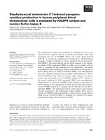
A decrease of proangiogenic molecule was observed
in tumor tissue treated with modified IFNs
In the Western blot of pro-angiogenic molecule, the
expression level of VEGF-β was decreased in the si-IFN1,
si-IFN2, peginterferon α-2a and IFN-α treated groups. The
decrease of the expression level was observed particularly
in si-IFN1, si-IFN2, and peginterferon α-2a compared to
the control (Fig. 6).
Discussion
IFN-α, the first cytokine to be produced by recombinant
48 Jun-Sung Kim et al.
Fig. 4. Effects of interferons on tumor growth inhibition. Mice were inoculated with 5 × 10
5
colon 26 adenocarcinoma cells in RPMI
1640 with 10% FBS subcutaneously. Seven days after tumor cell inoculation, the tumor size was measured. The mice were then treate
d
with different interferons by intratumoral injection. IFN-α was injected every day for 3 weeks, the others were injected at 1 week, 2
weeks and 3 weeks. Tumor volume was estimated for the largest (a) and smallest (b) diameter, and the tumor volume was calculated usin
g
V= ab
2
/2. Each point represents the mean ± SD. *Significantly different from control (p < 0.05). **Significantly different from control
(p < 0.01).
Fig. 5. Change of pro-apoptotic molecules in tumor tissue treated with 50 × groups. Protein samples were extracted from the tumor
tissues of control, si-IFN1, si-IFN2, peginterferon α-2a and IFN-α treated groups at 50 × concentrations. Protein sample were prepare
d
for Western blot using antibodies to mouse cytochrome c, caspase 9, caspase 6, and caspase 3, p53, p21, Bax and Bad. Each band was
further analyzed by densitometer. Each number on the figure represents the density compared control.
DNA technology, has been identified as a pivotal regulator
of cellp growth, differentiation, cell to cell communication
and signal pathway [4]. Although there are many advan-
tages in the use of IFN-α in various diseases, clinical trials
Tumor-inhibitory effects of modified IFN-α 49
Fig. 6. Change of angiogenesis-related molecules in tumor tissue
of mice. Protein samples were extracted from the tumor tissues o
f
control, si-IFN1, si-IFN2, peginterferon α-2a and IFN-α treate
d
groups in 50 ×. Protein samples were prepared for Western
b
lot
using antibodies to mouse VEGF-β and FGF-2. Each band was
further analyzed by densitometer program. Each number on the
figure represents the density compared to the control.
have been limited due to their disadvantages such as their
short half-life in in vivo systems. To overcome this dis-
advantage, various trials have been carried out using them
in combination with other agents and making modifications
to recombinant IFN-α. The present paper reports that a
once-a-week injection of serum immunoglobulin fused
IFN-α into athymic nude mice bearing tumor cells can
inhibit tumor growth effectively. IFN-α is a multifunctional
regulatory cytokine regulating cell function and proliferation.
In the present experiments, serum immunoglobulin fused
IFN-α named si-IFN1 and si-IFN2 was used. The results
showed that at 21 days after tumor cells inoculation, the
mean tumor volumes of si-IFN1, si-IFN2, peginterferon α
-2a and IFN-α treated with 50 × were significantly decreased
compared to the control. In the biochemical tests of the 50
× group, a decrease of BUN was observed in the si-IFN2,
peginterferon α-2a and IFN- α group comparing to the
damage but also dehydration. ALT is a predominantly
hepatocyte enzyme and increase of ALT activity is known
to be highly associated with liver damage. However, the
current study is an initial report on the screening tests for
the potential effects of various IFN types as antitumor
drugs. These tests are based on various blood or serum
measurements: (a) total white blood cell counts; (b) BUN
for kidney toxicity and (c) ALT for liver toxicity. On the
basis of the limited parameters tested, although varying in
sensitivity, our data seem to correlate with the reduced
toxicity and antitumor effects of the IFNs tested. However,
the final validation of these tests, especially the BUN and
ALT, will require both further study of the histopathologic
effects and correlation with the results from further
efficacy trials over an extended period. Further studies of
this type are essential.
The pivotal events of tumor growth inhibition are
apoptosis and anti-angiogenesis. Apoptosis is orchestrated
by various molecules including extrinsic and intrinsic
pathways. Also, a recent study reported that the IFN-
family increased apoptosis in vitro [2]. To confirm the
effect of serum immunoglobulin fused IFN-α to the
pro-apoptotic function, we performed a Western Blot test.
In the extrinsic pathway related to the death receptor, an
increase of cytochrome c, caspase 3, 6, and 9 was observed
in si-IFN1 and si-IFN2. Cytochrome c is located in the
mitochondria of all aerobic cells and is involved in the
electron transport system that functions in oxidative
phosphorylation. In addition to its role in oxidative phosphory-
lation, the release of cytochrome c from the mitochondrial
intermembrane space results in nuclear apoptosis. Binding
of Apaf-1 to cytochrome c allows Apaf-1 to form a ternary
complex with, and activate, the initiator procaspase-9 [8].
In the intrinsic pathway associated with mitochondria, an
increase of p53, p21 and Bad was also detected. Takaoka et
al. [7] demonstrated that transcription of the p53 gene was
induced by IFN-α, accompanied by an increase in p53
protein levels and they provided examples in which p53
gene induction by IFN-α indeed contributed to tumor
suppression. Our study also confirmed that si-IFN1 and
si-IFN2 showed strong anti-tumor activity similar to that of
peginterferon α-2a.
The expansion of the tumor masses depends on neovascu-
larization and the formation of new vasculature involves
multiple, interdependent steps [3]. In addition, the onset of
angiogenesis involves a change in the local equilibrium
between pro-angiogenic and anti-angiogenic molecules
[3,9]. The key molecules associated with pro-angiogenesis
include fibroblast growth factor 2 (FGF2) and vascular
endothelial cell growth factor beta (VEGF-β). FGF2 is a
wide-spectrum mitogenic, angiogenic, and neurotrophic
factor that is expressed at low levels in many tissues and
cell types and reaches high concentrations in the brain and
pituitary. FGF2 has been implicated in a multitude of
physiologic and pathologic processes, including limb
development, angiogenesis, wound healing, and tumor
growth. In our study, to evaluate the efficiency of serum
immunoglobulin fused IFN-α to anti-angiogenesis, the
expression level of VEGF-β and FGF2 was measured by
Western Blot. Our results showed that the expression level
of VEGF-β was decreased in the si-IFN1, si-IFN2, pegin-
terferon α-2a and IFN-α treated groups. The decrease of
the expression level was observed prominently in the
si-IFN2 and peginterferon α-2a treated groups compared
to the control. These data suggest that inhibition of tumor
growth may be also due to the anti-angiogenic effect of
si-IFN1 and si-IFN2.
In conclusion, serum immunoglobulin fused IFN-α,
si-IFN1 and si-IFN2 are able to inhibit the tumor growth of
athymic mice bearing colon 26 adenocarcinoma cells and
50 Jun-Sung Kim et al.
the inhibitory effects may be associated with facilitating
apoptosis and suppressing angiogenesis.
References
1. Chen SA, Sawchuk RJ, Brundage RC, Horvath C,
Mendenhall HV, Gunther RA, Braeckman RA. Plasma
and lymph pharmacokinetics of recombinant human inter-
leukin-2 and polyethylene glycol-modified interleukin-2 in
pigs. J Pharmacol Exp Ther 2000, 293, 248-259.
2. de Luj
án Alvarez M, Ronco MT, Ochoa JE, Monti JA,
Carnovale CE, Pisani GB, Lugano MC, Carrillo MC.
Interferon
α-induced apoptosis on rat preneoplastic liver is
mediated by hepatocytic transforming growth factor
β
1
.
Hepatology 2004, 40, 394-402.
3. Fidler IJ. Regulation of neoplastic angiogenesis. J Natl
Cancer Inst Monogr 2000, 28, 10-14
4. Gutterman JU. Cyt okine therapeutics: lessons from inter-
feron
α. Proc Natl Acad Sci USA 1994, 91, 1198-1205.
5. Lee CW, Hong DH, Han SB, Jung SH, Kim HC, Fine RL,
Lee SH, Kim HM. A novel stereo-selective sulfonylurea,
1-[1-(4-aminobenzoyl)-2,3-dihydro-1H-indol-6-sulfonyl]-
4-phenyl-imidazolidin-2-one, has antitumor efficacy in in
vitro and in vivo tumor models. Biochem Pharmacol 2002,
64, 473-480.
6. Morinaga Y, Suga Y, Ehara S, Harada K, Nihei Y, Suzuki
M. Combination effect of AC-7700, a novel combretastatin
A-4 derivative, and cisplatin against murine and human tu-
mors in vivo. Cancer Sci 2003, 94, 200-204.
7. Takaoka A, Hayakawa S, Yanai H, Stoiber D, Negishi H,
Kikuchi H, Sasaki S, Imai K, Shibue T, Honda K,
Taniguchi T. Integration of interferon-
α/β signalling to p53
responses in tumour suppression and antiviral defence.
Nature 2003, 424, 516-523.
8. Tsunoda S, Ishikawa T, Yamamoto Y, Kamada H,
Koizumi K, Matsui J, Tsutsumi Y, Hirano T, Mayumi T.
Enhanced antitumor potency of polyethylene glycolylated tu-
mor necrosis factor-
α: a novel polymer-conjugation techni-
que with a reversible amino-protective reagent. J Pharmacol
Exp Ther 1999, 290, 368-372.
9. von Marschall Z, Scholz A, Cramer T, Sch
äfer G,
Schirner M, Oberg K, Wiedenmann B, H
öcker M,
Rosewicz S. Effects of interferon alpha on vascular endothe-
lial growth factor gene transcription and tumor angiogenesis.
J Natl Cancer Inst 2003, 95, 437-448.
10. Zhang F, Lu W, Dong Z. Tumor-infiltrating macrophages
are involved in suppressing growth and metastasis of human
prostate cancer cells by INF-beta gene therapy in nude mice.
Clin Cancer Res 2002, 8, 2942-2951.