Báo cáo khoa học: "Comparison of cardiac function and coronary angiography between conventional pigs and micropigs as measured by multidetector row computed tomography" docx
Bạn đang xem bản rút gọn của tài liệu. Xem và tải ngay bản đầy đủ của tài liệu tại đây (1.5 MB, 6 trang )
JOURNAL OF
Veterinary
Science
J. Vet. Sci. (2008), 9(2), 121
126
*Corresponding author
Tel: +82-62-530-2831; Fax: 82-62-530-2809
E-mail:
†
First two authors contributed equally to this study.
Comparison of cardiac function and coronary angiography between
conventional pigs and micropigs as measured by multidetector row
computed tomography
Young Keun Ahn
2,3,4,†
, Jung Min Ryu
1,†
, Hea Chang Jeong
2
, Yun Hyeon Kim
5
, Myung Ho Jeong
2,3,4
, Min Young
Lee
1
, Sang Hun Lee
1
, Jae Hong Park
1
, Seung Pil Yun
1
, Ho Jae Han
1,
*
1
College of Veterinary Medicine, Biotherapy Human Resources Center, Chonnam National University, Gwangju 500-757,
Korea
2
The Heart Center,
3
Cardiovascular Research Institute,
4
Clinical Trial Center, and
5
Department of Radiology, Chonnam
National University Hospital, Gwangju 501-757, Korea
Pigs are the most likely source animals for cardiac xeno-
transplantation. However, an appropriate method for
estimating the cardiac function of micropigs had not been
established. Computed tomography (CT) analysis aimed
at estimating cardiac function and assessing the coronary
arteries has not been carried out in micropigs. This study
determined the feasibility of evaluating cardiac function in
a micropig model using multidetector row computed
tomography (MDCT) and compared the cardiac function
values with those of conventional pigs. The mean age of
the conventional pigs and micropigs was approximately 80
days and approximately 360 days, respectively. The mean
body weight in the conventional pigs and micropigs was
29.70
±
0.73 and 34.10
±
0.98 kg, respectively. Cardiac
MDCT detected ejection fractions of 52.93
±
3.10% and
59.00
±
5.56% and cardiac outputs of 1.46
±
0.64 l/min
and 1.21
±
0.24 l/min in conventional pigs and micropigs,
respectively. There were no significant differences in
cardiac function between conventional pigs and micropigs
in the reconstructed CT images. There were also no
differences in the coronary angiographic images obtained
by MDCT. It is expected that the results of this study will
help improve understanding of cardiac function in micro-
pigs. The data presented in this study suggest that MDCT
is a feasible method for evaluating cardiac function in
micropigs.
Keywords: cardiac function, coronary angiography, MDCT,
micropig, multidetector row computed tomography
Introduction
Transplantation is often used to treat fulminant organ
failure. However, severe shortages in the availability of
suitable human donors have limited the volume of heart
transplants [15]. This shortage of donors has stimulated in-
terest in the possibility of using animal organs for trans-
plantation into humans. Animal-to-human transplantation
(or xenotransplantation) would offer an unlimited supply
of organs and tissue for transplantation. Both non-human
primates and non-primate mammals have been used as
sources for heart transplants. Non-human primates such as
chimpanzees and baboons are closely related to humans
phylogenetically and share many immunological proper-
ties with humans [3,5,25]. However, non-human primates
are unlikely to be a fruitful source of organs in the future for
the following reasons: slow growth rate, limited offspring,
difficulty breeding in captivity, and smaller size [27]. Pigs
are now considered to be the most likely source animals for
human xenotransplantation in the future. They have sev-
eral advantages over non-human primates. The heart size
of miniature swine including the micropig is compatible
with the human heart, and cardiovascular function and he-
modynamic parameters are also similar [2]. In addition, the
reproduction-related features of pigs, such as early sexual
maturity, short gestation time, and generation of large lit-
ters, allow for a potentially large pool of animal donors for
xenotransplantation [37].
One essential question in xenotransplantation is whether
the animal organ can be an effective physiological proxy
for the human organ. In order to answer to this question, the
functional and anatomical compatibility of a pig heart and
its human counterpart has been investigated [35]. The car-
diac output of porcine and human hearts of similar size was
found to be comparable [19], and their action potentials
122 Young Keun Ahn et al.
were also similar [35]. Despite previous study, most phys-
iological incompatibilities and species-specific differ-
ences between mammalian species remain unknown.
Moreover, even individuals of one species or strain may
exhibit slight genetically-derived metabolic differences
[13]. These differences have the potential to create prob-
lems for support of the human cardiovascular system after
xenotransplantation.
It is clinically important to measure cardiac function in in-
dividual micropigs used for heart transplantation for pur-
poses of diagnosis and prognosis. The indices of a healthy
heart are verified through assessment of ejection fraction
(EF), end systolic volume (ESV), end diastolic volume
(EDV), cardiac output (CO), and coronary artery angiog-
raphy [32,36]. However, there is no reliable method for
evaluating these parameters in micropigs. Therefore, we
studied the feasibility of evaluating cardiac function in mi-
cropigs using multidetector row computed tomography
(MDCT) and compared the parameters with those of con-
ventional pigs.
In recent years, major technological improvements have
been achieved in computed tomography (CT). The most
significant development has been the introduction of
MDCT, which has brought about substantial improve-
ments in spatial and temporal resolution [12,17]. This
study is the first to show MDCT to be a reliable method for
assessing cardiac function in micropigs and for selecting
suitable donor pigs for heart xenotransplantation.
Materials and Methods
Animals
All experimental procedures were approved by the Ethics
Committee of Chonnam National University Hospital.
Studies were performed using mixed-breed, conditioned
Yucatan micropigs and Landrace breed conventional pigs,
all of which were provided by the animal breeding house of
Chonnam National University Research Institute of Medi-
cal Sciences. The pigs were housed individually indoors in
cages, fed dry pig food, and provided with water. The mean
age was approximately 80 days for conventional pigs and
approximately 360 days for micropigs. The mean body
weight for the conventional pigs and micropigs was 29.70
± 0.73 kg and 34.10 ± 0.98 kg, respectively.
Radiological assessment of cardiac function
After premedication with azaperone (0.5 mg/kg, intra-
muscular) and xylazine (8 mg/kg, intramuscular), normal
saline with midazolam (0.2 mg/kg) was infused through a
20-G venous access line placed in an ear vein. CT examina-
tions were performed using a two-phase, contrast-en-
hanced, ECG-gated, MDCT scanner (SOMATOM Sensa-
tion Cardiac 64; Siemens, Germany) set at a 0.75-mm sec-
tion thickness, with a gantry rotation time of 330 msec and
a kernel value of B25f. The tube current was 800 mAs at
120 kVP. The pitch, which is defined as the ratio of the ta-
ble feed in a single rotation over the detector coverage in
the transverse direction, was determined to be 0.2. Serial
CT scanning in the axial plane, together with an ECG-trig-
gered examination, was performed from the level of the left
ventricular apex after a bolus injection of 60 ml of non-ion-
ic contrast media (Ultravist 370; Schering, Germany) fol-
lowed by a 60 ml saline bolus injection through the ear
vein. Both were injected at a flow rate of 4 ml/sec. Axial
images were reconstructed at multiple phases that covered
the cardiac cycle in 10% increments of the RR interval be-
tween 5% and 95%. Multiphase reconstruction was per-
formed with commercially available software (Argus;
Siemens, Germany) by using short axis slices from the
base of the heart to the apex. The end-diastole and end-sys-
tole were defined as the maximal and minimal left ven-
tricular volume, respectively. The EDV, ESV, LVEF, stroke
volume (SV), CO, and myocardial mass were compared
between the two groups. A 7 Fr. arterial sheath was placed
in the left carotid artery after achieving local anesthesia
with 2% lidocaine, and a cutdown was made. After infus-
ing 10,000 units of heparin, a 7 Fr. coronary artery guiding
catheter was placed within the ostia of the left and right cor-
onary arteries under fluoroscopic guidance using a mobile
C-arm (Phillips BV-25 Gold; Phillips, Netherlands). Coro-
nary angiography demonstrated left and right coronary ar-
teries with branches. The coronary angiograms for the left
and right coronary arteries and their branches were com-
pared with the images of the CT angiogram. During the ex-
periment, oxygen and normal saline were supplied con-
tinuously, and anesthesia was maintained with an addi-
tional infusion of midazolam. Continuous ECG monitor-
ing was performed in order to confirm that the pigs had a
normal ST segment at baseline.
Statistical analysis
Statistical analysis was carried out with Statistical
Package for Social Sciences software (SPSS 12.0 for
Windows; SPSS, USA). Continuous variables with normal
distributions were compared using an unpaired Student's
t-test and are expressed as mean ± SD. Where appropriate,
categorical variables were compared using a Wilcoxon
test. A p-value < 0.05 was considered significant.
Results
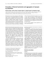
The end-systolic and end-diastolic phase images were
displayed (Figs. 1 and 2), and EDV, ESV, EF, SV, CO, and
myocardial mass were calculated automatically (Tables 1
and 2). The EDV and ESV were 48.80 ± 23.30 ml and
22.97 ± 11.30 ml in conventional pigs and 36.70 ± 9.36 ml
and 13.30 ± 7.43 ml in micropigs under a pre-medicated
condition, respectively. There were no significant differ-
Assessment of cardiac function in micropigs 123
Fig. 1. Left ventriculogram by computerized tomography (A:
end-systolic phase, B: end-diastolic phase) in a conventional pig.
Fig. 2. Left ventriculogram by computerized tomography (A:
end-systolic phase, B: end-diastolic phase) in a micropig.
Table 1. Cardiac parameters in conventional pigs and micropigs
as measured by multidetector row computed tomography
Conventional Micropig
p
pig (n = 3) (n = 3)
End diastolic
48.80 ± 23.30 36.70 ± 9.36 0.451
volume (ml)
End systolic
22.97 ± 11.30 13.30 ± 7.43 0.283
volume (ml)
Stroke volume
25.83 ± 12.11 23.40 ± 2.26 0.750
(ml)
Cardiac
1.46 ± 0.64 1.21 ± 0.24 0.567
output (l/min)
Myocardial
41.83 ± 19.47 37.03 ± 6.89 0.602
mass (g)
Table 2. Comparison of ejection fraction (EF) detected by multi-
detector row computed tomography (MDCT) and echocardio-
graphy in conventional pigs and micropigs
EF (%)
MDCT Echocardiography
(n = 3) (n = 5)
*
Conventional pig 52.93 ± 3.10 65.47 ± 5.17
Micropig 59.00 ± 5.56 58.40 ± 8.18
*
Cited from Lee et al. [22].
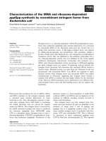
Fig. 3. Coronary circulation by computerized tomography in a
conventional pig (A) and a micropig (B). Arrow; right circumfle
x
artery (RCA). Arrowhead; left anterior descending artery (LAD).
Arrow with dotted line; left circumflex artery (LCX).
ences in MDCT-measured parameters between conven-
tional pigs and micropigs, including SV (25.83 ± 12.11 ml
vs. 23.40 ± 2.26 ml), CO (1.46 ± 0.64 l/min vs. 1.21 ± 0.24
l/min), and myocardial mass (41.83 ± 19.47 g vs. 37.03 ±
6.89 g) (Table 1). The values measured in conventional
pigs had broader variation relative to those of micropigs.
This is likely due to the fact that the conventional pigs used
in the present experiment were mixed-breed and were not
fully grown. There was no significant difference in the
measured EF, which was 52.93 ± 3.10% in conventional
pigs and 59.00 ± 5.56% in micropigs. Furthermore, the EF
measured by MDCT was not significantly different than
the EF value detected by echocardiography (65.47 ± 5.17%
vs. 58.40 ± 8.18%) (Table 2). The small differences be-
tween the MDCT and echocardiography values are likely
due to differences in individual characteristics, body
weights, ages, conditions of evaluation, and modalities.
The coronary circulation, as revealed by CT angiogram,
was similar between conventional pigs and micropigs (Fig.
3).
Discussion
Cardiac mortality is closely related to cardiac volumes
and global left ventricular function, which is expressed as
the LVEF [36]. An accurate assessment of these parame-
ters is essential for determining prognosis in micropigs
used for xenotransplantation, as well as in individual
patients. Measures of cardiac function should be carried
out using modalities that provide quick, noninvasive im-
ages with superior temporal and spatial resolution. To date,
124 Young Keun Ahn et al.
cardiac functional assessment has been performed using
various noninvasive modalities, such as echocardiography
[7,29], nuclear medicine [6], single-detector row helical
CT [26], MDCT [12,17], electron beam CT [23], and mag-
netic resonance imaging (MRI) [28].
Over the past few years, MDCT has become more popular
for noninvasive cardiac imaging [1,30]. Initially, MDCT
was used to detect coronary artery stenosis and to assess
cardiac morphology [14,31]. Because data acquisition in
MDCT is continuous, retrospective ECG-gating allows
image reconstruction in any phase of the cardiac cycle.
Therefore, an end-systolic and end-diastolic image can be
produced to assess the ventricular volume and function.
The LV volume measurement from the ECG-gated MDCT
image shows a good correlation with cardiac MRI, which is
accepted as the reference method for a precise quantitative
LV functional analysis [24,38].
In this study, there were no significant differences in left
ventricular end systolic volume (LV-ESV) or end diastolic
volume (LV-EDV) between conventional pigs and micro-
pigs. This result correlates well with a previous study that
compared the cardiac function between the two strains us-
ing echocardiography and radiography [22]. In previous
evaluations of cardiac function, MDCT with a temporal
resolution of 125-250 ms was shown to be comparable to
echocardiography [9,34], single photon emission CT [38],
and MRI. Over the last few years, rapid technical develop-
ments in scanner hardware have led to improvements in
spatial and temporal resolution, as well as led to signifi-
cantly faster cardiac scans. Consequently, MDCT has be-
come an attractive option for evaluating coronary artery
obstruction and assessing ventricular function. According
to a previous study that estimated the EF in humans using
MDCT, MRI, and echocardiography, the EF by echo-
cardiography, MDCT, and MRI was 54.6 ± 16.7%, 50.7 ±
16.0%, and 51.8 ± 15.9%, respectively [38]. This suggests
that there are no significant differences in cardiac function
between humans and micropigs.
A few studies comparing MDCT LV functional measure-
ments with ventriculography, MRI, and transthoracic
echocardiography (TTE) have reported a good correlation
among these modalities [10,11,18]. Moreover, in studies
focused on visual analysis and quantification of regional
LV function [8,16,33], wall-motion anomalies were accu-
rately identified with regard to TTE and MR. The reprodu-
cibility of global function parameters in MDCT appears to
be comparable to that seen in other modalities. The re-
ported inter-observer variability has ranged from 2% to
11% for LV-EDV and from 6% to 9% for LV-ESV. The cor-
responding values for MR are 2% to 6% [4]. MDCT allows
imaging of the coronary arteries with a high temporal and
spatial resolution. In recent years, there has been con-
tinuous improvement in the capability of both technologies
for the visualization of the coronary lumen and the detec-
tion of coronary artery stenosis. The sensitivity of MDCT
for the detection of significant stenosis ranges from 72% to
95%, with specificity ranging from 84% to 97% [1,20,21].
In conclusion, MDCT is a reliable modality for evaluating
cardiac function in healthy micropigs.
Acknowledgments
The authors wish to thank Drs. Yong Sook Kim, Jin Sook
Kwon, Jae Young Cho, Ki Hong Lee, Woo Seok Lee, and
Dae Ho Jeong for their technical support in acquiring the
cardiac MDCT images and data analysis. This work was
supported by a grant (code # 20070401034006) from the
BioGreen 21 Program, Rural Development Administra-
tion, Korea. The authors acknowledge a graduate fellow-
ship provided by the Ministry of Education, Science and
Technology through the Brain Korea 21 project, Korea.
References
1. Achenbach S, Giesler T, Ropers D, Ulzheimer S, Derlien
H, Schulte C, Wenkel E, Moshage W, Bautz W, Daniel
WG, Kalender WA, Baum U. Detection of coronary artery
stenoses by contrast-enhanced, retrospectively electrocar-
diographically-gated, multislice spiral computed tomogra-
phy. Circulation 2001, 103, 2535-2538.
2. Appel JZ 3rd, Buhler L, Cooper DK. The pig as a source of
cardiac xenografts. J Card Surg 2001, 16, 345-56.
3. Balner H, van Leeuwen A, van Vreeswijk W, Dersjant H,
van Rood JJ. Leukocytes antigens of chimpanzees and their
relation to human HL-A antigens. Transplant Proc 1970, 2,
454-462.
4. Barkhausen J, Ruehm SG, Goyen M, Buck T, Laub G,
Debatin JF. MR evaluation of ventricular function: true fast
imaging with steady-state precession versus fast low-angle
shot cine MR imaging: feasibility study. Radiology 2001,
219, 264-269.
5. Barnes AD, Hawker RJ. Leukocyte antigens in baboons: a
preliminary to tissue typing for organ grafting. Transplant
Proc 1972, 4, 37-42.
6. Bavelaar-Croon CDL, Kayser HWM, van der Wall EE,
de Roos A, Dibbets-Schneider P, Pauwels EKJ, Germano
G, Atsma DE. Left ventricular function: correlation of quan-
titative gated SPECT and MR imaging over a wide range of
values. Radiology 2000, 217, 572-575.
7. Buck T, Hunold P, Wentz KU, Tkalec W, Nesser HJ,
Erbel R. Tomographic three-dimensional echocardiogra-
phic determination of chamber size and systolic function in
patients with left ventricular aneurysm: comparison to mag-
netic resonance imaging, cineventriculography, and two-di-
mensional echocardiography. Circulation 1997, 96, 4286-
4297.
8. Dirksen MS, Bax JJ, de Roos A, Jukema JW, van der
Geest RJ, Geleijns J, van der Wall EE, Lamb HJ. Images
in cardiovascular medicine. Dynamic multislice computed
tomography of left ventricular function. Circulation 2004,
109, e25-26.
Assessment of cardiac function in micropigs 125
9. Dirksen MS, Bax JJ, de Roos A, Jukema JW, van der
Geest RJ, Geleijns K, Boersma E, van der Wall EE, Lamb
HJ. Usefulness of dynamic Multislice computed tomog-
raphy of left ventricular function in unstable angina pectoris
and comparison with echocardiography. Am J Cardiol 2002,
90, 1157-1160.
10. Fisher MR, von Schulthess GK, Higgins CB. Multiphasic
cardiac magnetic resonance imaging: normal regional left
ventricular wall thickening. AJR Am J Roentgenol 1985,
145, 27-30.
11. Germano G, Erel J, Lewin H, Kavanagh PB, Berman DS.
Automatic quantitation of regional myocardial wall motion
and thickening from gated technetium-99m sestamibi my-
ocardial perfusion single-photon emission computed tomo-
graphy. J Am Coll Cardiol 1997, 30, 1360-1367.
12. Grude M, Juergens KU, Wichter T, Paul M, Fallenberg
EM, Muller JG, Heindel W, Breithardt G, Fischbach R.
Evaluation of global left ventricular myocardial function
with electrocardiogram-gated multidetector computed to-
mography: comparison with magnetic resonance imaging.
Invest Radiol 2003, 38, 653-661.
13. Hammer C, Thein E. Determining significant physiologic
incompatibilities. Graft 2001, 4, 108-110.
14. Hoffmann U, Moselewski F, Cury RC, Ferencik M, Jang
IK, Diaz LJ, Abbara S, Brady TJ, Achenbach S. Predic-
tive value of 16-slice multidetector spiral computed tomog-
raphy to detect significant obstructive coronary artery dis-
ease in patients at high risk for coronary artery disease: pa-
tient-versus segment-based analysis. Circulation 2004, 110,
2638-2643.
15. Hosenpud JD, Bennett LE, Keck BM, Fiol B, Boucek
MM, Novick RJ. The registry of the International Society
for Heart and Lung Transplantation: fifteenth official re-
port-1998. J heart Lung Transplant 1998, 17, 656-668.
16. Juergens KU, Fischbach R. Left ventricular function stud-
ied with MDCT. Eur Radiol 2006, 16, 342-357.
17. Juergens KU, Grude M, Maintz D, Fallenberg EM,
Wichter T, Heindel W, Fischbach R. Multi-detector row
CT of left ventricular function with dedicated analysis soft-
ware versus MR imaging: initial experience. Radiology
2004, 230, 403-410.
18. Juergens KU, Maintz D, Grude M, Boese JM, Heimes B,
Fallenberg EM, Heindel W, Fischbach R. Multi-detector
row computed tomography of the heart: does a multi-seg-
ment reconstruction algorithm improve left ventricular vol-
ume measurements? Eur Radiol 2005, 15, 111-117.
19.
Kirkman R. Of swine and man: organ physiology in differ-
ent species. In: Hardy M (ed.). Xenograft. vol. 25. pp.
124-136, Elsevier, Amsterdam, 1989.
20. Knez A, Becker CR, Leber A, Ohnesorge B, Becker A,
White C, Haberl R, Reiser MF, Steinbeck G. Usefulness
of multislice spiral computed tomography angiography for
determination of coronary artery stenoses. Am J Cardiol
2001, 88, 1191-1194.
21. Kopp AF, Schroeder S, Kuettner A, Baumbach A, Georg
C, Kuzo R, Heuschmid M, Ohnesorge B, Karsch KR,
Claussen CD. Non-invasive coronary angiography with
high resolution multidetector-row computed tomography.
Results in 102 patients. Eur Heart J 2002, 23, 1714-1725.
22. Lee MY, Lee SH, Lee SG, Park SH, Lee CY, Kim KH,
Hwang SH, Lim SY, Ahn YK, Han HJ. Comparative anal-
ysis of heart functions in micropigs and conventional pigs us-
ing echocardiography and radiography. J Vet Sci 2007, 8,
7-14.
23. Lipton MJ, Higgins CB, Farmer D, Boyd DP. Cardiac
imaging with a high-speed Cine-CT Scanner: preliminary
results. Radiology 1984, 152, 579-582.
24. Mahnken AH, Spuentrup E, Niethammer M, Buecker A,
Boese J, Wildberger JE, Flohr T, Sinha AM, Krombach
GA, Gunther RW. Quantitative and qualitative assessment
of left ventricular volume with ECG-gated multislice spiral
CT: value of different image reconstruction algorithms in
comparison to MRI. Acta Radiol 2003, 44, 604-611.
25. Metzgar RS, Seigler HF. Tissue antigens of man and chim-
panzees; their role in xenografting. Transplant Proc 1970, 2,
463-467.
26. Mochizuki T, Murase K, Higashino H, Koyama Y, Doi
M, Miyagawa M, Nakata S, Shimizu K, Ikezoe J. Two-
and three-dimensional CT ventriculography: a new applica-
tion of helical CT. AJR Am J Roentgenol 2000, 174, 203-
208.
27. Nairne P, Allen I, Brazier M, Forrester D, Heap B,
Kennedy I. Animal-to-human transplants: the ethics of xen-
otransplantation. pp. 1-123, Nuffield Council on Bioethics,
London, 1996.
28. Pattynama PM, Lamb HJ, van der Velde EA, van der
Wall EE, de Roos A. Left ventricular measurements with
cine and spin-echo MR imaging: a study of reproducibility
with variance component analysis. Radiology 1993, 187,
261-268.
29. Qin JX, Jones M, Shiota T, Greenberg NL, Tsujino H,
Firstenberg MS, Gupta PC, Zetts AD, Xu Y, Ping Sun J,
Cardon LA, Odabashian JA, Flamm SD, White RD,
Panza JA, Thomas JD. Validation of real-time three-di-
mensional echocardiography for quantifying left ventricular
volumes in the presence of a left ventricular aneurysm: in vi-
tro and in vivo studies. J Am Coll Cardiol 2000, 36, 900-907.
30. Raff GL, Gallagher MJ, O'Neill WW, Goldstein JA.
Diagnostic accuracy of noninvasive coronary angiography
using 64-slice spiral computed tomography. J Am Coll
Cardiol 2005, 46, 552-557.
31. Ropers D, Baum U, Pohle K, Anders K, Ulzheimer S,
Ohnesorge B, Schlundt C, Bautz W, Daniel WG,
Achenbach S. Detection of coronary artery stenoses with
thin-slice multi-detector row spiral computed tomography
and multiplanar reconstruction. Circulation 2003, 107, 664-
666.
32. Schoepf UJ, Savino G, Lake DR, Ravenel JG, Costello P.
The age of CT pulmonary angiography. J Thorac Imaging
2005, 20, 273-279.
33. Schuijf JD, Bax JJ, Jukema JW, Lamb HJ, Vliegen HW,
Salm LP, de Roos A, van der Wall EE. Noninvasive an-
giography and assessment of left ventricular function using
multislice computed tomography in patients with type 2
diabetes. Diabetes Care 2004, 27, 2905-2910.
34. Schuijf JD, Bax JJ, Salm LP, Jukema JW, Lamb HJ, van
der Wall EE, de Roos A. Noninvasive coronary imaging
and assessment of left ventricular function using 16-slice
126 Young Keun Ahn et al.
computed tomography. Am J Cardiol 2005, 95, 571-574.
35. Stankovicova T, Szilard M, De Scheerder I, Sipido KR. M
cells and transmural heterogeneity of action potential config-
uration in myocytes from the left ventricular wall of the pig
heart. Cardiovasc Res 2000, 45, 952-960.
36. White HD, Norris RM, Brown MA, Brandt PW,
Whitlock RM, Wild CJ. Left ventricular end-systolic vol-
ume as the major determinant of survival after recovery from
myocardial infarction. Circulation 1987, 76, 44-51.
37. Wilmut I, Schnieke AE, McWhir J, Kind AJ, Campbell
KH. Viable offspring derived from fetal and adult mamma-
lian cells. Nature 1997, 385, 810-813.
38. Yamamuro M, Tadamura E, Kubo S, Toyoda H, Nishina
T, Ohba M, Hosokawa R, Kimura T, Tamaki N, Komeda
M, Kita T, Konishi J. Cardiac functional analysis with mul-
ti-detector row CT and segmental reconstruction algorithm:
comparison with echocardiography, SPECT, and MR
imaging. Radiology 2005, 234, 381-390.