Báo cáo khoa học: "Aquaporin 1 expression in tissues of canines possessing inherited high K+ erythrocytes" pptx
Bạn đang xem bản rút gọn của tài liệu. Xem và tải ngay bản đầy đủ của tài liệu tại đây (732.68 KB, 3 trang )
JOURNAL OF
Veterinary
Science
J. Vet. Sci. (2008), 9(2), 203
205
Short Communication
*Corresponding author
Tel: +81-42-769-2108; Fax: +81-42-754-9930
E-mail:
Aquaporin 1 expression in tissues of canines possessing inherited high
K
+
erythrocytes
Hideharu Ochiai
1,
*
, Nobuya Hishiyama
2
, Shin Hisamatsu
3
, Nobuyuki Kanemaki
4
1
Research Institute of Biosciences,
2
Laboratory of Pathobiochemistry, School of Veterinary Medicine, and
3
Laboratory of
Environmental Chemistry, College of Environmental Health, and
4
Veterinary Teaching Hospital, Azabu University,
Kanagawa 229-8501, Japan
We investigated the expression of aquaporin 1 (AQP1) in
tissues from canines with an inherited anomaly that causes
their erythrocytes to have high K
+
. Northern blot analysis
revealed abundant AQP1 expression in lung and kidney,
though little expression was found in spleen. Using anti-
C-terminus for dog AQP1, abundant expression was
shown in kidney, trachea, and eye, but little expression
was shown in pancreas and cerebrum, indicating that
AQP1 expression in canine tissues is similar to that noted
in other mammals.
Keywords: aquaporin 1 expression, canine, erythrocyte
Aquaporins (AQP) are expressed in a variety of wa-
ter-transporting epithelia and in many other tissues, in
which they play an important role in facilitating water
transport across the cell membrane. The AQP1 water chan-
nel was first isolated from human red blood cells (RBCs)
[2] and was characterized to function as a water channel
with high osmotic water permeability [15]. In human er-
ythroleukemia HEL and K562 cells, AQP1 expression has
been induced by sodium butyrate, which is a strong inducer
of erythroid differentiation [16]; a putative butyrate-re-
sponse element has been identified in the promoter se-
quence of the human AQP1 gene. AQP1 expression has
been induced by dimethyl sulfoxide and corticosteroids in
mouse erythroleukemia MEL cells [12]. Although a great
deal of information is known about AQP1 expression in
humans and rodents [1,8], information is quite limited in
canines. Previously, we determined the cDNA sequence in
canine erythroblasts and undertook functional analysis of
canine AQP1 using Xenopus oocytes [5]. Mature RBCs
from carnivores usually lack a Na
+
-K
+
- ATPase, and their
cation composition is high Na
+
and low K
+
(LK), just like
plasma. However, some dogs in the Japanese Shiba dog
family have been found to possess a Na
+
-K
+
pump, and
their RBC cation composition is high K
+
(HK) and low
Na
+
, like other mammals [10]. We previously reported that
the K
+
-Cl
co-transporter plays an important role in regu-
latory volume decrease (RVD) in HK RBCs when they are
swollen in hypo-osmotic condition; the Na
+
-Ca
2+
ex-
changer plays the same role in LK RBCs [3]. In each case,
water permeation mediated by AQP1 may cooperate with
each transporter to achieve RVD. In this study, we inves-
tigated AQP1 expression in tissues from canines with in-
herited HK erythrocytes using Northern blot and Western
blot analyses. We then compared ours results with those
found in normal LK dogs and other animals.
All experiments met the guidelines of the Laboratory
Animal Care Committee of Azabu University. For the
Northern blot analysis, 10 μg of mRNA from each tissue
sample, purified with oligo-(dT) cellulose, was subjected
to standard electrophoresis on 1% agarose gels containing
1 × MOPS buffer with formaldehyde. The gels were trans-
ferred to a Hybond-N filter (GE Healthcare Bio-Sciences,
Japan) and hybridized with a probe containing the coding
sequence of the dog AQP1 from nt428-816. The DNA
fragment used as a probe was amplified by RT-PCR with
the primer set listed in Table 1. Radioactivity was vi-
sualized by autoradiography using the FLA-2000 digital
imaging system (Fuji Film, Japan). The dog glycer-
aldehyde-3-phosphate-dehydrogenase (GAPDH) fragment
was used as a control for RNA integrity. Signal intensity
for each sample was standardized using that of GAPDH.
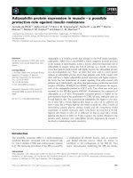
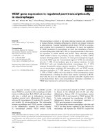
Fig. 1 shows the Northern blot of AQP1 in HK dog tissues
(A). Lung, heart, and kidney demonstrated an intense sig-
nal compared with other tissues. Each sample represented
the major transcripts of approximately 3.1 kb and/or 1.4 kb
signals. Skeletal muscle and small intestine composed the
predominant signal in the 1.4 kb band. Signal intensity of
GAPDH varied among tissues, despite loading of an equal
amount of mRNA across tissues (B). Therefore, relative
204 Hideharu Ochiai et al.
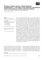
Table 1. Sequences of oligonucleotides used for RT-PCR
Transcript Primers Location Sequence (5'-3') Accession No.
Oligonucleotide for Northern blot
AQP1 5' 428-447 TCGAGATCATTGGCACCCTG AB011373
3' 793-816 CTACTTGGGCTTCATCTCCACCCG
GAPDH 5' 260-281 ATGCTGGTGCTGAGTATGTTGT AB038240
3' 637-657 GATGACCTTGCCCACAGCCTT
Fig. 1. Northern blot analysis of AQP1 expression in dog tissues
(A). Each 10 μg sample of mRNA was purified, electropho-
resed, and blotted. Hybridization of this blot with glyce-
raldehyde-3-phosphate dehydrogenase (GAPDH) to ensure
RNA integrity is also shown (B). AQP1 expression of each tissue
was standardized using the signal intensity of GAPDH (C).
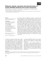
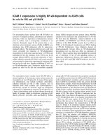
Fig. 2. Immunoblotting of membranes isolated from various H
K
and LK dog tissues. Membrane protein samples (10 μg) were
electrophoresed on 12% polyacrylamide gels and immuno-
blotted with anti-dog AQP1 serum.
AQP1 expression was standardized by that of GAPDH in
each tissue. Standardization revealed abundant AQP1 ex-
pression in lung and kidney, but little in spleen (C).
There were some differences in the mRNA transcriptional
pattern between high K dogs and rats. Unlike rat tissues,
there was no 4.2 kb band in any HK dog tissue preparation.
The 1.4 kb band was predominant in skeletal muscle and
small intestine of HK dogs, whereas only skeletal muscle
exhibited a predominant 1.4 kb band in rats [13]. In rats,
AQP1 expression was clearly detected in spleen [13],
though AQP1 expression in HK dog spleen was unpro-
nounced. To investigate AQP1 protein expression in vari-
ous HK dog tissues, anti-dog AQP1 serum was prepared
AQP1 in HK canine tissues 205
with the peptide antigen designed according to the C-ter-
minus amino acid sequence of dog AQP1 (RVKVWTS-
GQVEEYEL; residues 243-257) [5].
The membrane of each tissue was prepared for Western
blot as reported by Denker et al. [2]. Protein concentration
was determined by the BCA method, and the protein was
used for Western blot analysis.
Fig. 2 shows the distribution of AQP1 in HK and LK
tissues. We found that AQP1 was very abundant in kidney,
lung, trachea, and eye, but was scarce in pancreas and
cerebrum. This finding is, as a whole, consistent with that
of reported ribonuclease protectin and Western blot assays
[14,17]. The strong Western blot signal in spleen, which
was weak on Northern blot, was considered to be due to
abundance of RBC membrane proteins in the spleen. There
was no significant difference in AQP1 expression between
the HK and LK tissues examined (Fig. 2A).
In this report, we investigated the expression of AQP1 in
canines possessing an inherited trait that causes their eryth-
rocytes to have high K
+
. We previously reported the high
incidence of HK dogs in some breeds in Korea and Japan,
but no HK dogs have been found in other areas of East Asia
[4]. Interestingly, these HK cells exhibit characteristics dif-
ferent from normal LK cells in several ways. Firstly, HK
cells have activated Na
+
-dependent amino acid transport
due to the Na
+
driving force created by the Na
+
-K
+
pump.
This results in abnormal accumulation of three amino acids
(Asp, Glu, and Gln) and glutathione [6,11]. The volume of
HK cells is greater than that of LK cells, the lifetime of the
HK cells is half that of LK cells, and some of the glycolytic
enzymes exhibit an immature type of isozyme [7]. These
characteristics have been shown to be inherited in an auto-
somal recessive manner [9]. The above abnormalities sug-
gest that there are defects in the differentiation or matura-
tion of HK cells. This dimorphism in RBC intracellular
cation composition causes the differential regulatory vol-
ume decrease seen when the cells are swollen in a hy-
po-osmotic environment, despite the fact that there is no
difference in AQP1 expression between HK and LK dogs.
Still, the reason why the Na
+
-K
+
pump is retained on HK
RBCs is unknown. Analysis of HK dogs may shed light on
the evolution of carnivore erythrocytes. Further inves-
tigation in HK dogs possessing unique RBCs will provide
more insight into the physiology of water homeostasis in
canines.
References
1. Burg MB, Kwon ED, Kültz D. Regulation of gene ex-
pression by hypertonicity. Annu Rev Physiol 1997, 59, 437-
455.
2. Denker BM, Smith BL, Kuhajda FP, Agre P. Identifica-
tion, purification, and partial characterization of a novel Mr
28,000 integral membrane protein from erythrocytes and re-
nal tubules. J Biol Chem 1988, 263, 15634-15642.
3. Fujise H, Higa K, Kanemaru T, Fukuda M, Adragna NC,
Lauf P. GSH depletion, K-Cl cotransport, and regulatory
volume decrease in high-K/high-GSH dog red blood cells.
Am J Physiol Cell Physiol 2001, 281, C2003-2009.
4. Fujise H, Higa K, Nakayama T, Wada K, Ochiai H,
Tanabe Y. Incidence of dogs possessing red blood cells with
high K in Japan and East Asia. J Vet Med Sci 1997, 59, 495-
497.
5. Higa K, Ochiai H, Fujise H. Molecular cloning and ex-
pression of aquaporin 1 (AQP1) in dog kidney and erythro-
blasts. Biochim Biophys Acta 2000, 1463, 374-382.
6. Inaba M, Maede Y. Increase of Na
+
gradient-dependent
L-glutamate and L-aspartate transport in high K
+
dog eryth-
rocytes associated with high activity of (Na
+
, K
+
)-ATPase. J
Biol Chem 1984, 259, 312-317.
7. Inaba M, Maede Y. Inherited persistence of immature type
pyruvate kinase and hexokinase isozymes in dog erythro-
cytes. Comp Biochem Physiol B 1989, 92, 151-156.
8. Jenq W, Cooper DR, Bittle P, Ramirez G. Aquaporin-1 ex-
pression in proximal tubule epithelial cells of human kidney
is regulated by hyperosmolarity and contrast agents.
Biochem Biophys Res Commun 1999, 256, 240-248.
9. Maede Y, Inaba M. Energy metabolism in canine eryth-
rocytes associated with inherited high Na
+
- and K
+
-stimu-
lated adenosine triphosphatase activity. Am J Vet Res 1987,
48, 114-118.
10. Maede Y, Inaba M, Taniguchi N. Increase of Na-K-
ATPase activity, glutamate, and aspartate uptake in dog er-
ythrocytes associated with hereditary high accumulation of
GSH, glutamate, glutamine, and aspartate. Blood 1983, 61,
493-499.
11. Maede Y, Kasai N, Taniguchi N. Hereditary high concen-
tration of glutathione in canine erythrocytes associated with
high accumulation of glutamate, glutamine, and aspartate.
Blood 1982, 59, 883-889.
12. Moon C, King LS, Agre P. Aqp1 expression in eryth-
roleukemia cells: genetic regulation of glucocorticoid and
chemical induction. Am J Physiol 1997, 273, C1562-1570.
13. Moon C, Preston GM, Griffin CA, Jabs EW, Agre P. The
human aquaporin-CHIP gene. Structure, organization, and
chromosomal localization. J Biol Chem 1993, 268, 15772-
15778.
14. Nielsen S, Smith BL, Christensen EI, Agre P. Distribution
of the aquaporin CHIP in secretory and resorptive epithelia
and capillary endothelia. Proc Natl Acad Sci USA 1993, 90,
7275-7279.
15. Preston GM, Carroll TP, Guggino WB, Agre P. Appear-
ance of water channels in Xenopus oocytes expressing red
cell CHIP28 protein. Science 1992, 256, 385-387.
16. Umenishi F, Verkman AS. Isolation of the human aqua-
porin-1 promoter and functional characterization in human
erythroleukemia cell lines. Genomics 1998, 47, 341-349.
17. Yamamoto T, Sasaki S. Aquaporins in the kidney: Emerg-
ing new aspects. Kidney Int 1998, 54, 1041-1054.