Báo cáo khoa học: " Prevalence of feline herpesvirus 1, feline calicivirus and Chlamydophila felis in clinically normal cats at a Korean animal shelter" ppt
Bạn đang xem bản rút gọn của tài liệu. Xem và tải ngay bản đầy đủ của tài liệu tại đây (383.39 KB, 3 trang )
JOURNAL OF
Veterinary
Science
J. Vet. Sci. (2008), 9(2), 207
209
Short Communication
*Corresponding author
Tel:
+82-2-450-4140; Fax: +82-2-444-4396
E-mail:
Prevalence of feline herpesvirus 1, feline calicivirus and Chlamydophila
felis in clinically normal cats at a Korean animal shelter
Byeong-Teck Kang, Hee-Myung Park
*
Department of Veterinary Internal Medicine, College of Veterinary Medicine, Konkuk University, Seoul 143-701, Korea
The prevalence of feline herpesvirus-1 (FHV-1), feline
calicivirus (FCV), and Chlamydophila (C.) felis was stud-
ied in cats of an animal shelter in Korea. Total 78 cats
without ocular and upper respiratory tract disease were
examined. Specimens were obtained from ocular con-
junctiva and oropharynx. Using multiplex polymerase
chain reaction (PCR) and reverse transcription PCR,
three pathogens were simultaneously detected. In exam-
ined 78 cats, 49 (63%) cats were positive for FHV-1.
However, all specimens were negative for C. felis and
FCV. In conclusion, many cats recovered from FHV-1 in-
fection remain subclinical carriers in shelter environment.
Keywords: Chlamydophila felis, feline calicivirus, feline herpes-
virus-1
Feline herpesvirus type 1 (FHV-1) is the most frequent
cause of conjunctivitis, and it also induces corneal ulcers,
stromal keratitis, corneal sequestration and keratocon-
junctivitis sicca in cats [14]. Chlamydophila (C.) felis
(previously called Chlamydia psittaci) is another major
conjunctival pathogen [13]. Feline calicivirus (FCV) is an
unlikely and minor primary conjunctival pathogen [9]. It is
considered to be the most common upper respiratory tract
disease (URTD) associated pathogen in cats [16].
The prevalence of FHV-1, FCV and C. felis in cats through-
out the world has been frequently reported [1-3,5,6,12,
15,16]. However, there have been relatively fewer of these
studies for clinically normal cats in a shelter environment
[1,5,11] and no such research has been done in Korea. The
purpose of this study is to identify the prevalence of
FHV-1, FCV and C. felis in clinically normal cats of a
Korean animal shelter by performing multiplex reverse
transcription-polymerase chain reaction (RT-PCR)/PCR.
Samples were collected from one animal shelter (Yangju,
Korea) during January 2005. The animals were held at least
30 days and euthanasia was performed because of illness or
injury, or insufficient available space. The shelter was
traditional with no quarantine for incoming cats, but the
cats having diseases were separated from the main shelter.
A total of 78 cats were examined before they underwent
euthanasia. Based on their history and ophthalmic (using
slit lamp biomicroscopy) and physical examinations, they
had clinically normal eyes and they had no specific clinical
signs of URTD. All the cats were domestic short-haired or
long-haired. The number of male cats examined was 40
and 38 were females. The cats’ age and neuter and vaccina-
tion status could not be exactly determined. For each cat,
specimens were obtained using three sterile cotton tipped
swabs in the conjunctival sac of both eyes and the oropha-
rynx, respectively. These were preserved in 2 ml phosphate
buffered saline (PBS, 0.01 M NaPO
4
, pH 7.0) and they
were immediately sent to the laboratory within 2 h and then
stored at 70
o
C until they were assayed. Before the sub-
sequent nucleic acid extraction, the specimens that were
separately obtained from the three sites were thoroughly
mixed.
One commercial vaccine (Felocell CVR-C; Pfizer Ani-
mal Health, USA) was used as a positive control. It was the
only licensed vaccine for preventing FHV-1, FCV and C.
felis in Korea at the time of the study. Sterile distilled water
without nucleic acids was used as a negative control. Both
the controls were subjected to nucleic acid extraction and
PCR. The nucleic acids were extracted from specimens by
using the Viral Gene-spin kit (Intron Biotechnology,
Korea) according to the manufacturer’s instructions.
Three pairs of oligonucleotide primers were used for the
amplifying reaction. We used the previous designed primer
sequences by Sykes et al. [16]. HerpF (5'-GACGTGGT
GAATTATCAGC-3') and HerpR (5'-CAACTAGATTT
CCACCAGGA-3') amplify a 292-base pair (bp) region in
the thymidine kinase (TK) gene of FHV-1. ChlaF (5'-
ATGAAAAAACTCTTGAAATCGG-3') and ChlaR (5'-
CAAGATTTTCTAGACTTCATTTTGTT-3') amplify a
1069-bp fragment in the outer membrane protein gene of
208 Byeong-Teck Kang et al.
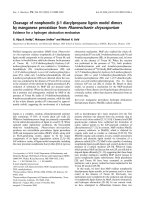
Fig. 1. Multiplex RT-PCR/PCR amplication of FHV-1, C. felis
and FCV from the specimens (S1 and S2) collected from two
cats. Molecular size standard markers (M) are shown as
b
ase
p
airs (bp) on the left for comparison. The positive control (P) ex
-
tracted from commercial vaccine strains shows three bands, an
d
their sizes are 292 bp (for FHV), 673 bp (for FCV) and 1069
b
p
(for C. felis). S1 and S2 show positive reactions for FHV-1, an
d
they are negative for FCV and C. felis. The negative control (N)
is shown on the right.
C. felis. CalcapF (5'-TTCGGCCTTTTGTGTTCC-3') and
CalcapR (5'-TTGAGAATTGAACACATCAATAGATC-3')
amplify a 673-bp conserved region in the capsid protein
gene of FCV.
Multiplex RT-PCR/PCR was performed according to a
previous study [16]. Each 15 μl of reaction products was
electrophoresed through a 1.5% agarose gel and the
proteins were stained with ethidium bromide; the appro-
priate molecular weight markers (100-bp DNA ladder;
Bioneer, Korea) are adjacent to them. The positive control
included the extracted nucleic acid of the commercial
strains in the vaccine and the negative control consisted of
all the RT-PCR/PCR reagents except the nucleic acid;
these were included in each reaction.
When 78 clinically normal cats were examined, 49 (63%)
were positive for FHV-1. Of the 40 male cats examined,
FHV-1 was detected in 23 cats (58%) and 26 of the 38
female cats (68%) were positive to FHV-1. However, C.
felis and FCV were negative in all the specimens we
obtained. The products resulting from amplification of
FHV-1, C. felis and FCV in two specimens are shown in
Fig. 1.
Many studies have reported on the prevalence of FHV-1,
C. felis and FCV in clinically normal or abnormal cats
[1-3,5,6,8,12,15,16]. There were various detection rates of
FHV-1 according to the breeding sites, the countries and
the cats’ clinical status. The prevalence of FHV-1 was
ranged from 0 to 52% for clinically normal cats of a
breeding cattery or shelter in Sweden [6], the USA [2,
11,15] and European countries [5]. The prevalence of
FHV-1 for client-owned normal cats was 5.9% [10] and
this was 12% [15] in the USA. The percentage of cats with
diseases at a breeding cattery or shelter in European
countries [5] and the USA [1] ranged from 0 to 41%. For
client-owned abnormal cats, FHV-1 was detected in 4.5%
to 76.3% of the cats in Japan [3,9], the USA [2,3,10,15],
Australia [16] and Italy [12]. Based on these findings, the
FHV-1 positive rates of the shelter cats were generally
higher than those of the client owned-cats or the breeding
cattery cats. Especially, many shelter cats without diseases
had a higher prevalence than did the clinically abnormal
client-owned cats. We think that the results are due to that
many shelter cats have been infected with FHV-1 and they
remain subclinical carriers after recovery. At least 80% of
infected cats remain latently infected and 29% of them
shed virus spontaneously [4]. Thus, FHV-1 has been
frequently detected in latently infected cats that are without
clinical signs. Also, those cats that live in a colony like an
animal shelter, an animal hospital and a multi-cat house-
hold can be more frequently exposed to FHV-1.
Among the shelter cats around the world, those in Korea
have the highest prevalence of FHV-1 (63%). This result
may be due to some reasons. The first is the difference in
the frequency and level of stressful events that cats are
exposed to in an animal shelter. Stress as simple as moving
a cat into a new environment can convert a latent infection
to an active infection in a few days [11]. Virus shedding
continues for 1-2 weeks around 1 week after a stressful
event [6]. Thus, in this study, cats may have the greatest
risk of infection through more exposure to reactivated
virus from carriers because the Korean housing enviro-
nment may be more stressful than that in other countries.
The second reason is there is a high shedding rate for
incoming cats at the time of first entry to the shelter.
However, in other study, the shedding rate was much lower
than the rate after 1 week in the shelter [11]. To certify this
possibility, it will be necessary to investigate the pro-
portion of cats that already shed FHV-1 at the time they are
relinquished.
It was previously reported that the prevalence of C. felis in
clinically normal cats of a breeding cattery or shelter was
only 3% in European countries [5]. For cats with diseases
and that are in the same environment, the prevalence of C.
felis ranged from 0 to 10% in the USA [1] and European
countries [5], whereas that of client owned abnormal cats
was 11.5%, 20% and 59.1% in Australia [16], Italy [12]
and Japan [3], respectively. According to these previous
reports, clinically normal cats had a much lower pre-
valence of C. felis than that of abnormal cats. Unlike
FHV-1, those cats in a shelter or a breeding cattery had a
low detection rate of C. felis. These results indicate that the
Prevalence of FHV-1, FCV and C. felis in an animal shelter 209
clinical signs of URTD or ocular disease are correlated
with infection of C. felis and they are not related to the
housing environment. C. felis was not detected in our
study.
Generally, normal cats without disease have a lower
detection rate of FCV, ranging from 1 to 29% [5,8,9,11],
than abnormal cats, ranging from 0 to 47% [1,3,5,9,16].
FCV was rarely detected in two studies that focused on
clinically normal cats (1% in Japan and 2.6% in Sweden)
[6,9]. One previous study even showed that FCV was not
detected from clinically abnormal cats in some shelters [1].
In our present study, FCV was not detected at all. This
result may be due to several reasons. First is a possibility
that all the examined cats were not truly infected with FCV.
The second possible reason is that the chronic infected cats
were not shedding virus. Last, it has been reported that the
lower detection rate of FCV is due to some problems for
detecting virus on in the feline mucosal swabs, that is, the
very small number of virus particles, the presence of
RNase in the mucosa and the FCV’s genetic variability
[16]. We can not exactly determine how these factors
contribute to a low detection rate.
This study demonstrated that many cats of an animal
shelter in Korea, as well as other countries, remain latently
infected with FHV-1. These cats would be sources of
infection after returning to society. Thus, proper manage-
ment by veterinarians, cleaning protocols, a low stress
environment and proper cage design are necessary in an
animal shelter. Especially, it may be important to make a
less stressful environment through decreasing the extent of
crowding. Although vaccination with FHV-1 and FCV
cannot lead to prevention of infection and viral shedding,
this may reduce the overall severity of disease [16]. Thus,
it is necessary to routinely vaccinate the individual shelter
cats and to keep incoming cats from infected sources such
as shelters and breeding catteries.
Acknowledgments
This work was supported by the SRC/ERC program of
MOST/KOSEF (R11-2002-103).
References
1. Bannasch MJ, Foley JE. Epidemiologic evaluation of mul-
tiple respiratory pathogens in cats in animal shelters. J Feline
Med Surg 2005, 7, 109-119.
2. Burgesser KM, Hotaling S, Schiebel A, Ashbaugh SE,
Roberts SM, Collins JK. Comparison of PCR, virus iso-
lation, and indirect fluorescent antibody staining in the de-
tection of naturally occurring feline herpesvirus infections. J
Vet Diagn Invest 1999, 11, 122-126.
3. Cai Y, Fukushi H, Koyasu S, Kuroda E, Yamaguchi T,
Hirai K. An etiological investigation of domestic cats with
conjunctivitis and upper respiratory tract disease in Japan. J
Vet Med Sci 2002, 64, 215-219.
4. Gaskell RM, Povey RC. Experimental induction of feline
viral rhinotracheitis virus re-excretion in FVR-recovered
cats. Vet Rec 1977, 100, 128-133.
5. Helps CR, Lait P, Damhuis A, Bj
örnehammar U, Bolta D,
Brovida C, Chabanne L, Egberink H, Ferrand G,
Fontbonne A, Pennisi MG, Gruffydd-Jones T, Gunn-
Moore D, Hartmann K, Lutz H, Malandain E, M
östl K,
Stengel C, Harbour DA, Graat EA. Factors associated with
upper respiratory tract disease caused by feline herpesvirus,
feline calicivirus, Chlamydophila felis and Bordetella bron-
chiseptica in cats: experience from 218 European catteries.
Vet Rec 2005, 156, 669-673.
6. Holst BS, Berndtsson LT, Englund L. Isolation of feline
herpesvirus-1 and feline calicivirus from healthy cats in
Swedish breeding catteries. J Feline Med Surg 2005, 7, 325-
31.
7. Hoover EA, Kahn DE. Experimentally induced feline cal-
icivirus infection: clinical signs and lesions. J Am Vet Med
Assoc 1975, 166, 463-468.
8. Maggs DJ, Lappin MR, Nasisse MP. Detection of feline
herpesvirus-specific antibodies and DNA in aqueous humor
from cats with or without uveitis. Am J Vet Res 1999, 60,
932-936.
9. Mochizuki M, Kawakami K, Hashimoto M, Ishida T.
Recent epidemiological status of feline upper respiratory in-
fections in Japan. J Vet Med Sci 2000, 62, 801-803.
10. Nasisse MP, Glover TL, Moore CP, Weigler BJ. Detec-
tion of feline herpesvirus 1 DNA in corneas of cats with eosi-
nophilic keratitis or corneal sequestration. Am J Vet Res
1998, 59, 856-858.
11. Pedersen NC, Sato R, Foley JE, Poland AM. Common vi-
rus infections in cats, before and after being placed in shel-
ters, with emphasis on feline enteric coronavirus. J Feline
Med Surg 2004, 6, 83-88.
12. Rampazzo A, Appino S, Pregel P, Tarducci A, Zini E,
Biolatti B. Prevalence of Chlamydophila felis and feline
herpesvirus 1 in cats with conjunctivitis in northern Italy. J
Vet Intern Med 2003, 17, 799-807.
13. Shewen PE, Povey RC, Wilson MR. A survey of the con-
junctival flora of clinically normal cats and cats with con-
junctivitis. Can Vet J 1980, 21, 231-233.
14. Stiles J. Treatment of cats with ocular disease attributable to
herpesvirus infection: 17 cases (1983-1993). J Am Vet Med
Assoc 1995, 207, 599-603.
15. Stiles J, McDermott M, Bigsby D, Willis M, Martin C,
Roberts W, Greene C. Use of nested polymerase chain re-
action to identify feline herpesvirus in ocular tissue from
clinically normal cats and cats with corneal sequestra or
conjunctivitis. Am J Vet Res 1997, 58, 338-342.
16. Sykes JE, Allen JL, Studdert VP, Browning GF. Detec-
tion of feline calicivirus, feline herpesvirus 1 and Chlamydia
psittaci mucosal swabs by multiplex RT-PCR/ PCR. Vet
Microbiol 2001, 81, 95-108.