Báo cáo khoa học: "The expression and localization of inhibin isotypes in mouse testis during postnatal development" pdf
Bạn đang xem bản rút gọn của tài liệu. Xem và tải ngay bản đầy đủ của tài liệu tại đây (1.73 MB, 5 trang )
JOURNAL OF
Veterinary
Science
J. Vet. Sci. (2008), 9(4), 345
349
*Corresponding author
Tel: +82-62-530-2838; Fax: +82-62-530-2841
E-mail:
The expression and localization of inhibin isotypes in mouse testis
during postnatal development
Yujin Kim
1
, Joong-Sun Kim
1
, Myoung-Sub Song
1
, Heung-Sik Seo
1
, Jong Choon Kim
2
, Chun-Sik Bae
3
,
Seungjoon Kim
4
, Taekyun Shin
5
, Sung-Ho Kim
1
, Changjong Moon
1,
*
Departments of
1
Veterinary Anatomy,
2
Veterinary Toxicology,
3
Veterinary Surgery, College of Veterinary Medicine and
Veterinary Medical Research Center, Chonnam National University, Gwangju 500-757, Korea
4
Department of Veterinary Obstetrics, College of Veterinary Medicine, Kyungpook National University, Daegu 702-701,
Korea
5
Department of Veterinary Anatomy, College of Veterinary Medicine and Applied Radiological Science Research Institute,
Cheju National University, Jeju 690-756, Korea
Inhibin, which is important for normal gonadal function,
acts on the pituitary gonadotropins to suppress follicle-
stimulating hormone (FSH) secretion. The level and cellular
localization of the inhibin isotypes,
α
,
β
A
and
β
B
, in the testis
of mice were examined during postnatal development in
order to determine if inhibin expression is related to
testicular maturation. Mouse testes were sampled on
postnatal days (PNDs) 1, 3, 6, 18, 48 and 120, and analyzed
by Western blotting and immunofluorescence. Western blot
analysis showed very low levels of inhibin
α
,
β
A
and
β
B
expression in the testes at days 1 to 6 after birth. The levels
then increased gradually from PND 18 to 48-120, and there
were significant peaks at PND 48. Inhibin
α
,
β
A
and
β
B
were
detected in testicular cells during postnatal development
using immunohistochemistry. The immunoreactivity of
inhibin
α
was rarely observed in testicular cells during PND
1 to 6, or in the cytoplasmic process of Sertoli cells
surrounding the germ cells and interstitial cells during PND
18 to 120. Inhibin
β
A
and
β
B
immunoreactivity was rarely
observed in the testis from PND 1 to 6. On the other hand, it
was observed in some spermatogonial cells, as well as in the
interstitial space between PND 48 and PND 120. We
conclude that the expression of inhibin isotypes increases
progressively in the testis of mice with increasing postnatal
age, suggesting that inhibin is associated with a negative
feedback signal for FSH in testicular maturation.
Keywords:
inhibin, mouse, postnatal development, Sertoli cell,
testis
Introduction
Inhibin is a glycoprotein hormone that is produced
principally by the gonads. It is a disulfide linked dimer of
two different subunits, a common α isotype and a β
A
isotype
forming inhibin A subunit or a β
B
isotype forming inhibin
B subunit [21]. Although five distinct β isotypes have been
isolated, which are termed β
A
to β
E
, only the biological
activity of β
A
and β
B
has been demonstrated [11]. Inhibin
belongs to the transforming growth factor β superfamily of
growth and differentiation factors, which are important for
normal gonadal function. Previous studies reported expression
of inhibin in the testis of various mammals including humans
[7], primates [20], rats [26], mice [23], hamsters [9], and pigs
[8]. Inhibin acts on pituitary gonadotropins to suppress
follicle-stimulating hormone (FSH) secretion [5] and to
reduce spermatogonial numbers [25].
The pattern of inhibin expression is associated with the two
distinct phases of rat Sertoli cells [10]. The first phase is
related to an increase in circulating FSH levels [10], which
induce Sertoli cell proliferation. The second phase is related
to the increasing levels of FSH that are present during
pubertal maturation [2,10,24]. Inhibin provides a negative
feedback signal that downregulates the secretion of FSH
[5,17]. In addition, inhibin α isotype knockout mice show
testicular stromal tumors and arrest of gametogenesis
[12,18]. On the other hand, transgenic mice overexpressing
the inhibin A subunit or the inhibin α isotype have small
testes and a reduced level of spermatogenesis [13]. This
suggests that inhibin isotypes may regulate testicular
maturation along with FSH. The secretion of inhibin is
restricted primarily to Sertoli cells in rat testis [16].
Spermatogenic cells in the seminiferous tubules are capable
of modulating the expression of inhibin in Sertoli cells both
in vitro [4,19] and in vivo [1,6]. Therefore, differential
346 Yujin Kim et al.
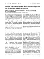
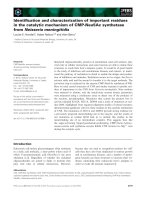
Fig. 1. Light micrographs of the mouse testes at postnatal day
(PND) 1 (A), PND 18 (B), and PND 48 (C). The arrows in
A
indicate gonocytes in undifferentiated seminiferous epithelium.
The asterisk in C indicates the defined lumens of the tubules
including mature sperm cells. H&E stain. Scale bars = 40 μm.
expression of inhibin isotypes might be observed in
seminiferous tubules in mice during testicular development.
This study examined the level and cellular localization of
inhibin isotypes, α, β
A
and β
B
, in the testis of mice during
postnatal development in order to determine if inhibin is
associated with testicular maturation.
Materials and Methods
Animals and tissue preparation
ICR mice used in this experiment were obtained from the
animal center at the Korea Research Institute of Bioscience
and Biotechnology. Mice were housed in a room maintained
under the following conditions: a temperature of 23 ± 2
o
C,
a relative humidity of 50 ± 5%, with artificial lighting from
08:00 to 20:00 and 13-18 air changes per h. The mice were
fed a standard animal diet. Three mice at postnatal days
(PNDs) 1, 3, 6, 18, 48 and 120 were obtained from the same
litters.
Mice were sacrificed and testes were immediately
removed (n = 3). A sample of the testes was embedded in
paraffin wax after routine fixation in 10% buffered
formalin. Paraffin sections (5 μm thick) were used in all
immunostaining experiments. The opposite testis was
snap-frozen and stored for immunoblot analysis. All
experiments were carried out in accordance with the
National Research Council’s Guide for the Care and Use of
Laboratory Animals (USA).
Antisera
Rabbit polyclonal anti-inhibin α (H-134), β
A
(H-120) and
β
B
(H-110) antibodies were obtained from Santa Cruz
Biotechnology (USA). Mouse monoclonal anti-beta-actin
and vimentin antibodies were purchased from Sigma
(USA) and Neomarkers (USA), respectively.
Western blot analysis
Testes tissues were immersed quickly in buffer H (50 mM
β-glycerophosphate, 1.5 mM EGTA, 0.1 mM Na
3
VO
4
, 1
mM DTT, 10 μg/ml aprotinin, 2 μg/ml pepstatin, 10 μg/ml
leupeptin, 1 mM PMSF, pH 7.4), and sonicated for 10 sec.
The homogenate was transferred to microtubes and
centrifuged at 19,340 × g for 10 min. The supernatant was
then harvested. For the immunoblot assay, the supernatant
was loaded into individual lanes of 10% sodium dodecyl
(lauryl) sulfate-polyacrylamide gels, electrophoresed and
immunoblotted onto polyvinylidene difluoride membranes
(Immobilon-P; Millipore, USA). The residual binding sites
on the membrane were blocked by incubation with 5%
nonfat milk in phosphate-buffered saline (PBS, pH 7.4) for
1 h. Subsequently, the membrane was incubated overnight
at 4
o
C with rabbit polyclonal anti-inhibin-α, β
A
and β
B
antibodies (1 : 1,000 dilution). After extensive washing and
incubation with horseradish peroxidase-conjugated goat
anti-rabbit antibody (1 : 20,000 dilution; Pierce, USA),
signals were visualized using chemiluminescence (Super
Signal West Pico; Pierce, USA). For normalization purposes,
membranes were re-probed with antibodies against
beta-actin (1 : 20,000 dilution; Sigma, USA). Several exposure
times were used to obtain signals in the linear range. The
bands were quantified using Scion Image Beta 4.0.2 for
Windows XP software (Scion, USA). The data were analyzed
using one-way ANOVA followed by a Student-Newman-Keuls
post hoc test for multiple comparisons. In all cases, a p value
< 0.05 was considered significant.
Immunofluorescence
Paraffin-embedded sections of testes (5 μm) were
deparaffinized, treated with a citrate buffer (0.01 M, pH
6.0) in a microwave for 20 min, and then treated with 0.3%
hydrogen peroxide in methyl alcohol for 20 min to block
endogenous peroxidase activity. After three washes with
Inhibin isotypes in mouse testicular maturation 347
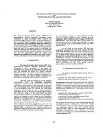
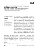
Fig. 2. Expression of inhibin isotypes α, β
A
and β
B
in mouse testis increased progressively with postnatal age. Photographs: Representativ
e
p
hotographs of Western blots for inhibin isotypes α, β
A
and β
B
and beta-actin (A). Arrowheads indicate the positions of the inhibin isotype
s
(40∼47 kDa) and beta-actin (45 kDa). Minor bands at various molecular weights were detected on the immunoblots for the inhibin isotypes
α, β
A
and β
B
. Bar graph: The results of densitometric data analysis (mean ± SE, n = 3 mice/group). The relative expression levels of th
e
inhibin isotypes were calculated after normalization to the beta-actin band from three different samples. The value for the testis at postnatal
day (PND) 1 was arbitrarily defined as 1 (B, C and D, graphs). *p < 0.05, **p < 0.01 vs. PND 1-6.
PBS, sections were incubated with 10% normal goat
serum, and then incubated with rabbit monoclonal inhibin
α, β
A
and β
B
(1 : 100 dilution) for 1 h at room temperature.
The immunoreactivity was visualized using fluorescein
isothiocyanate (FITC)-labeled goat anti-rabbit IgG (1 : 50
dilution; Sigma, USA). Cell phenotypes of inhibin α, β
A
and β
B
expression were examined by double label
immunofluorescence using cell-type-specific markers,
including vimentin (1 : 500 dilution) for the Sertoli and
interstitial cells. First, the paraffin sections were reacted
with primary rabbit anti-inhibin α, β
A
and β
B
followed by
FITC-labeled goat anti-rabbit IgG (1 : 50 dilution; Sigma,
USA). Slides were then incubated with mouse vimentin
followed by tetramethyl rhodamine isothiocyanate-labeled
goat anti-mouse IgG (1 : 50 dilution; Sigma, USA).
Results
Histological finding of the mouse testis during
postnatal development
The testis at PND 48-120 showed an increase in the height
of the seminiferous epithelium and the defined lumens of the
tubules including mature sperm cells (Fig. 1C), while the
tubules at PND 1-18 were largely undifferentiated (Figs. 1A
and B). As shown in Fig. 1C, there was an abundant
population of interstitial cells in the testis at PND 48. The
seminiferous tubules contained primary spermatocytes,
spermatids and Sertoli cells at various stages. This suggests
that sexual maturation in this experimental animal occurs
between PND 18 and 48.
348 Yujin Kim et al.
Fig. 3. Immunofluorescent localization of inhibin α, β
A
, and β
B
isotypes in mouse testis at postnatal days 48. (A and B)
Double-immunofluorescent staining in the same section showe
d
the co-localization of inhibin α with vimentin in cell bodies o
f
Sertoli cells (arrowheads), the cytoplasmic process of Sertoli
cells (arrows) and in interstitial spaces (asterisks). (C)
Immunofluorescent localization of the inhibin β
A
subunit was
observed in the cell membrane of some spermatogenic cells
(arrows) as well as in the interstitial cells (asterisk). (D)
Immunofluorescent localization of the inhibin β
B
subunit was
observed mainly in cell membranes of interstitial cells (asterisk)
as well as in some spermatogonia (arrows). Scale bars = 30 μm.
Temporal expression pattern of the inhibin isotypes
α, β
A
and β
B
during the postnatal development of
mouse testis
The protein levels of the inhibin α, β
A
and β
B
isotypes in
the testes during postnatal development were analyzed
semiquantitatively by Western blotting to determine the
developmental changes in the inhibin isotypes.
As shown in Fig. 2, a low intensity signal for inhibin α
expression was detected in the testis at days 1-6 after birth.
The level gradually increased at PND 18 to 120, and there
was a significant peak (approximately 2 fold, p < 0.01 vs.
PND 1-6) at PND 48 (Figs. 2A and B). A low level of inhibin
β
A
expression was observed in the early phase of
development (PND 1-6). The level increased and showed a
significant peak (approximately 2 fold, p < 0.05 vs. PND
1 and 6) at day 48 after birth (Figs. 2A and C). A low intensity
signal for inhibin β
B
expression was detected in the testis at
PND 1-6. The level increased at PND 48-120, and there were
substantial levels at both PND 48 (approximately 1.5 fold,
p < 0.01 vs. PND 1-18) and PND 120 (approximately 1.6
fold, p < 0.05 vs. PND 1-18) (Figs. 2A and D).
Immunofluorescent detection of inhibin α, β
A
and
β
B
in mice testis
At PND 1-6, there was little immunoreactivity for inhibin
α, β
A
and β
B
subunits in testicular cells (data not shown).
Inhibin α expression (Fig. 3A) was observed in
cytoplasmic processes of vimentin-positive Sertoli cells
surrounding spermatogenic cells (Fig. 3B) at PND 18-120.
Immunoreactivity for inhibin β
A
was observed in the
interstitial and spermatogenic cells (Fig. 3C) during PND
48-120. Inhibin β
B
immunoreactivity was observed mainly
in cell membranes of some spermatogonia in the
seminiferous tubules as well as in the interstitial cells after
PND 48 (Fig. 3D).
Discussion
This study shows a gradual increase in the expression of
inhibin isotypes, α, β
A
and β
B
, in the testis of mice during
postnatal development. Each inhibin isotype was localized
differentially in testicular cells of the testes between PNDs
18-120. However, expression of these isotypes were rarely
observed in testes during the early phase of postnatal
development (PND 1-6).
In this study, histological examination of the development
of mouse testis showed that sexual maturation is acquired
between PND 18 and 48. This suggests that the two major
functions of the sexually matured testis, spermatogenesis
and generation of sexual hormones, were accomplished
between PND 18 and 48. During this phase, protein levels
of the three isotypes of inhibin in the testis also increased.
The histological findings in the sexual maturation of
developing mouse testis are consistent with those of a
previous report [23].
In this study, protein levels of the inhibin isotypes (α, β
A
and β
B
), were analyzed by western blotting. Low intensities
of the isotypes were detected in the early phase, but the levels
increased gradually during sexual maturation (PND 18 to
48). Immunohistochemical results showed that expression
of inhibin isotypes increased gradually during postnatal
development of mouse testis, mainly in the Sertoli and
interstitial cells. Previously, it had not been reported that
mRNAs for the α, β
A
and β
B
isotypes were closely associated
with testicular maturation [14,22,23]. The level of FSH
increased in rats during pubertal maturation [2,10,24].
Inhibin provides a negative feedback signal that regulates
FSH secretion [5,17]. Therefore, the maturation of Sertoli
cells by FSH stimulation promotes the expression of inhibin
isotypes. Hence, inhibin regulates the development of
Sertoli cells and spermatogenesis in mouse testis.
In this study, inhibin α immunoreactivity was detected
mainly in Sertoli cells from puberty to adulthood, as
previously indicated for rat testis [16]. In addition, expression
of inhibin β
A
and β
B
subunits was detected in interstitial and
spermatogenic cells in the testes of mice from puberty to
adulthood. Several studies have reported that the differential
expression in various types of testicular cells depends on the
animal species [3,8,9,15,17]. Therefore, further studies will
be needed to determine the functional role of inhibin via local
or paracrine secretion among testicular cells.
In conclusion, expression of the inhibin isotypes α, β
A
and
β
B
, in the testes of mice gradually increased during postnatal
development. Each isotype was localized differentially in
testicular cells during maturation. The expression of inhibin
isotypes in the testis of mice increased progressively with
Inhibin isotypes in mouse testicular maturation 349
postnatal age, which suggests that inhibin is associated with
a negative feedback signal for FSH during testicular
maturation.
Acknowledgments
This work was supported by the Grant of the Korean Ministry
of Education, Science and Technology (The Regional Core
Research Program/Biohousing Research Institute). This
work was supported by the Biohousing Research Center.
References
1. Allenby G, Foster PM, Sharpe RM. Evidence that
secretion of immunoactive inhibin by seminiferous tubules
from the adult rat testis is regulated by specific germ cell
types: correlation between in vivo and in vitro studies.
Endocrinology 1991, 128, 467-476.
2. Au CL, Robertson DM, de Kretser DM. Measurement of
inhibin and an index of inhibin production by rat testes
during postnatal development. Biol Reprod 1986, 35, 37-43.
3. Buzzard JJ, Loveland KL, O’Bryan MK, O’Connor AE,
Bakker M, Hayashi T, Wreford NG, Morrison JR, de
Kretser DM. Changes in circulating and testicular levels of
inhibin A and B and activin A during postnatal development
in the rat. Endocrinology 2004, 145, 3532-3541.
4. Clifton RJ, O’Donnell L, Robertson DM. Pachytene
spermatocytes in co-culture inhibit rat Sertoli cell synthesis
of inhibin
β B-subunit and inhibin B but not the inhibin α-
subunit. J Endocrinol 2002, 172, 565-574.
5. de Kretser DM, Robertson DM. The isolation and
physiology of inhibin and related proteins. Biol Reprod
1989, 40, 33-47.
6. Guitton N, Touzalin AM, Sharpe RM, Cheng CY,
Pinon-Lataillade G, M
éritte H, Chenal C, Jégou B.
Regulatory influence of germ cells on Sertoli cell function in
the pre-pubertal rat after acute irradiation of the testis. Int J
Androl 2000, 23, 332-339.
7. Illingworth PJ, Groome NP, Byrd W, Rainey WE,
McNeilly AS, Mather JP, Bremner WJ. Inhibin-B: a likely
candidate for the physiologically important form of inhibin
in men. J Clin Endocrinol Metab 1996, 81, 1321-1325.
8. Jin W, Arai KY, Herath CB, Kondo M, Ishi H, Tanioka
Y, Watanabe G, Groome NP, Taya K. Inhibins in the male
G
öttingen miniature pig: Leydig cells are the predominant
source of inhibin B. J Androl 2001, 22, 953-960
9. Jin W, Wada S, Arai KY, Kishi H, Herath CB, Watanabe
G, Suzuki AK, Groome NP, Taya K. Testicular secretion
of inhibin in the male golden hamster (Mesocricetus
auratus). J Androl 2001, 22, 207-211.
10. Lee VW, de Kretser DM, Hudson B, Wang C. Variations
in serum FSH, LH and testosterone levels in male rats from
birth to sexual maturity. J Reprod Fertil 1975, 42, 121-126.
11. Mason AJ. Human inhibin and activin: Structure and
recombinant expression in mammalian cells. In: Burger HG,
de Kretser D, Findlay J, Igarashi M (eds.). Inhibin-Non-Ste-
roidal Regulation of Follicle Stimulating Hormone Secretion.
pp. 42-77, Raven Press, New York, 1987.
12. Matzuk MM, Finegold MJ, Su JG, Hsueh AJ, Bradley A.
α-inhibin is a tumour-suppressor gene with gonadal speci-
ficity in mice. Nature 1992, 360, 313-319.
13. McMullen ML, Cho BN, Yates CJ, Mayo KE. Gonadal
pathologies in transgenic mice expressing the rat inhibin
α-subunit. Endocrinology 2001, 142, 5005-5014.
14. Mellor SL, Richards MG, Pedersen JS, Robertson DM,
Risbridger GP. Loss of the expression and localization of
inhibin
α-subunit in high grade prostate cancer. J Clin
Endocrinol Metab 1998, 83, 969-975.
15. Nagata S, Tsunoda N, Nagamine N, Tanaka Y, Taniyama
H, Nambo Y, Watanabe G, Taya K. Testicular inhibin in
the stallion: cellular source and seasonal changes in its
secretion. Biol Reprod 1998, 59, 62-68.
16. Noguchi J, Hikono H, Sato S, Watanabe G, Taya K,
Sasamoto S, Hasegawa Y. Ontogeny of inhibin secretion in
the rat testis: secretion of inhibin-related proteins from fetal
Leydig cells and of bioactive inhibin from Sertoli cells. J
Endocrinol 1997, 155, 27-34.
17. O’Connor AE, de Kretser DM. Inhibins in normal male
physiology. Semin Reprod Med 2004, 22, 177-185.
18. Pierson TM, Wang Y, DeMayo FJ, Matzuk MM, Tsai
SY, O’Malley BW. Regulable expression of inhibin A in
wild-type and inhibin
α null mice. Mol Endocrinol 2000, 14,
1075-1085.
19. Pineau C, Sharpe RM, Saunders PT, G
érard N, Jégou B.
Regulation of Sertoli cell inhibin production and of inhibin
α-
subunit mRNA levels by specific germ cell types. Mol Cell
Endocrinol 1990, 72, 13-22.
20. Plant TM, Marshall GR. The functional significance of
FSH in spermatogenesis and the control of its secretion in
male primates. Endocr Rev 2001, 22, 764-786.
21. Robertson DM, Cahir N, Findlay JK, Burger HG,
Groome N. The biological and immunological characteriza-
tion of inhibin A and B forms in human follicular fluid and
plasma. J Clin Endocrinol Metab 1997, 82, 889-896.
22. Schmitt JF, Millar DS, Pedersen JS, Clark SL, Venter
DJ, Frydenberg M, Molloy PL, Risbridger GP. Hyper-
methylation of the inhibin
α-subunit gene in prostate
carcinoma. Mol Endocrinol 2002, 16, 213-220.
23. Seok OS, Ahn JM, Mayo KE, Cho BN. Developmental
changes in inhibin-
α gene expression in the mouse testis.
Mol Cells 2004, 17, 67-72.
24. Sharpe RM, Turner KJ, McKinnell C, Groome NP,
Atanassova N, Millar MR, Buchanan DL, Cooke PS.
Inhibin B levels in plasma of the male rat from birth to
adulthood: effect of experimental manipulation of Sertoli
cell number. J Androl 1999, 20, 94-101.
25. van Dissel-Emiliani FM, Grootenhuis AJ, de Jong FH, de
Rooij DG. Inhibin reduces spermatogonial numbers in testes
of adult mice and Chinese hamsters. Endocrinology 1989,
125, 1899-1903.
26. Woodruff TK, Besecke LM, Groome N, Draper LB,
Schwartz NB, Weiss J. Inhibin A and inhibin B are
inversely correlated to follicle-stimulating hormone, yet are
discordant during the follicular phase of the rat estrous cycle,
and inhibin A is expressed in a sexually dimorphic manner.
Endocrinology 1996, 137, 5463-5467.