Báo cáo khoa học: "Radioprotective effects of fucoidan on bone marrow cells: improvement of the cell survival and immunoreactivity" doc
Bạn đang xem bản rút gọn của tài liệu. Xem và tải ngay bản đầy đủ của tài liệu tại đây (848.4 KB, 7 trang )
JOURNAL OF
Veterinary
Science
J. Vet. Sci. (2008), 9(4), 359
365
*Corresponding author
Tel: +82-64-754-3379; Fax: +82-64-756-3354
E-mail:
Radioprotective effects of fucoidan on bone marrow cells: improvement
of the cell survival and immunoreactivity
Yun-Young Byon
1,2
, Mi-Hyoung Kim
1
, Eun-Sook Yoo
2
, Kyu-Kye Hwang
1
, Youngheun Jee
1,3
, Taekyun Shin
1,3
,
Hong-Gu Joo
1,4,
*
1
College of Veterinary Medicine,
2
Department of Pharmacology, College of Medicine,
3
Applied Radiological Science Research Institute,
4
The Research Institute for Subtropical Agriculture and Biotechnology,
Cheju National University, Jeju 690-756, Korea
Fucoidan is a sulfated polysaccharide purified from brown
algae including Fucus vesiculosus and has a variety of
biological effects including mobilization of hematopoietic
progenitor cells. Recently, we demonstrated that fucoidan
stimulates the antigen-presenting functions of dendritic
cells. In this study, we investigated the radioprotective
effects of fucoidan on bone marrow cells (BMCs), which are
the main cellular reservoir for the hematopoietic and
immune system. To evaluate the effects of fucoidan, we
assayed cell viability and immune responses. In a viability
assay, fucoidan significantly increased the viability of
BMCs. Based on the results of flow cytometric analysis, the
increased viability of fucoidan-treated BMCs was attributed
to the inhibition of radiation-induced apoptosis. Furthermore,
fucoidan altered the production of immune-related cytokines
from BMCs and increased the capability of BMCs to induce
proliferation of allogeneic splenocytes. Taken together, our
study demonstrated that fucoidan has radioprotective effects
on BMCs with respect to cell viability and immunoreactivity.
These results may provide valuable information, useful in
the field of radiotherapy.
Keywords: bone marrow cells, fucoidan, immunoreactivity,
radioprotection
Introduction
Bone marrow contains a variety of cells that are required
to maintain the hematopoietic and immune systems. On
exposure to ionizing radiation, bone marrow is profoundly
damaged and severe immunosuppression occurs. Therefore,
protection of bone marrow against gamma radiation is
extremely important in individuals in danger of radiation
exposure.
To protect the host against the harmful effects of radiation
exposure, radioprotective agents have been developed over
several decades [5]. Although the radioprotective agent
amifostine has been used in clinical settings [17,22], it
generates serious side effects, including nausea, probably
due to its synthetic nature [15]. Recent studies have focused
on the development of radioprotective agents derived from
natural sources and that display minimal side effects on
normal cells [1]. For example, some polysaccharides purified
from herbs have been shown to have radioprotective and
immunostimulating effects on host immune cells [9,21].
Fucoidan is a sulfated polysaccharide purified from brown
algae, such as Fucus vesiculosus, and has been shown to have
a variety of biological effects [2,3]. Previous studies have
indicated that fucoidan stimulates the mobilization of
hematopoietic progenitor cells (HPCs) from their niche
within bone marrow to peripheral blood via inhibition of the
selectin-mediated adhesion of HPCs [3]. In addition,
fucoidan-induced mobilization of HPCs is associated with
increased plasma levels of stromal-derived factor-1 in vivo
[19]. Recently, we demonstrated that fucoidan stimulates
multiple functions of dendritic cells (DCs), which are potent
antigen-presenting cells (APCs) in the immune system [10].
Although the biological functions of fucoidan in hemato-
poietic and immune systems have been studied for many
years, its radioprotective effects on bone marrow cells
(BMCs) have not been elucidated. In this study, we
investigated the protective effects of fucoidan on irradiated
BMCs. Specifically, we quantified the viability of cells,
cytokine production, and allostimulatory capability of this
agent.
Materials and Methods
Animal and reagents
C57BL/6 and Balb/c mice were purchased from Orient Bio
360 Yun-Young Byon et al.
(Korea) and maintained at our animal facility. Seven to 12
week-old female mice were used in this study. All
experiments using animal were performed based on the
institutional guideline of Cheju National University for
laboratory animal use and care (Approval No: 20070005).
Fucoidan, originated from Fucus vesiculosus, was obtained
from Sigma (USA) and dissolved in phosphate-buffered
saline (Invitrogen, USA). The endotoxin level of the fucoidan
preparation was less than 0.1 EU/ml from QCL-1000
Chromogenic LAL endpoint assay (Lonza Walkersville, USA)
according to the manufacturer’s protocol.
Preparation of cells and measurement of cell viability
BMCs were harvested from the femurs and tibias of mice
of C57BL/6 mice as described in previous report [9]. Any
contaminated red blood cells were eliminated by ammonium
chloride-potassium carbonate lysis buffer. To culture BMCs,
RPMI 1640 media containing 5% fetal bovine serum (FBS),
2 mM L-glutamine, and 100 U/ml penicillin/ streptomycin
(Invitrogen, USA) was used. For the cell viability assay,
BMCs were cultured at a concentration of 2 × 10
5
cells/well
in 96-well plates and treated with fucoidan before
irradiation. The cultured wells were incubated with 10 μl/
well of Cell Counting Kit-8 solution (CCK-8 solution;
Dojindo, USA) for 4 h and the optical density (O.D.) of wells
was measured at 450 nm by using a microplate reader
(Molecular Devices, USA).
Gamma irradiation
The BMCs were irradiated using a
60
Co γ-ray source (MDS
Nordion C-188 standard source) established in the Applied
Radiological Science Research Institute, Cheju National
University (Korea). Irradiation on cells was performed
once at 1 Gy for this study.
Determination of interleukin-12 (IL-12) and tumor
necrosis factor-alpha (TNF-alpha) production
Fucoidan (50 μg/ml) was administrated in 1 × 10
6
cells/ml
of BMCs for 24 h and then single-exposed to gamma
irradiation. After 24 h, the supernatants were harvested and
used for the determination of IL-12 and TNF-alpha, the
representative cytokines of cell-mediated and innate immunity.
The cytokine concentration of supernatant were measured
by enzyme-linked immunosorbent assay (ELISA) using
CytoSet antibody pairs (Biosource International, USA)
based on the manufacturer’s manual.
Flow cytometric analysis
BMCs were cultured at a concentration of 1 × 10
6
cells/ml
in 6-well plates and treated with fucoidan (50 μg/ml) for 24
h and then irradiated once (1 Gy). After 24 h, the cells were
harvested and stained for flow cytometric analysis as
described in our previous report [9]. Briefly, we used
biotin-labeled anti-Gr-1, anti-I-A
b
(MHC II), anti-CD86
(B7.2) monoclonal antibody as the primary antibody and
streptavidin-fluorescein isothiocyanate (FITC) as the
secondary antibody (BD Biosciences, USA). For the
measurement the percentage of apoptotic cells, cells were
stained by using annexin V-FITC/propidium iodide (PI) kit
(Biosource International, USA) as described in the
manufacturer’s instructions. For the measurement of
mitochondrial membrane potential, cells were incubated
with 10 μg/ml rhodamine 123 (Sigma, USA) for 30 min.
All stained cells were analyzed by using FACSCalibur and
CellQuest (Beckton Dickinson, USA).
Mixed lymphocyte reaction (MLR)
BMCs were cultured and treated as we described in flow
cytometric analysis. Splenocytes obtained from Balb/c mice
were used as allogeneic responder cells for the co-culture
with BMCs of C57BL/6 mice as described in previous report
[9]. 2 × 10
5
cells/well allogeneic splenocytes were co-cultured
with 1,852-5 × 10
4
cells/well BMCs in 96-well culture plates
for 5 days. To minimize the proliferation of BMCs
themselves in total value, all BMCs were irradiated
immediately prior to co-culture. The culture medium was
RPMI 1640 medium containing 10% FBS, 0.1 mM
non-essential amino acid, 1 mM sodium pyruvate, 2 mM
L-glutamine, 100 IU/ml penicillin/ streptomycin, and 50
μM 2-mercaptoethanol (Invitrogen, USA). The co-cultures
were pulsed with 1 μCi/well
3
H-thymidine (PerkinElmer,
USA) for last 18 hrs and the incorporated radioactivity in
cells was measured by a liquid scintillation counter (Wallac
Microbeta TriLux; PerkinElmer, USA).
Western blot analysis
BMCs were cultured and treated as previously described
in flow cytometric analysis section. The cells were
harvested and then used for Western blot analysis. The
lysates of treated BMCs were obtained and the Western
blot analysis was performed as described in a previous
report [8]. Briefly, proteins blotted on nitrocellulose
membrane were probed with anti-Bcl-2, Bcl-xL, Bax
antibody (Santa Cruz Biotechnology, USA), anti-cIAP-1,
cIAP-2 antibody (Millipore, USA), and anti-β-actin antibody
(Sigma, USA) and sequentially appropriate horseradish
peroxidase-labeled secondary antibodies. The blot was
developed by SuperSignal West Pico Cheminluminescent
Substrate (Pierce Biotechnology, USA).
Statistical analysis
Most of data were obtained from three experiments and
analyzed with Turkey-Kramer multiple comparison tests.
A p value <0.05 was recognized as statistically significant.
Radioprotective effects of fucoidan 361
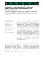
Fig. 1. Fucoidan increases bone marrow cell (BMC) viability.
Asterisks (*, **, ***) indicate p < 0.05, 0.01, 0.001 vs Non-
irradiation (NO-IR) control (fucoidan 0 μg/ml) and ##, ###
indicate p < 0.01, 0.001 vs irradiation (IR) control (fucoidan 0
μg/ml), respectively.
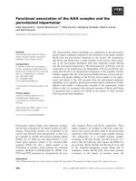
Fig. 2. Inhibition of bone marrow cell (BMC) apoptosis by fucoidan treatment. (A) Numbers indicate the cell percentage of quadrant. (B
)
N
umbers indicate the mean fluorescence intensity of all cells and brackets include the percentage of high expressed cells (M1).
Results
Fucoidan significantly increases BMC viability
To examine the effects of fucoidan on BMC viability, we
cultured BMCs at a concentration of 2 × 10
5
cells/well in
96-well plates and treated them with fucoidan before
irradiation (Fig. 1). After culturing the cells, CCK-8 solution
was added and the O.D. of wells was measured. In the absence
of fucoidan, control BMCs showed significantly higher
viability than irradiated BMCs (p<0.05). Fucoidan
significantly increased the viability of control BMCs within
the range of concentration (2-50 μg/ml) and in irradiated
BMCs (10-50 μg/ml). BMCs were cultured without
cytokines, which may maintain their survival in vivo. Thus,
it was likely that this manipulation would lead to cytokine
withdrawal-induced cell death. Irradiation significantly
decreased the BMC viability. Together, these results suggest
that fucoidan may protect BMCs from cytokine withdrawal-
induced cell death and irradiation.
Fucoidan treatment decreases cell death in irradiated
BMCs
For this test, BMCs were cultured at a concentration of 1 ×
10
6
cells/ml in 6-well plates and treated with fucoidan (50
μg/ml) for 24 h and then irradiated once (1 Gy). After 24 h,
cells were harvested and used for the cell death measurements.
Annexin V-FITC/PI staining and rhodamine 123 staining
were used to confirm the protective effects of fucoidan on
the viability of irradiated BMCs (Fig. 2). With the annexin
V-FITC/PI staining (Fig. 2A), we found that fucoidan
consistently increased the number of viable cells, the
double-negative cells (annexin V-/PI-). Specifically, cell
viability was only 15% with irradiation, which increased to
62% with fucoidan treatment. In addition, fucoidan
decreased the ratio of late apoptotic cells (annexin V+/PI+).
Mitochondria are known to play a critical role in the
apoptotic process of cells. Thus, we performed rhodamine
123 staining to determine mitochondrial potential as
362 Yun-Young Byon et al.
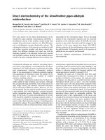
Fig. 3. The altered expression of apoptosis-related molecules in
fucoidan-treated bone marrow cells. (A) Western blot. (B) Densitometr
y
of western
b
lot.
cellular apoptosis decreases the stability of mitochondrial
potential (Fig. 2B). Our results indicated that irradiation
decreased mitochondrial potential; however, this was
recovered by fucoidan treatment. Taken together, our
results demonstrate that fucoidan increases BMC viability
following irradiation. These effects may be the result of
apoptotic inhibition in irradiated BMCs.
Fucoidan treatment alters the expression of apoptosis-
related molecules in BMCs
To further investigate the functional role of fucoidan, we
examined the expression levels of apoptosis-related
molecules in BMCs. Western blot analysis (Fig. 3) indicated
that the expression levels of Bcl-2, Bax, and cIAP-1 were
increased in fucoidan-treated BMCs. In densitometry
results (B), Bcl-2 and Bax expression was increased in all
fucoidan-treated BMCs whereas cIAP-1 expression was
increased only in fucoidan-treated BMCs. However Bcl-xL
was not detected in these experiments.
Fucoidan treatment increases the expression of
surface markers in BMCs
BMCs are one of the main immune cell reservoirs in
hosts. To determine the recovery effects of fucoidan on the
immune function of irradiated BMCs, we measured the
expression levels of some important cell surface markers.
By flow cytometric analyses (Fig. 4), we found that a
phenotypic marker for granulocytes, Gr-1 expression, was
significantly increased by fucoidan treatment. In contrast,
the expression of immune-related markers, B7.2 (data not
shown) and MHC II, were not significantly altered. Our
results demonstrated that fucoidan significantly increases
the expression of a granulocyte marker on BMCs,
suggesting that this agent may facilitate proportional
changes of cell types in BMCs and preferentially protect
granulocytes in BMCs from growth factor-withdrawal or
irradiation induced cell death.
Fucoidan treatment enhances BMC cytokine production
We investigated whether fucoidan-treated BMCs produce
high levels of cytokines related to immune responses. IL-12
and TNF-alpha were selected for this study as they are
representative cytokines which are involved in cell-mediated
and innate immunity, respectively. In ELISA (Fig. 5),
fucoidan significantly increased the production of IL-12 in
fucoidan-treated BMCs compared to control BMCs and
increased the production of TNF-alpha in fucoidan-treated
BMCs and fucoidan-treated irradiated BMCs compared to
control BMCs and irraditated BMCs respectively. However,
any significant increase of IL-12 was not detected in fucoidan-
treated irradiated BMCs. These results suggest that fucoidan
may alter cytokine production as well as cell viability.
Increased allogeneic splenocyte proliferation activated
by fucoidan-treated BMCs
The capacity of BMCs to activate the proliferation of
allogeneic splenocytes was examined to determine whether
fucoidan alters these immune responses. BMCs include
various types of APCs as precursors or matured cells. Using
MLR assays, BMCs of C57BL/6 mice were used as
stimulators and splenocytes of Balb/c mice were used as
allogeneic responder cells. To measure the proliferation of
allogeneic splenocytes alone, all BMCs were irradiated
immediately prior to co-culture. Fucoidan-treated BMCs
and fucoidan-treated irradiated BMCs were found to have
a significantly increased capability to enhance the proliferation
of allogeneic responder cells compared to control BMCs and
irradiated BMCs respectively (Fig. 6). These results suggest
that fucoidan treatment significantly up-regulates APC
functions of BMCs.
Discussion
Fucoidan is known to have a variety of biological
Radioprotective effects of fucoidan 363
Fig. 4. Surface marker expression was up-regulated on
b
one
marrow cells. (A) Numbers indicate the percentage of high
expressed cells (M1) compared to that of fluorescence control.
(B) Percentages of high expressed cells were analyzed
statistically. An asterisk (*) indicate p < 0.05 vs control. A sharp
(
#
)
indicate
p
< 0.05 vs IR control.
Fig. 5. Fucoidan treatment enhances cytokine production of bone marrow cells (BMCs). The supernatants of BMCs were collected an
d
ELISA was performed to quantify cytokine concentrations. N/D: non-detectable level of cytokine. Asterisks (***) indicate p < 0.001
vs control. Sharps (###) indicate p < 0.001 vs IR control.
364 Yun-Young Byon et al.
Fig. 6. Fucoidan-treated bone marrow cells (BMCs) increases the
p
roliferation of allogeneic splenocytes. Allogeneic splenocytes
(2 × 10
5
cells/well) were co-cultured with BMCs in 96-well culture
p
lates. Asterisks (**, ***) indicate p < 0.01, 0.001 vs control,
respectively. Sharps (###) indicate p < 0.001 vs IR control.
functions, and the immunomodulatory activity of this
agent has been studied extensively for many years. Recent
studies demonstrated that fucoidan has significantly
important biological effects on natural killer cells [13],
hematopoietic stem cells [6], endothelial cells [11], and
DCs [10]. Although the effects of fucoidan on the
mobilization of HPCs in bone marrow have been studied
well, there are few studies about the direct effects of
fucoidan on BMCs. In this study, we investigated the
radioprotective effects of fucoidan on BMCs by examining
cell viability and immunostimulatory activity.
Research on the development of radioprotective agents
has focused on natural plant-derived compounds as
potential candidates; medicinal herbs, including ginseng,
are the main sources for the purification of effective
candidates [7,18]. In a previous study, we demonstrated
that ginsan, a polysaccharide from Panax ginseng, has
radioprotective effects on BMCs [9]. Some biological
response modifiers, including those consisting of
polysaccharides, have been shown to have radioprotective
and immunostimulatory effects. Immunostimulatory
signaling is thought to transduce the survival signal in
immune cells and is then manifested as radioprotective
activity.
On flow cytometric analysis, fucoidan-treated BMCs
showed higher levels of surface Gr-1 expression than
controls. As bone marrow is the main cellular source for
the hematopoietic and immune systems, it contains large
numbers of lymphocytes, granulocytes, and stromal cells
as precursors and mature cells. Our results suggested that a
specific population of BMCs, such as Gr-1
+
granulocytes,
may selectively survive in response to fucoidan treatment
following irradiation. Future research should focus on
determining which cell types are selected by fucoidan and
the mechanism of action of this agent.
To investigate how fucoidan protects BMCs from cell
death, the expression levels of apoptosis-related molecules
were measured by Western blot analysis. Among the
molecules belonging to the Bcl-2 family, Bcl-2, Bcl-xL, and
Bax were selected due to their connection to apoptotic
pathways involving mitochondria [4,14]. In addition, the
levels of expression of cIAP-1 and cIAP-2, other anti-
apoptotic molecules [12,16,20] were examined in the same
experiments. We found that the levels of expression of Bcl-2,
Bax, and cIAP-1 were higher in fucoidan-treated BMCs as
compared with controls; there were no differences in cIAP-2
expression between treated and control BMCs. Bcl-2 and
Bax proteins show anti-apoptotic and pro-apoptotic effects,
respectively. However, both were upregulated in the present
study. It is possible that enhanced Bcl-2 expression may
compensate for the activity of Bax and other anti-apoptotic
molecules, such as cIAP-1 also may support the process.
And also, there is another possibility that fucoidan may
induce the cell death of some specific cell types whereas it
enhances the survival of other cell types of BMCs. The
effects of fucoidan on specific cell types of BMCs need to
be investigated in future work.
Our findings indicated that fucoidan-treated BMCs have
an increased capability to stimulate allogeneic splenocytes
as compared with controls. As fucoidan increased cytokine
production, but not immune-related surface markers, it is
postulated that the observed up-regulation of immune
responses may be due primarily to increased cytokine
production and a higher percentage of Gr-1
+
cells, including
APCs within stimulator cells.
Taken together, our results demonstrate that the sulfated
polysaccharide fucoidan has radioprotective effects on
BMCs. These effects include aspects of cell viability and
immunomodulatory activity. In conclusion, the results of
this study may facilitate the development of new
radioprotective agents with reduced toxicity.
Acknowledgments
This work was supported by the Korea Research Foundation
Grant funded by the Korean Government (MOEHRD)
(KRF-2004-202-E00184) and performed under the program
of Basic Atomic Energy Research Institute which is a part
of the Nuclear R&D Programs funded by the Ministry of
Science & Technology of Korea.
References
1. Arora R, Gupta D, Chawla R, Sagar R, Sharma A,
Kumar R, Prasad J, Singh S, Samanta N, Sharma RK.
Radioprotection by plant products: present status and future
prospects. Phytother Res 2005, 19, 1-22.
2. Choi EM, Kim AJ, Kim YO, Hwang JK. Immunomodulating
activity of arabinogalactan and fucoidan in vitro. J Med Food
Radioprotective effects of fucoidan 365
2005, 8, 446-453.
3. Frenette PS, Weiss L. Sulfated glycans induce rapid hema-
topoietic progenitor cell mobilization: evidence for se-
lectin-dependent and independent mechanisms. Blood 2000,
96, 2460-2468.
4. Gross A, McDonnell JM, Korsmeyer SJ. BCL-2 family
members and the mitochondria in apoptosis. Genes Dev
1999, 13, 1899-1911.
5. Hosseinimehr SJ. Trends in the development of radio-
protective agents. Drug Discov Today 2007, 12, 794-805.
6. Irhimeh MR, Fitton JH, Lowenthal RM. Fucoidan in-
gestion increases the expression of CXCR4 on human
CD34+ cells. Exp Hematol 2007, 35, 989-994.
7. Ivanova T, Han Y, Son HJ, Yun YS, Song JY. Antimutagenic
effect of polysaccharide ginsan extracted from Panax
ginseng. Food Chem Toxicol 2006, 44, 517-521.
8. Joo HG, Goedegebuure PS, Sadanaga N, Nagoshi M, von
Bernstorff W, Eberlein TJ. Expression and function of ga-
lectin-3, a
β-galactoside-binding protein in activated T
lymphocytes. J Leukoc Biol 2001, 69, 555-564.
9. Kim HJ, Kim MH, Byon YY, Park JW, Jee Y, Joo HG.
Radioprotective effects of an acidic polysaccharide of Panax
ginseng on bone marrow cells. J Vet Sci 2007, 8, 39-44.
10. Kim MH, Joo HG. Immunostimulatory effects of fucoidan
on bone marrow-derived dendritic cells. Immunol Lett 2008,
115, 138-143.
11. Lake AC, Vassy R, Di Benedetto M, Lavigne D, Le Visage
C, Perret GY, Letourneur D. Low molecular weight fucoi-
dan increases VEGF165-induced endothelial cell migration
by enhancing VEGF165 binding to VEGFR-2 and NRP1. J
Biol Chem 2006, 281, 37844-37852.
12. Liston P, Fong WG, Korneluk RG. The inhibitors of apop-
tosis: there is more to life than Bcl2. Oncogene 2003, 22,
8568-8580.
13. Maruyama H, Tamauchi H, Iizuka M, Nakano T. The
role of NK cells in antitumor activity of dietary fucoidan
from Undaria pinnatifida sporophylls (Mekabu). Planta Med
2006, 72, 1415-1417.
14. Opferman JT, Korsmeyer SJ. Apoptosis in the develop-
ment and maintenance of the immune system. Nat Immunol
2003, 4, 410-415.
15. Rades D, Fehlauer F, Bajrovic A, Mahlmann B, Richter
E, Alberti W. Serious adverse effects of amifostine during
radiotherapy in head and neck cancer patients. Radiother
Oncol 2004, 70, 261-264.
16. Rothe M, Pan MG, Henzel WJ, Ayres TM, Goeddel DV.
The TNFR2-TRAF signaling complex contains two novel
proteins related to baculoviral inhibitor of apoptosis proteins.
Cell 1995, 83, 1243-1252.
17. Santini V, Giles FJ. The potential of amifostine: from cyto-
protectant to therapeutic agent. Haematologica 1999, 84,
1035-1042.
18. Subramanian M, Chintalwar GJ, Chattopadhyay S.
Antioxidant and radioprotective properties of an Ocimum
sanctum polysaccharide. Redox Rep 2005, 10, 257-264.
19. Sweeney EA, Lortat-Jacob H, Priestley GV, Nakamoto
B, Papayannopoulou T. Sulfated polysaccharides increase
plasma levels of SDF-1 in monkeys and mice: involvement
in mobilization of stem/progenitor cells. Blood 2002, 99,
44-51.
20. Uren AG, Pakusch M, Hawkins CJ, Puls KL, Vaux DL.
Cloning and expression of apoptosis inhibitory protein ho-
mologs that function to inhibit apoptosis and/or bind tumor
necrosis factor receptor-associated factors. Proc Natl Acad
Sci USA 1996,
93, 4974-4978.
21. Wang ZW, Zhou JM, Huang ZS, Yang AP, Liu ZC, Xia
YF, Zeng YX, Zhu XF. Aloe polysaccharides mediated ra-
dioprotective effect through the inhibition of apoptosis. J
Radiat Res 2004, 45, 447-454.
22. Wrembel-Wargocka J, Jab
ł
o
ń
ska H, Chomiczewski K.
Clinical use of amifostine (WR-2721) as a preparation pro-
tecting healthy tissues from the cytotoxic effects of chemo-
therapy and radiation therapy. Przegl Lek 1996, 53, 820-825.