Báo cáo khoa học: "Two different types of malignant fibrous histiocytomas from pet dogs" docx
Bạn đang xem bản rút gọn của tài liệu. Xem và tải ngay bản đầy đủ của tài liệu tại đây (4.88 MB, 3 trang )
JOURNAL OF
Veterinary
Science
Case Report
J. Vet. Sci. (2009), 10(2), 169
171
DOI: 10.4142/jvs.2009.10.2.169
*Corresponding author
Tel: +82-53-950-5975; Fax: +82-53-950-5955
E-mail:
Two different types of malignant fibrous histiocytomas from pet dogs
Sun Hee Do
1
, Il-Hwa Hong
2
, Jin-Kyu Park
2
, Ae-Ri Ji
2
, Tae-Hwan Kim
2
, Dong-Mi Kwak
2
, Kyu-Shik Jeong
2,
*
1
Department of Clinical Pathology, College of Veterinary Medicine, Konkuk University, Seoul 143-701, Korea
2
Department of Pathology, College of Veterinary Medicine, Kyungpook National University, Daegu 702-701, Korea
We describe 2 cases of malignant fibrous histiocytomas
(MFHs) that spontaneously developed in young pet dogs.
To classify these tumors, we applied a panel of antibodies
(vimentin, desmin,
α
-SMA, and ED1) and Azan staining
for collagen. The MFHs were most consistent with osteoclast-
like giant and inflammatory cell types. The first case had
positive staining for ED1 and vimentin, and given the
osteoclast-like giant cells, calcification sites accompanying
peripheral giant cell infiltrates. The latter case, the
inflammatory cell type, exhibited a storiform-pleomorphic
variant of neoplastic cells, including an ossifying matrix.
MFHs are among the most highly aggressive tumors
occurring in soft tissue sarcomas in elderly dogs; however,
MFHs have been poorly studied from a diagnostic point of
view. Herein, we describe the histologic and immunohistologic
features of MFHs in detail, thus classifying the subtypes of
these tumors.
Keywords:
dog, inflammatory, malignant fibrous histiocytoma,
osteoclast-like giant cell
Malignant fibrous histiocytomas (MFHs) are mesenchymal
tumors frequently occurring in skeletal muscles and
cutaneous regions in elderly humans; the visceral form is
most common in young immunodepressed humans [6,7,10].
Based on histologic and immunohistologic studies, most of
the tumors have been shown to originate from fibroblasts
and/or myofibroblasts, presumably from undifferentiated
mesenchymal cells [2,3,5]. MFHs have been diagnostic
dilemmas because of the histologic variation from plexiform
fibrohistiocytic to infiltrative subcutaneous fibroblast-like
spindle cell types [7,10].
The neoplasm is characterized by a mixture of neoplastic
fibroblasts, histiocytes, and multi-nucleated giant cells that
interlace in tight bundles [8]. Large giant cells that resemble
osteoclasts are generally present [1]. The primary tumor
cells are pleomorphic, vary in appearance from fusiform-to-
round, and have a nucleus with one or two prominent and
irregular nucleoli [8]. Extracellular amorphous eosinophilic
material may be prominent and likely represents reactive
collagen production by the neoplastic cells [8].
The osteoclast-like giant cell type of MFH is rare. The
characteristic features of the osteoclast-like giant cell type
have not been described in detail in domestic animals. Thus,
we studied the histopathologic features of osteoclast-like
giant cell type MFH with calcifications in a young healthy
dog and compared of the findings to another case of
inflammatory MFH.
A two-month old female Shih-tzu was brought to a local
veterinary clinic with a raised subcutaneous mass in the left
dorsal region. Because of the rapid growth of the mass, the
veterinarian performed a surgical excision. The mass was
2.5 cm in diameter and had well-defined boundaries without
involving the surrounding tissues. The tumor was circumscribed
with an incomplete capsule, was firm and gritty, and
included focal calcifications of dense portions.
The second case involved a 6-year old male Pointer. The
dog had a history of a mass at the same site 3 years earlier
with a red-to-brown blood-filled nodule, approximately 2
cm in diameter, in the left ultimate costa. The clinical
examination revealed that the nodule extended into the
muscle layer.
Biopsies from each dog were submitted to Kyungpook
National University for pathologic evaluation. The masses
were fixed in 10% neutral-buffered formalin, and paraffin-
embedded tissues were sectioned at 4 μm and stained with
hematoxylin and eosin (H&E) and Azan for collagen
activity in the neoplastic cells. Immunohistochemical
evaluation was performed using commercially available
antibodies. The source and dilution of the primary
antibodies were as follows: anti-vimentin (clone V9, 1:
100; DAKO, USA), anti-desmin (clone D33, 1 : 100;
DAKO), anti-α-smooth muscle actin (α-SMA: clone 1A4,
1 : 800; Sigma, St. Louis, MO, USA), and anti-monocytes/
macrophages antibody (clone ED-1, 1:100; Chemicon,
USA). Histologically, the two masses extended from the
subcutaneous tissue into the dermis. The first mass was
170 Sun Hee Do et al.
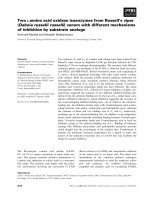
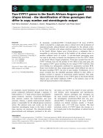
Fig. 1. Malignant fibrous histiocytoma (MFH), subtype osteoclast-like giant cell type (A-E). Osteoclast-like giant cells (arrow head)
resembling normal osteoclast are scattered throughout the lesion, especially around the calcification sites. In addition to these features
,
p
eripheral giant cells (arrow) appeared (A). Immunostaining for vimentin, desmin, α-SMA, and ED1, respectively (B-E). Tumor cells
were positive for vimentin (B), and negative for α-SMA (D) and ED1 (E). Note the strong positive for desmin of smooth muscle cells
of blood vessels vs. negative staining of tumor cells (C). Osteoclast-like giant cells showed positive for ED1 (E). Inflammatory cell typ
e
of MFH (F-H). This type of MHF was characterized by infiltration of various inflammatory cells (F) and ossifying matrix (G). Note th
e
bone matrix surrounding the neoplastic fibroblasts (H). A, F and G: H&E stain, B, C, D and E: ABC method counterstained with
hematoxylin, H: Azan stain. A-E: ×66, F and G: ×132, H: ×33.
composed of spindle-shaped neoplastic cells with a storiform
pattern intermixed with blood vessels and a few inflammatory
cells. Giant cells were scattered throughout the lesion,
especially around the calcification site, and resembled
normal osteoclasts (Fig. 1A). In addition, peripheral giant
cells were seen in association with epitheloid cells, which
is a common feature in giant cell type MFH. Multinucleated
giant cells engulfed calcifying material at the site of
calcification. Areas of necrosis and hemorrhage were seen
in the adjacent fat and muscle tissue. The tumor cells were
positive for vimentin (Fig. 1B), and negative for desmin
(Fig. 1C) and α-SMA (Fig. 1D). The osteoclast-like giant
cells, the majority of giant cells in this case, were positive
for ED1 on immunohistochemical staining (Fig. 1E). The
histologic appearance of this lesion was a salient feature of
the osteoclast-like giant cell type of MFH. The animal was
very young compared to most animals in which such tumors
have been reported.
The tumor consisted of neoplastic histiocytes, hyperplastic
fibroblasts, and an ossifying matrix, including spindle cell
areas showing an occasional storiform pattern in the second
case. The undifferentiated sarcoma was diagnosed as an
inflammatory cell type of MFH because of mixed cellularity
with a heavy infiltration of lymphocytes, plasma cells, and
neutrophils (Fig. 1F). An ossifying matrix was also detected
as the result of continuous and reactive hyperplasia of
fibroblasts (Fig. 1G). At the center of the tumor, areas of
osteocentral fibrous histiocytomas, characterized by bone
matrix formation, were present (Fig. 1H). The neoplasm also
included neovascularization, necrosis of affected muscle
cells, and involvement of the surrounding tissue.
MFH is one of the most common soft tissue sarcomas in
elderly humans and animals in which the cells have
morphologic features of both fibroblasts and histiocytes.
Generally, the storiform-pleomorphic type of MFH exhibits
a cartwheel (storiform) pattern of fibroblast-like cells and
Two different types of malignant fibrous histiocytomas 171
histiocytoid cells. Inflammatory-type tumors showed bizzare
histiocytoid cells concealed by inflammatory cells, including
lymphocytes, plasma cells, eosinophils, and neutrophils.
Tumors of the giant type had multi-nucleated giant cells,
spindle cells, and mononuclear histiocytic cells. Human
MFHs have been classified into 5 subtypes based on the
pattern and predominance of the cell types: storiform-
pleomorphic, inflammatory, giant cell, myxoid, and
angiomatoid, but only the first three types have been
reported in non-human animals [2,5].
The origin of histiocyte-like cells in MFH remains
controversial. In some reports, three possible cells have been
proposed: 1) facultative histiocytic cells that are able to
differentiate into fibroblasts, 2) fibroblasts, and 3) primitive
mesenchymal cells that are able to differentiate into
fibroblasts and histiocytes [1,4]. In most cases of MFH,
immunohistochemical studies have not supported the
hypothesis that these tumor cells are derived from true
histiocytes [8,9]. Several other studies have suggested that
the immunohistologic heterogenity of MFH tumor cells
indicate that these tumors are indeed from a primitive cell
type or are the end result of “differentiation” of several
different types of sarcomas [4,8]. The undifferentiated
mesenchymal cell-origin tumors in which giant cells are
usually present have also been described as fascial sarcomas,
epithelioid sarcomas, malignant histiocytomas, reticulum
cell sarcomas, and giant cell tumors [8].
The specimens of the first case included the osteoclast- like
giant cell type of MFH, a rare case in non-human animals,
with large numbers of osteoclast-like giant cells and
multi-nucleated giant cells engulfing calcified materials.
This tumor had immunoreactivity for vimentin. However,
the tumor showed no immunoreactivity for desmin and α-
SMA. The osteoclast-like giant cell type of MFH rarely
occurs in young animals. In addition, the second case
showed typical findings of the inflammatory cell type of
MFH with an ossifying matrix resulting from continuous
fibroblast reactive hyperplasia and bone matrix surrounding
neoplastic fibroblasts in the central region of the lesion.
The current report may be important in identifying MFHs
based on histologic and immunohistologic findings and
classifying the subtypes of these tumors that have atypical
features occurring in pet dogs.
Acknowledgments
This research was supported by a grant (A081039) from
Health&Medical Technology R&D program, Health &
Korean Industry Development Institute in Korea.
References
1. Carip C, de Beaumont T. Malignant histiocytofibroma of the
small intestine in a young immune deficient patient. Presse
Med 2002, 31, 214-216.
2. Folpe AL, Morris RJ, Weiss SW. Soft tissue giant cell tumor
of low malignant potential: a proposal for the reclassification
of malignant giant cell tumor of soft parts. Mod Pathol 1999,
12, 894-902.
3. Gregory M, Barbara EP, Dennis M, Stephen JW. Small
Animal Clinical Oncology. 3rd ed. pp. 283-287, Saunders,
Pennsylvenia, 2001.
4. Lew W, Lim HS, Kim YC. Cutaneous metastatic malignant
fibrous histiocytoma. J Am Acad Dermatol 2003, 48, S39-40.
5. Morris JS, McInnes EF, Bostock DE, Hoather TM,
Dobson JM. Immunohistochemical and histopathologic
features of 14 malignant fibrous histiocytomas from Flat-
Coated Retrievers. Vet Pathol 2002, 39, 473-479.
6. Orlandi A, Bianchi L, Ferlosio A, Innocenzi I, Spagnoli
LG. The origin of osteoclast-like giant cells in atypical
fibroxanthoma. Histopathology 2003, 42, 407-410.
7. P
érez-Martínez C, García Fernández RA, Reyes Avila LE,
P
érez-Pérez V, González N, García-Iglesias MJ. Malignant
fibrous histiocytoma (giant cell type) associated with a
malignant mixed tumor in the salivary gland of a dog. Vet
Pathol 2000, 37, 350-353.
8. Rebecca B, John HL. Color Atlas of Cytology of the Dog and
Cat. p. 46, Mosby, Missouri, 2000.
9. Kaddu S, McMenamin ME, Fletcher CD. Atypical fibrous
histiocytoma of the skin: clinicopathologic analysis of 59 cases
with evidence of infrequent metastasis. Am J Surg Pathol 2002,
26, 35-46.
10. Wiriosuparto S, Krassilnik N, Gologan A, Cohen JM,
Wenig B. Malignant fibrous histiocytoma, giant cell type, of
the breast mimicking metaplastic carcinoma. A case report.
Acta Cytol 2003, 47, 673-678.