Báo cáo khoa học: "Factors involved in Pinus radiata D. Don. micrografting" pot
Bạn đang xem bản rút gọn của tài liệu. Xem và tải ngay bản đầy đủ của tài liệu tại đây (563.94 KB, 7 trang )
M.F. Fraga et al.Optimisation of Pinus radiata micrografting
Original article
Factors involved in Pinus radiata D. Don. micrografting
Mario F. Fraga
a
*, Maria Jesús Cañal
a,b
, Ana Aragonés
c
, Roberto Rodríguez
a,b
a
Lab. Fisiología Vegetal, Dpto. B.O.S., Facultad de Biología Universidad de Oviedo,
C/ Catedrático Rodrigo Uría s/n, 33071, Oviedo, Spain
b
Instituto de Biotecnología de Asturias (asociado al CSIC), 33071, Oviedo, Spain
c
Instituto Vasco de Investigación y Desarrollo Agrario (Neiker), Arcaute, s/n, Vitoria, Spain
(Received 1 December 2000; accepted 25 September 2001)
Abstract – A series of micrografting conditions using needle fascicles from trees of different ages as scions have been evaluated for
Pinus radiata D. Don. to increase success of in vitro propagation. Micrografting success depended on the quality of the graft process as
well as age, location and development stage of the scion and tree age. 11-month-old scions, taken in January from terminal portions of
basal branches showthe best micrografting-induced response. Responsivenessof scions decreases with thedonor tree age, although this
could be overcome by optimising micrografting conditions.
reinvigoration / micrografting / maturation / vegetative propagation / Pinus radiata / in vitro culture
Résumé – Facteurs impliqués dans le micro-greffage de Pinus radiata D. Don. Différentes conditions de micro-greffage, utilisant
comme greffons des brachyblastes provenant d’arbres d’âges différents, ont été comparées afin d’évaluer les possibilités d’améliorer la
propagation in vitro de Pinus radiata. Le succès du micro-greffage dépend toutautantdelaqualitéduprocessusdegreffagequedel’âge,
de la localisation et du stade de développement du greffon, ou que de l’âge de l’arbre. Des greffons de 11 mois prélevés en janvier sur la
portion terminale de branches de la base de l’arbre donnentles meilleures réponses au micro-greffage. Cette réponse diminue avec l’âge
de l’arbre sur lequel ils sont prélevés, bien que ceci puisse en partie être surmonté en optimisant les conditions du micro-greffage.
vigueur / micro-greffage / maturation / multiplication végétative / Pinus radiata / culture in vitro
Abbreviations
BA: benzyladenine
IBA: indolebutyric acid
MS: Murashige and Skoog culture medium
NAA: naphtalenacetic acid
QL: Quoirin and Lepoivre culture medium
QLP: elongation culture medium
QLS1: stimulation culture medium
QLY: high proliferation culture medium
QL1: proliferation culture medium.
1. INTRODUCTION
Maximizing gains from genetic improvement pro-
grams in forestry requires propagation of genotypes. Un-
fortunately, the maturation and ageing processes which
affect the expression of additive and non-additive desir-
able characteristics, also hinders the exploitation of trees
by traditional methods and biotechnological techniques
Ann. For. Sci. 59 (2002) 155–161 155
© INRA, EDP Sciences, 2002
DOI: 10.1051/forest:2002002
* Correspondence and reprints
Tel. 985104834; Fax. 985104867; e-mail:
since morphogenic competence is generally lost. Practi-
cal benefits from vegetative multiplication are possible
when effective methodologies that allow the multiplica-
tion of mature trees are available.
Mature conifer trees are generally cloned in vivo by
grafting whereas propagation of juvenile individuals is
done via rooted cuttings [1,16]. Unless scionsor cuttings
are taken from very juvenile plants of specific clones, the
explants recovered generally retain undesirable charac-
teristics of the mature state, such as reduced growth and
increased plagiotropism [7]. Traditional methods of veg-
etative propagation have not been very successful in the
Pinaceae, and particularlyin Pinus radiata[18]. The suc-
cess declines during the juvenile-mature phase change.
Reinvigoration of explants from mature selections
that have lost their vegetative propagation ability could
allow in vitro establishment of mature radiata pine. Al-
though in vitro multiplication of radiata pine was previ-
ously reviewed [18], no study of effects of serial
propagation on propagation success and in vitro estab-
lishment of mature radiata pine material through
micrografting has been published, unlike in other
Pinaceae such as larch [5].
Micrografting is used for both practical applications
and basic research [9, 12]. It has becoming an acceptable
methodology for the cloning of several mature species,
as Sequoiadendron giganteum [11], Pinus pinaster [4]
and Pinus nigra [14].
The practical interest of micrografting mature selec-
tions onto juvenile rootstocks arises from the potential of
this technique to facilitate in vitro establishment and,
therefore, cloning of selected mature materials [6, 8].
Although the advantages of this technique are clear,
micrografting is a very complex procedure because dif-
ferent factors contribute to the final success. Manipula-
tion of scions, physiological state and scion age were
studied. This provides a basis for the definition of opti-
mal conditions for micrografting Pinus radiata and so,
for the in vitroestablishmentof selected mature material.
2. MATERIALS AND METHODS
2.1. Plant material
Different genotypes of Pinus radiata D. Don. were
used from the genetic improvement program developed
by the Environmental Research Centre NEIKER
(Vitoria, Spain).
One-year-old (P1) and four-year-old (P4) plants from
controlled pollinated seeds (68 of “Iurre” × 40 of
“Orozko”) were tested as juvenile trees.
Four types of mature trees were used: C1, grafted
from a 30-year-old selected tree (clone 7); C3, three con-
secutive grafts from C1; NF, grafted from a 32-year-old
selected tree (clone 32) and NR, grafted from a 30-year-
old selected tree (clone 45). In all cases, 1-year-old seed-
lings were used as rootstocks. Chronological age of the
treated trees when collection was 8-year-old except C3
that was 3-year-old. Also a series of non-treated trees at
age varying between 15and40-year-old were used (AA).
2.2. Micrografting technique
Micrografts were carried out as indicated (fig-
ures 1a–f) by apical grafting of needle fascicle scions to
microshoot rootstocks. To prepare the scions, the needle
sheath was removed and the needle was cut just above
needle base (figures 1a, b). After 2 slanted cuts of 3 mm
in the basal portion (figure 1c), the scion was inserted in-
side a cut (3 mm) in the apical part of the rootstock (fig-
ures 1d, e). Contact among the surfaces of the rootstock-
scion was assured by elastic silicone rings (figure 1f).
2.3. Rootstocks
Pinus radiata microshoots (25–30 mm length) iso-
lated from in vitro proliferationseries started fromyoung
seedlings were used as rootstock. Multiplication of
microshoots was as previously reported [17].
2.4. Scion collection types and factors analysed
Terminal parts of the shoots were taken from the se-
lected trees, sealed with Parafilm
to avoid drying and
stored at 4 ºC for a maximum of 40 days until tested. Just
prior to sterilisation, needles were removed and the
brachyblasts were kept to avoid dehydration.
Isolated needles prepared as indicated were used as
scions. For theevaluation of the treeage, scions collected
in January from all the selected trees were used.
The evaluation of the scion chronological and physio-
logical age was developed using isolated needles of trees
in three stages of maturation: b1, b11 and b13. The index
156 M.F. Fraga et al.
indicates months of development starting from active
growth (1 month; b1) to mature developed needles
(11 months; b11) and completely mature needles
(13 months; b13).
The effect of the season when tissues are collected
was assayed using as scions needles taken from basal
portions of different aged trees (14–40 years of age, AA)
in summer, autumn, winter and spring.
Tree architecture and branch scionposition were eval-
uated by using b11 scions taken in January from mature
trees. Scions used were selected from basal and apical
levels in the tree. Scions taken from three different
Optimisation of Pinus radiata micrografting 157
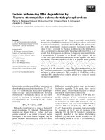
Figure 1. Micrografting technique steps. (a) needle fascicleexcisedfromthemacroblast(seeneedle sheath in the basal portion). (b) nee-
dle without brachyblast (5 × ). (c) needle with two longitudinal cuts (3 ×). (d) cleft of the rootstock (4 × ). (e) scion-rootstock assembly
(4 × ). (f) maintenance of the structure with anelastic silicone ring (4 × ). (g) formation of the scion-rootstockcallus (30 ×). (h) develop-
ment and elongation ofneedles from the axillary budof the scion. (i) mature radiatapine in vitro established after reinvigoration.(j) Ma-
ture radiata pine microshoots.
positions (basal, middle and apical) along the annual
growth of macroblast were also analysed. Needles used
as scionswere collected in different branches in thebasal
portion of the tree.
In order to study the effect of the apical dominance in
the micrografting response of the b11 scions, the termi-
nal bud of basal branches of mature trees (AA) was re-
moved in October1998, and the closedb11 needles to the
end of the branch were collected and micrografted in
February 1999.
2.5. Sterilisation
Scions composed of basal parts of needles containing
an axillary bud (≈ 40 mm) were sterilised by dipping
into 70% ethanol (in sterile conditions) for 30 s. These
were washed with sterile water, dipped into a solution of
Tween 20, 2.5% (v/v) and sodium hypochlorite for
15 min and then washed four times with sterile water.
The b1 explants were sterilised whole, without remov-
ing their bracts. Due to the high sensitivity of the scion
to the sterilisation process, several ranges of sodium
hypochlorite (1, 5, 12.5 and 25 g L
–1
) were tested.
2.6. Culture conditions
In all the cases, the different steps of micrografting
were carried out in sterile tubes (20 × 150 mm), containing
10 ml of culture media, at 25 ± 2
o
C, 70–80 µmol m
–2
s
–1
light intensity and a 16:8 (day/light) photoperiod. The
micrografts were cultured far 10 d in a stimulation cul-
ture medium called QLS1 composed of 1/3 diluted
macroelements of QL medium [15]; microelements; Fe
2+
and vitaminsof MSmedium [13]; 30 g L
–1
sucrose, 0.8%
agar and pH 5.8. In addition, the medium was supple-
mented with 2.69 mm naphtalenacetic acid (NAA) and
22.19 mM benzyladenine (BA). Later, micrografting
systems were transferred to development medium (QLP)
for 30 days. QLP composition was QLS1 but without
phytohormone supplementation.
Proliferation of microshoots was achieved in a QLY,
QL1, QLP sequence culture medium. QL1 was com-
posed of QLS1 salts supplemented with 0.1 mg L
–1
indolebutyric acid (IBA), 0.2 mg L
–1
BA and 3 g L
–1
of
activated charcoal. QLY medium was composed of
QLS1 salts supplemented with 0.1 mg L
–1
IBA and
1mgL
–1
BA.
2.7. Quantification of results
Micrografting response was quantified according to
the following four criteria: (1) establishment (callus for-
mation after 10 days culture) (figure 1g), (2) consolida-
tion, or vascular formation between scion and rootstock
(non-necrotic scions after 30 days culture), (3) develop-
ment (outgrowth after 45 days) (figure 1h) and (4) the
ability to initiate serial culture (figure 1i, j).
2.8. Statistical
Results correspond to 15 micrografts for each treat-
ment. Results were processed with a SPSS
package us-
ing the contingency analysis utility for each qualitative
variable. χ
2
tests (P < 0.05) were performed for each
variable. At a later stage and once the significant differ-
ences between variables were proved, a comparison of
these variables in pairs with the χ
2
test (P < 0.05) was
carried out.
3. RESULTS AND DISCUSSION
Success of micrografting selected P. radiata elite
trees is strongly influenced by the handling procedure
both before, during and after surface sterilisation has
taken place.
To ensure micrografting success the needle sheath
was removed (figure 1b) just prior to surface sterilisa-
tion, and a small piece of brachyblast near the base of the
scion was retained.In addition, aftersurface sterilisation,
basal tissues must be removed. As it was previously re-
ported for Pinus nigra [14], these actions increase scion
viability byeliminating phenol exudation and necrosisof
tissues normallyassociated with sterilising agents. It was
shown that 5 g L
–1
was the optimal sodium hypochlorite
concentration (table I). Other concentrations decreased
scion viability.
158 M.F. Fraga et al.
Table I. Effect of the sodium hypochlorite concentration on the
explant viability (n = 15).
[sodium hypochlorite] (g L
–1
) Contamination (%) Necrosis (%)
1 68±15 29±2
5 18±5 28±6
12.5 20 ± 10 62 ± 12
25 13±7 85±2
In Pinus radiata high concentrations of auxins and
cytokinins were required for early development of the
micrograft in vitro. This differed from Sequoia, in which
exogenous gibberelin and cytokinins do not influence the
reinvigoration effect of the rootstock on the scion [8].
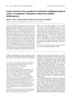
We followed theperformanceof differently agedtrees
(P1 and P4; C3, C1, NF, NR and AA) to ascertain the ef-
fect of maturation on micrograft production (figure 2).
Scions taken from juvenile trees (P1 and P4) easily and
quickly underwent all the micrografting steps. Close to
90% of the scions grew and could then be used for serial
propagation.
At first, few micrografts from scions from adult trees
(C3, C1, NF, NR and AA) reached the goal of elongation
but their progress depended on the morphogenic compe-
tence of the tree (figure 2).
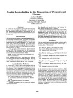
Once the tree age effect was demonstrated, we pro-
ceeded to analyse several factors involved on the suc-
cessful micrograft production. The first one was needle
developmental stage (figure 3). It was observed that b11
needles showed the highest outgrowth and shoot devel-
opment. Needles older than 11 months, collected just be-
fore the spring growth, showed high establishment and
consolidation responses (60–70%) however, no develop-
ment was observed. This shows that inductiveness does
not guarantee further development.
The second factor studied was the seasonal period of
collection. This was of paramount importance for
success in micrografting of mature scions (figure 4). We
verified thatthe winter periodrepresents the timeat which
the scions are most receptive to being micrografted. This
may be the result from the physiological status of the do-
nor plant and hormone levels at the time of excision.
Optimisation of Pinus radiata micrografting 159
Figure 2. Micrografting response of different
aged and reinvigorated state trees (see text for
definition of plant code). Differentletters for the
same variable indicate significant differences
(χ
2
test with P < 0.05).
Figure 3. Micrografting response of 1-month-old (b1), 11-
month-old (b11) and 13-month-old (b13) scions taken from ma-
ture trees (AA). Different letters for the same variable indicate
significant differences (χ
2
test with P < 0.05).
Figure 4. Incidence of time collection onmicrograftingdevelop-
ment of scions taken frommaturetrees.Resultscorrespondtothe
mean value of 15 experiments and its standard deviation.
Figure 5. Micrografting response of scions taken from apical
and basal parts of mature trees. Different letters for the same
variable indicate significant differences (χ
2
test with P < 0.05).
The scions location within the tree can also influence
micrografting. An average of 50% of scion outgrowth
was achieved when needles (b11) were taken from the
basal branches (figure 5) whereas, only 10% was ob-
served when scions were isolated from the apical parts.
Finally, scion location along the annual growth of the
macroblast (figure 6) also affected the micrografting re-
sponse. It was shown that the most reactive scions were
those located at the apical terminal end. A gradual de-
crease on micrografting development was observed as
scion position became moredistantfrom the lateral apex.
Among other factors, the apical dominance [3, 10]
could be the reason of the location-related scion re-
sponse. It was described that the auxin synthesised in the
apical bud inhibits the growth of the axillary buds [2],
and so the location of the scion into the tree becomes de-
cisive for the micrografting success.
Using optimal micrografting conditions, we studied
effect of true age on grafting success (figure 7). In vitro
establishment ability using micrografting depends on the
tree age since outgrowth decreases during ageing. But
the development of the micrografts also depends on a cu-
mulative amount of parameters; among them, ex vitro
graft (C3) further increases the levels reached by the in
vitro technique. Results show a higher ability of NF over
NR to initiateserialcultures, which seems toindicatethat
more than the chronological age, the morphogenic state
of the donor tree is critical for the micrografting-induced
response.
Despite the higher micrografting responses of ex vitro
reinvigorated materials, consecutive grafting is a tedious
and long-time technique, being usually necessary more
than 5 years in order to obtain enough reinvigoration to
allow vegetative propagation. However, there are other
possibilities, which allow the improvement ofthe mature
micrografting response: when the apical bud was
160 M.F. Fraga et al.
Figure 6. Incidence of the lack of close links between the apical
bud and the scion on the micrografting response. Different let-
ters for the same variable indicate significant differences (χ
2
test
with P < 0.05).
Figure 7. Micrografting response and ability to initiate serial cultures of terminal b11 scions taken in January from basal portions of
different aged trees. Different letters for same variable indicate significant differences (χ
2
test with P < 0.05).
Figure 8. Effect of the apical dominance elimination on the
micrografting response of b11 scions taken from mature trees.
Different letters for same variable indicate significant differ-
ences (χ
2
test with P < 0.05).
excised, the needles located just below it showed the
highest development response (figure 8) (80%), as op-
posed to 50% development of controls.
Finally it is important to remark that, as the
micrografting technique allowsthein vitro establishment
of adult trees, the mature in vitro established material
(figure 1j) showed similar growth rates to the juvenile
ones at the end of 6 months (data not presented).
Acknowledgements: We wish to thank the Environ-
mental Research Institute Neiker and specially Dr. E.
Ritter and Dr. S. Espinel in Vitoria (Spain) for supplying
the plant material used in this work. Critical reading is
gratefully acknowledged to Prof. Belén Fernández. This
research and the fellowshipsofM.F.F. were supported by
the UE (CE-96-FAIR-CT-1445).
REFERENCES
[1] Bonga J.M., von Aderkas P., Rejuvenation of tissues
from mature conifers and its implications for propagation in vi-
tro, in: Ahuja M.R., Libby W.J. (Eds.), Clonal Forestry I, Gene-
tics and Biotechnology, Springer-Verlag, Berlin, Heidelberg,
1993, pp. 182–199.
[2] Cline M.G., The role of hormones in apical dominance.
New approaches to an old problem in plant development, Phy-
siol. Plant. 90 (1994) 230–237.
[3] Cline M.G., Concepts and terminology of apical domi-
nance, Am. J. Bot. 84 (1997) 1064–1069.
[4] Dumas E., Franclet A., Monteuuis O., Microgreffe de
méristèmes primaires caulinaires de pins maritimes (Pinus pi-
naster Ait.) âgés sur jeunes semis cultivés in vitro, C.R. Acad.
Sci. 3 (1989) 723–728.
[5] Ewald D., Kretzschmar U., The influence of micrograf-
ting in vitro on tissue culture behavior and vegetative propaga-
tion of old European larch trees, Plant Cell Tissue Organ. Cult.
44 (1996) 249–252.
[6] Franclet A., Rajeunissement par culture in vitro et pra-
tique sylvicole, Bull. Soc.Bot.Fr.,130,Actual.Bot.2(1983) 87.
[7] Gleed J.A., Development of plantings and stecklings of
radiata pine, in: Ahuja M.R., Libbby W.J. (Eds.), Clonal forestry
II, Genetics and Biotechnology, Springer-Verlag, Berlin, Hei-
delberg, 1993, pp. 141–158.
[8] Huang L.C., Lius S., Huang B.L., Murashige T., Mahdi
F.M., van Gundy R., Rejuvenation of Sequoia sempervirens by
repeated grafting of shoot tips onto juvenile rootstocks in vitro,
Plant Physiol. 98 (1992) 166–173.
[9] Jonard R., Micrografting and its applications to tree im-
provement, in: Bajaj Y.P.S. (Ed.), Biotechnology in agriculture
and forestry, Springer-Verlag, Berlin, Heilderberg, New York,
1986, pp. 31–48.
[10] Lang G., Dormancy: a new universal terminology,
HortScience 22 (1990) 817–820.
[11] Monteuuis O., Microgreffage de points végétatifs de
Sequoiadendron giganteum Buchholz séculaires sur de jeunes
semis cultivés in vitro, C. R. Acad. Sci. Par. 302 (1986) 223–225.
[12] Monteuuis O., Effect of technique and darkness on the
success of meristem micrografting of Picea abies, Silvae Genet.
42 (1994) 2–3.
[13] Murashige T., Skoog F., A revised medium for rapid
growth and bioassays with tobacco tissue culture, Physiol. Plant.
15 (1962) 473–497.
[14] Pacheco J., Revigorización de material adulto de Pinus
nigra ARN: criteriosmorfológicos y moleculares, Doctoral The-
sis, Universidad de Oviedo, 1995.
[15] Quoirin M., Lepoivre P., Études de milieux adaptés aux
cultures in vitro de prunes, Acta Hortic. 78 (1977) 437–442.
[16] Rodríguez R., Sánchez-Tamés R., Durzan D.J., Plant
Aging. Basic and Applied Approaches, Plenum Press, New
York, 1990.
[17] Rodríguez R.,Centeno M.L.,Cañal M.J.,Rodríguez A.,
Fernández B., Fraga M.F., Physiological basis of plant ageing.
Problems and solutions for micropropagation of gymnosperms
and angiosperms selected mature trees, in: Espinel S., Ritter E.
(Eds.), Applications of Biotechnology to Forest Genetics, Dipu-
tación Foral de Álava, Vitoria, 2000, pp. 411–424.
[18] Smith D.R., The role of in vitro methods in pine planta-
tion establishment: the lesson from New Zeeland, Plant Tissue
Cult. Biotech. 3 (1997) 63–73.
Optimisation of Pinus radiata micrografting 161