Báo cáo y học: "Familial Polycythemia Caused by a Novel Mutation in the Beta Globin Gene: Essential Role of P50 in Evaluation of Familial Polycythemi" potx
Bạn đang xem bản rút gọn của tài liệu. Xem và tải ngay bản đầy đủ của tài liệu tại đây (882.19 KB, 5 trang )
Int. J. Med. Sci. 2007, 4
232
International Journal of Medical Sciences
ISSN 1449-1907 www.medsci.org 2007 4(4):232-236
©Ivyspring International Publisher. All rights reserved
Short Research Communication
Familial Polycythemia Caused by a Novel Mutation in the Beta Globin Gene:
Essential Role of P50 in Evaluation of Familial Polycythemia
Neeraj Agarwal
1
, Mariluz P. Mojica-Henshaw
2
, Elizabeth. D. Simmons
3
, Dottie Hussey
4
, Ching N. Ou
5
,
Josef T. Prchal
6
1. Department of Internal Medicine, University of Utah, Salt Lake City, Utah, USA
2. Department of Internal Medicine, University of Utah, Salt Lake City, Utah, USA
3. Department of Hematology and Oncology, Kaiser Permanente, Southern California, USA
4. ARUP Laboratories, Salt Lake City, Utah, USA
5. Department of Pathology, Texas Children's Hospital. Houston, Texas, USA
6. Department of Internal Medicine, University of Utah, Salt Lake City, Utah & ARUP Laboratories, Salt Lake City, Utah,
USA
Correspondence to: Josef T. Prchal, MD. 5C 310, SOM, 50 North Medical Drive, Salt Lake City, Utah, 84132.
Received: 2007.08.03; Accepted: 2007.10.03; Published: 2007.10.04
Two polycythemic subjects from a family with multiple polycythemic subjects were evaluated. Estimation of
oxygen affinity of Hb from venous blood gas parameters (P50) revealed low P50 suggesting a high affinity Hb
variant. Further work up, which included beta globin gene sequencing, revealed a novel mutation changing a
codon to the previously reported high affinity Hb - Hb Johnstown (beta109 Val->Leu). Polycythemic subjects
with high affinity Hb variant are asymptomatic with normal life expectancy. Their differentiation from
polycythemia vera (PV) is crucial to avoid therapy which is otherwise reserved for PV patients. We provide an
electronic version (in Microsoft excel program) of a previously reported mathematical formula for rapid
calculation of P50 from venous blood gases. Estimation of P50 is an essential initial step in the evaluation of a
subject with personal and family history of polycythemia.
Key words: High affinity Hb, Polycythemia, Estimation of P50
1. Background
Polycythemia means many cells in the blood.
Polycythemia is clinically defined as elevated red
blood cell mass ( hemoglobin > 18.5 g/dL in men, 16.5
g/dL in women or other evidence of increased red cell
volume or
hemoglobin or hematocrit greater than 99th
percentile of method-specific reference range for age,
sex, altitude of residence or
hemoglobin greater than
17 g/dL in men, 15 g/dL in women if associated with a
documented and sustained increase of at least 2 g/dL
from an individual's baseline value that can not be
attributed to correction of iron deficiency, or
elevated
red cell mass greater than 25% above mean normal
predicted value [1].
Polycythemia can be further classified as primary
polycythemia, secondary polycythemia, or
polycythemia due to abnormal hypoxia sensing.
Primary polycythemias are caused by intrinsic
defects in the erythroid precursors that result in hyper
responsiveness to normal level of serum
erythropoietin (Epo). Secondary polycythemias are
driven by the factors (predominantly Epo but also
insulin growth factor 1 and cobalt) extrinsic to the
erythroid progenitor cells. Generally, in secondary
polycythemia, the increased red cell mass represents a
physiologic response to tissue hypoxia or abnormally
increased level of serum Epo [2].
Polycythemias due to abnormal hypoxia sensing
include Chuvash polycythemia, polycythemias
associated with von Hippel-Lindau mutations other
than the Chuvash polycythemia mutation, and
polycythemia due to proline hydroxylase mutation [3,
4]. Acquired conditions that lead to increased Epo
production, such as chronic hypoxia and a variety of
tumors, are the most common causes of secondary
polycythemias.
Secondary congenital polycythemia results from
inherited conditions that lead to increased Epo levels.
These include hemoglobin variants with high affinity
for oxygen, congenitally low erythrocyte 2, 3
biphosphoglycerate levels, and inherited
methemoglobinemias. All these conditions are
characterized by a left shift in Hb dissociation curve
which in turn leads to tissue hypoxia and a
physiologically appropriate increase in Epo levels.
Congenital cyanotic heart or lung disorders, which
lead to tissue hypoxia and increased Epo level are also
examples of secondary congenital polycythemias but
these are characterized by a normal Hb dissociation
curve.
2. Methods
Two subjects (mother and daughter), from a
family with multiple subjects with polycythemia, were
Int. J. Med. Sci. 2007, 4
233
available for evaluation:
Subject 1(mother): She is a 59 year old Caucasian
woman who presented with lifelong history of
polycythemia. Her medical history was remarkable for
1) multifocal ductal carcinoma in situ (DCIS) of the
breast in 2002, treated with simple mastectomy and 2)
colonic diverticula and internal hemorrhoids since
2005. Family history was remarkable for presence of
lifelong history of polycythemia in her mother and two
daughters. Physical examination was normal (no
hepatosplenomegaly). Laboratory parameters revealed
high Hb (16 gm%) , high hematocrit (48%), normal
MCV, normal platelet and white blood cell counts,
normal arterial oxygen saturation, and normal liver
function tests.
Subject 2 (daughter): She is a 30 year old Caucasian
woman who presented for further evaluation of
lifelong history of polycythemia. She had mild fatigue
but no other symptoms. Past medical history was
unremarkable. Family history was significant for
lifelong history of polycythemia in her mother,
maternal grandmother and her younger sister.
Physical examination was unremarkable. Laboratory
parameters were remarkable for elevated hemoglobin
(17.2 gm%) and hematocrit (51.4%) but normal MCV,
normal platelets and white blood cell counts, normal
arterial oxygen saturation, and normal liver function
tests.
Both subjects underwent following laboratory
tests: Serum Epo levels were normal in both subjects
(15 mIU/ml and 19 mIU/ml respectively, normal
range 4.1 - 19.5 mIU/ml). Venous blood gas
parameters were obtained which included partial
pressure of oxygen (venous pO2), venous pH and
venous oxygen saturation. Calculation of affinity of Hb
for oxygen (P50) was done using the mathematical
formula as described [5]. Routine Hb electrophoreses,
high performance liquid chromatography (HPLC) [6,
7], isoelectric focusing (IEF) in polyacrylamide gel,
globin chain analysis [8] and peptide mapping [9] were
performed. This was followed by beta globin gene
sequencing which was performed by ABI3730
96-capillary sequencer at the DNA Sequencing Core
Facility at the University of Utah School of Medicine
(primers and conditions available upon request).
3. Results and Discussion
The P50 was found to be low at 18 mm Hg
(normal range 22.6 to 29.4) suggesting increased
affinity of Hb for oxygen. Routine Hb electrophoresis
and HPLC failed to detect mutant hemoglobin. IEF
showed a band anodal to Hb A and globin chain
analysis by HPLC revealed an unidentified beta globin
variant in both subjects (Figure 1). Peptide mapping
showed an extra peak at 26.9 min but showed no
decrease in any peaks suggesting a mutation
somewhere in the core (Figure 2). Beta globin gene
sequencing revealed a novel mutation (G
TG->TTG ) of
codon 109 of exon 3 of beta globin gene. This mutation
leads to a previously reported high affinity Hb variant
known as Hb Johnstown (beta109 Val->Leu) [10-12];
however, this nucleotide change is novel and
previously unreported; and it leads to a previously
described amino acid substitution that was however,
caused by a different nucleotide missense mutation.
Hb Johnstown (beta109 (G11) Val->Leu) is a high
oxygen affinity Hb variant and there are three reports
in the literature. It was first reported by Jones and
colleagues in Oregon, in 1990, in a healthy
asymptomatic subject with mild erythrocytosis and
left-shifted hemoglobin-O2 dissociation curve[10]. As
with many other Hb variants, Hb Johnstown is silent
on standard hemoglobin electrophoretic analyses, and
was identified and isolated by reverse-phase HPLC of
individual globin chains. Structural analysis revealed
the substitution beta 109 (G11) Val ->Leu [10]. In 2000,
the underlying beta globin mutation [beta-globin
codon 109 (G
TG ->CTG )] was first reported by Ropero
and colleagues, in two unrelated families (total four
subjects) of Basque extraction in Spain [11]. In one of
these families, Hb Johnstown mutation was present in
double heterozygosity with another beta 0 thalassemia
mutation IVS-1-nt1 (G->A). In 2004, Feliu-Torres and
colleagues reported Hb Johnstown [beta-globin codon
109 (G
TG ->CTG )] in an eight year old girl, who had
been referred for evaluation of erythrocytosis, in
double heterozygosity with another beta globin
mutation [IVS-I-1(G->A)]. Her asymptomatic mother
was found to be heterozygous for Hb Johnstown
mutation [12]. However, these reports described a
causative G to C mutation that is different that
mutation we describe in our subject with Hb
Johnstown, namely G to T, both encoding the same
amino acid; i.e. leucine and present in subjects not
known to be of Spanish/Basque extraction.
Oxygenation and deoxygenation of hemoglobin
occur at the heme iron. The sigmoid shape of
Hb-oxygen dissociation curve is indicative of
cooperative interaction between heme and oxygen.
Oxygen affinity and Hb-oxygen dissociation is affected
by blood pH, 2, 3- biphosphoglycerate (2, 3 BPG) level
in the red cell and temperature, and globin structure
[13].
Affinity of Hb with oxygen is expressed as the
P50, which is the partial pressure of oxygen in blood at
which 50% of the Hb is saturated with oxygen. The
venous P50 can be measured directly using a
cooximeter which is no longer easily available in
routine and even reference laboratories. Lichtman and
colleagues have reported a mathematical formula
which can be used to calculate P50 reliably [5].
Calculating P50 using this formula requires the
following venous gas parameters: partial pressure of
oxygen (venous pO2), venous pH and venous oxygen
saturation, and uses anti-log mathematical function
that many clinicians find difficult to use for calculation
[5]. The P50 of a healthy person with normal Hb is 26 ±
1.3 mm Hg. The 99% confidence interval for individual
observations has been reported to be 22.6 to 29.4 mm
Hg. An abnormally low P50 reflects an increased
affinity of hemoglobin for oxygen and vice versa.
Elevations and reductions in 2, 3- BPG level in the
Int. J. Med. Sci. 2007, 4
234
erythrocyte will also lead to corresponding changes in
P50 values; however, in only reported subjects this
decrease was limited to a P50 value between 20 and 35
mm Hg. There should be high suspicion for the
presence of a high affinity Hb variant if P50 value is
<20 mm Hg [5].
During oxygenation and deoxygenation, there is
considerable movement along the interface of alpha 1
and beta 2 chains of the Hb tetramer. Several
hemoglobin variants have substitutions affecting this
interface. All these substitutions can affect the
cooperative nature of oxygen binding with heme, and
in turn, can change the affinity of Hb for oxygen. The
majority of mutations affecting oxygen affinity result
in high affinity Hb variants which result in leftward
shift of the dissociation curve and relative tissue
hypoxia [14]. There are 90 high affinity Hb variants,
listed on the globin server, known to be associated
with high affinity for oxygen
(
accessed on May 04, 2007) [15]. All these Hb variants
are inherited in an autosomal dominant manner. High
affinity Hb variants release oxygen in the tissue
relatively slowly and create relative tissue hypoxia.
This leads to increased production of Epo from
kidneys which results in increased red blood cell mass
and polycythemia. At an elevated level of increased
red blood cell mass (which depends upon the oxygen
affinity of a given variant) adequate oxygenation of the
tissue is reestablished and Epo production plateaus
and at this new steady state serum Epo is often at
normal level. This leads to stabilization of Hb level
after achieving a certain elevated level of hematocrit.
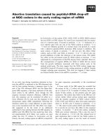
Figure 1. HPLC globin chain separation. Y axis denotes relative mass, X axis denotes the retention time in minutes. The unidentified
beta globin variant is shown as beta X.
Int. J. Med. Sci. 2007, 4
235
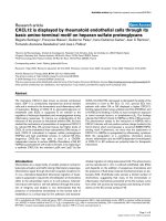
Figure 2. HPLC peptide mapping. Y axis denotes relative mass, X axis denotes the retention time in minutes. An extra peak at 26.9
min (X) was seen without decrease in any other peaks suggesting a mutation somewhere in the core.
4. Conclusion
Family history of polycythemia in a subject with
polycythemia should raise the suspicion for the
presence of a high affinity Hb variant [14]. A low P50
value (obtained from venous gas parameters) is
supportive of high oxygen affinity Hb variant or
decreased 2, 3 BPG level. In a polycythemic patient,
establishing a correct diagnosis of a high affinity Hb
variant is important as these patients have normal life
expectancy and do not require phlebotomy. The
therapies used for polycythemia vera such as
phlebotomy and chemotherapy should not be used in
patients who have polycythemia due to high affinity
Hb variants. With this report, we are providing an
electronic version (using Microsoft Excel program) of
the mathematical formula [5], with which P50 can be
calculated in few seconds, provided venous gas
parameters are available, without necessity of more
sophisticated calculations using antilog parameters.
With increased ease and rapidity of calculation using
our Excel program (Supplementary Material), we hope
that use of P50 will increase, leading to improved
detection of Hb variants in subjects with familial
polycythemia.
Supplementary Material
Excel program [
Acknowledgement
Ken J. Bulpitt, M.D, developed the electronic
program in Microsoft Excel program (Supplementary
Material) from the previous reported mathematical
formula for the calculation of P50. This work was
supported in part by National Institutes of Health
research Grants R01HL5007-09 (J.T.P.).
Conflict of interest
Financial disclosure: Consultant: Josef T. Prchal
for Astra Zeneca, Amgen; Honoraria: Josef T. Prchal
for Astra Zeneca, Amgen. The authors declared that no
other conflict of interest exists.
References
1. Tefferi A, Thiele J, Orazi A, et al. Proposals and rationale for
revision of the World Health Organization diagnostic criteria for
polycythemia vera, essential thrombocythemia, and primary
myelofibrosis: recommendations from an ad hoc international
expert panel. Blood 2007, 110:1092-1097.
2. Prchal J. Primary polycythemias. Curr Opinion Hematol 1995,
2:146–152.
3. Gordeuk V, Stockton DW, Prchal JT. Congenital polycythemias/
erythrocytoses. Haematologica 2005, 90:109-116.
4. Ang S, Chen H, Hirota K, Gordeuk VR, Jelinek J, Guan Y, Liu E,
Sergueeva AI, Miasnikova GY, Mole D, Maxwell PH, Stockton
DW, Semenza GL, Prchal JT. Disruption of oxygen homeostasis
underlies congenital Chuvash polycythemia. Nat Genet 2002,
32:614-621.
5. Lichtman M, Murphy MS, Adamson JW. Detection of mutant
hemoglobins with altered affinity for oxygen. A simplified
technique. Ann Intern Med 1976, 84:517–520.
6. Ou C, Buffone GJ, Reimer GL, Alpert AJ. High-performance
liquid chromatography of human hemoglobins on a new cation
exchanger. J Chromatogr 1983, 266:197-205.
Int. J. Med. Sci. 2007, 4
236
7. Ou C, Rognerud CL. Rapid analysis of Hb variants using a
cation-exchange HPLC method. Clin Chem 1993, 39:820-824.
8. Shelton JB, Shelton JR, Schroeder WA. High performance liquid
chromatographic separation of globin chains on a large-pore C4
column. J Liquid Chromatogr 1984;7:1969-77.
9. Schroeder W, Shelton JB, Shelton JR, Powars D. Separation of
peptides by high-pressure liquid chromatography for the
identification of a hemoglobin variant. J Chromatogr 1979,
174:385-392.
10. Jones R, Saiontz HI, Head C, Shih DT, Fairbanks VF. Hb
Johnstown [beta 109 (G11) Val Leu]: a new electrophoretically
silent variant that causes erythrocytosis.1. Hemoglobin 1990,
14(2):147-156.
11. Ropero P, Villegas A, Gonzalez AF, Anguita E, Sanchez J,
Carreno DL, Arrizabalaga B, Atuxta L. Hb Johnstown [beta 109
(G11) Val >Leu]: second case described and associated for the
first time with beta(0)-thalassemia in two Spanish families. Am J
Hematol 2000, 65(4):298-301.
12. Feliu-Torres A, Eberle SE, Roldan A, Gonzalez S, Sciuccati G. Hb
Johnstown [beta109(G11)Val >Leu]: A high oxygen affinity
variant associated with beta0-thalassemia. Hemoglobin 2004,
28(4):335-338.
13. Bunn H, Forget BG. Hemoglobin: Molecular, Genetic and
Clinical Aspects. Philadelphia: WB Saunders. 1986.
14. Wajcman H, Galacteros F. Hemoglobins with high oxygen
affinity leading to erythrocytosis. New variants and new
concepts. Hemoglobin 2005, 29:91-106.
15. Giardine B, van Baal S, Kaimakis P, Riemer C, Miller W, Samara
M, Kollia P, Anagnou NP, Chui DH, Wajcman H, Hardison RC,
Patrinos GP. HbVar database of human hemoglobin variants
and thalassemia mutations: 2007 update. Hum Mutat 2007,
28:206.