Báo cáo sinh học: " Severe osteomyelitis caused by Myceliophthora thermophila after a pitchfork injury'''' pot
Bạn đang xem bản rút gọn của tài liệu. Xem và tải ngay bản đầy đủ của tài liệu tại đây (349.35 KB, 5 trang )
BioMed Central
Page 1 of 5
(page number not for citation purposes)
Annals of Clinical Microbiology and
Antimicrobials
Open Access
Case report
Severe osteomyelitis caused by Myceliophthora thermophila after a
pitchfork injury
Lauren Destino
1
, Deanna A Sutton
2
, Anna L Helon
3
, Peter L Havens
1
,
John G Thometz
4
, Rodney E Willoughby Jr
1
and Michael J Chusid*
1
Address:
1
Department of Pediatrics, Medical College of Wisconsin and Children's Hospital of Wisconsin, USA,
2
Fungus Testing Laboratory,
Department of Pathology, University of Texas Health Sciences Center, San Antonio, TX, USA,
3
Pharmacy Department, Children's Hospital of
Wisconsin, USA and
4
Department of Orthopedic Surgery, Medical College of Wisconsin and Children's Hospital of Wisconsin, USA
Email: Lauren Destino - ; Deanna A Sutton - ; Anna L Helon - ;
Peter L Havens - ; John G Thometz - ; Rodney E Willoughby - ;
Michael J Chusid* -
* Corresponding author
Abstract
Background: Traumatic injuries occurring in agricultural settings are often associated with
infections caused by unusual organisms. Such agents may be difficult to isolate, identify, and treat
effectively.
Case report: A 4-year-old boy developed an extensive infection of his knee and distal femur
following a barnyard pitchfork injury. Ultimately the primary infecting agent was determined to be
Myceliophthora thermophila
, a thermophilic melanized hyphomycete, rarely associated with human
infection, found in animal excreta. Because of resistance to standard antifungal agents including
amphotericin B and caspofungin, therapy was instituted with a prolonged course of terbinafine and
voriconazole. Voriconazole blood levels demonstrated that the patient required a drug dosage
(13.4 mg/kg) several fold greater than that recommended for adults in order to attain therapeutic
blood levels.
Conclusion: Unusual pathogens should be sought following traumatic farm injuries.
Pharmacokinetic studies may be of critical importance when utilizing antifungal therapy with agents
for which little information exists regarding drug metabolism in children.
Myceliophthora thermophila
is a thermophilic phaeoid
mould found in pasture soil, wood chips, straw, mouldy
hay, compost piles and other environmental settings
where heat is generated. It is also found in the excreta and
rumen of cattle and is a pathogen of cultivated mush-
rooms [1]. A rare cause of invasive human infections, it
can be difficult to isolate and identify in clinical speci-
mens. We recently cared for a 4-1/2 year old boy who
developed osteomyelitis of the distal femur caused by
direct inoculation of Myceliophthora thermophila
via a
pitchfork injury to his knee. The patient demonstrated
severe destructive osseous and cartilaginous infection,
with slow clinical improvement, requiring the prolonged
use of multiple antifungal agents. Due to the limited
number of agents to which this organism was susceptible,
voriconazole therapy was instituted despite limited phar-
Published: 08 September 2006
Annals of Clinical Microbiology and Antimicrobials 2006, 5:21 doi:10.1186/1476-0711-5-
21
Received: 26 June 2006
Accepted: 08 September 2006
This article is available from: />© 2006 Destino et al; licensee BioMed Central Ltd.
This is an Open Access article distributed under the terms of the Creative Commons Attribution License ( />),
which permits unrestricted use, distribution, and reproduction in any medium, provided the original work is properly cited.
Annals of Clinical Microbiology and Antimicrobials 2006, 5:21 />Page 2 of 5
(page number not for citation purposes)
macokinetic data in children. Prolonged therapy with ter-
binafine, a drug generally employed for superficial
saprophytic infections of skin and nails also was utilized.
This case demonstrates the difficulties that can be encoun-
tered in identifying and treating this unusual but aggres-
sive fungal organism.
Case report
A 4-1/2 year old boy presented with a swollen right knee
after being impaled in that area by a pitchfork. The pitch-
fork was observed to be contaminated with cow manure
and hay. The knee was washed with soap and water. The
following morning the knee was swollen and the boy was
treated with orally administered antibiotics.
The mobility of the knee progressively decreased, and four
days later the child was admitted to the hospital. Bacterial
cultures of joint fluid yielded Bacillus
and Enterococcus
species. After a brief course of intravenous antibiotic ther-
apy, the boy was discharged to continue orally adminis-
tered antibiotics. At home, he developed increasing knee
pain with inability to walk. A magnetic resonance image
(MRI) obtained at transfer to our institution was consist-
ent with infection involving the synovium, the medial
femoral condyle and adjacent articular cartilage. Intrave-
nous antibacterial therapy was instituted with vancomy-
cin, piperacillin-tazobactam and amikacin.
Twenty seven days after the initial pitchfork injury, the
patient was returned to the operating room because of
persistent leg and knee swelling as well as increasing ele-
vation of inflammatory markers with an ESR of >100 and
a CRP of 6.5. An MRI revealed apparent osteomyelitis of
the medial femoral condyle. New bone cultures were
obtained which grew what was initially identified as a der-
matophyte. Orally administered terbinafine, 125 mg (6.7
mg/kg) daily, was initiated, and the patient began to
improve clinically. His CRP declined to a nadir of 1.8 with
absence of fever and better movement of his leg. However,
45 days following the initial injury and 14 days after
wound closure, an elevation in the CRP to 2.6, as well as
an increase of purulent drainage from the knee prompted
another surgical exploration of the distal femur and the
addition of intravenous Ambisome, 90 mg (4.8 mg/kg)
daily. At surgery, progressive bone loss was noted as well
as necrosis of knee cartilage. Fungal organisms with irreg-
ular branching hyphae were noted throughout the excised
cartilage, and fungus was recovered in culture two weeks
later.
Meanwhile, the fungal agent that had been isolated previ-
ously was forwarded to the Fungus Testing Laboratory at
the University of Texas Health Science Center in San Anto-
nio for identification and susceptibility testing and acces-
sioned into their stock collection as UTHSC 05-3365.
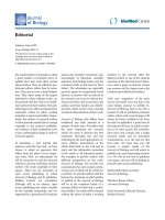
There, the organism was identified as Myceliophthora
thermophila, based upon observation of: 1) tan to brown
powdery colonies with ill-defined margins when grown
on potato flakes agar at 42°C; 2) more luxuriant growth
at elevated temperatures of 35°C and 42°C than at 26°C;
3) septate vegetative hyphae with conidial production
from ampulliform swellings; and 4) obovoid (inverted
egg shaped) or pyriform (pear-shaped) conidia measuring
4.5–11.0 × 3.0–4.5 µm that were hyaline and smooth
when immature, becoming darker and roughened at
maturity (Figure 1). Antifungal susceptibility testing was
performed according to the Clinical Laboratory Standards
Institute (CLSI) M38-A document for filamentous fungi
[2]. Although standardized susceptibility breakpoints
have not been established for this organism, the isolate
appeared resistant to a variety of standard antifungal
agents (Table 1). Based upon these susceptibilities, vorico-
nazole (4 mg/kg/12 hrs after a loading dose of 6 mg/kg/
12 hrs for 24 hours) was added to the patient's antifungal
regimen, and Ambisome was discontinued. Subsequently,
the patient underwent numerous debridement proce-
dures with gradual improvement in clinical picture and
CRP. A series of progressive increases in voriconazole dos-
age was required based upon periodic pharmacokinetic
studies to achieve appropriate blood levels (Table 2).
Conidia of Myceliophthora thermophila being produced from ampulliform swellingsFigure 1
Conidia of Myceliophthora thermophila being produced from
ampulliform swellings. Note both smooth, hyaline, immature
conidia, and darker, more mature roughened conidia. Lacto-
fuschin stain, approximately 1000×.
Annals of Clinical Microbiology and Antimicrobials 2006, 5:21 />Page 3 of 5
(page number not for citation purposes)
After his CRP had normalized, the patient was discharged
home receiving voriconazole, 250 mg (13.4 mg/kg)
orally, every 12 hours, and terbinafine 125 mg (6.7 mg/
kg) orally once daily. Anti-fungal therapy is to be contin-
ued for a total of one year. The leg wound has healed, but
the patient has had significant bone loss in his distal
femur with involvement of the growth plate, as well as
damage to the articular cartilage of the knee.
Discussion
Myceliophthora thermophila is a melanized filamentous
hyphomycete that initially grows as a white cottony col-
ony and subsequently turns pale brown and becomes
granular on a variety of media recommended for mould
identification, such as potato dextrose or 2% malt agar. Its
optimal growth is at 30–36°C. However, it also grows
well at 42°C, with maximal growth near 50°C. Thus it is
considered a thermophilic organism. Myceliophthora
thermophila is found in dry pasture soil, birch chips,
wood pulp, and straw compost [1]. Its cell wall contains
melanin resulting in dark pigmentation, and it is consid-
ered one of the etiologic agents of phaeohyphomycosis.
Phaeohyphomycosis includes those conditions in which
the pathogenic mould forms fungal elements which con-
tain melanin within their cell walls [3]. Despite the pres-
ence of melanin, cell walls of phaeoid moulds may appear
hyaline or clear upon routine microscopy. Hyphal ele-
ments usually demonstrate pigment when stained with
Masson-Fontana melanin stain, allowing identification of
a dark fungus [4].
A recent review reports that the number of publications
related to phaeohyphomycotic infections in the 1990's
numbered only 150 [5]. Phaeohyphomycosis most com-
monly manifests as a cutaneous infection, but deep infec-
tions with invasion of the sinuses, lungs, brain, blood,
and bone have also been reported [5]. Disseminated dis-
ease was reviewed by Revankar et al. who found 72 cases
reported between 1966 and 2001 [6]. Notably, the major-
ity of cases involving disseminated phaeohyphomycosis
were in immune-compromised patients. The mortality
rate in these individuals was high and many isolates were
resistant to amphotericin B. In immunocompetent
patients, most infections were associated with direct inoc-
ulation of the organism from an environmentally con-
taminated source.
There are just three previously reported cases of phaeohy-
phomycosis caused by Myceliophthora thermophila
(Table 3). In the first two patients, the source of the Myc-
eliophthora thermophila was uncertain, and both
patients died despite standard anti-fungal therapy with
amphotericin B. The most recently reported case of infec-
tion with this organism involved a 21-month-old boy
who sustained a penetrating head injury. A brain abscess
developed from which both Clostridium perfringens
and
Myceliophthora thermophila
were isolated. The patient
was treated successfully with en
bloc resection of the
lesion, six weeks of amphotericin B, and four months of
itraconazole [9].
Despite convincing evidence of progressive infection in
each of the three previously reported cases, it was not until
well into the clinical course or even after death that iden-
tification of the etiologic agent was confirmed. It is
unknown why recovery of Myceliophthora thermophila
from clinical specimens is so difficult. However, the situa-
tion may be analogous to mycotic infections with more
Table 1: Susceptibilities of Myceliophthora thermophila isolate
Drug MIC
Amphotericin B 2 resistant*
Fluconazole 8 resistant
Itraconazole 0.125 susceptible
Voriconazole 0.06 susceptible
Caspofungin 4 resistant
Terbinafine 1 susceptible
Griseofluvin >16 resistant
*There are no published breakpoints for this organism against any of
the antifungal agents tested. Interpretations are based upon normally
achievable concentrations of the drug using standard dosing regimens.
Table 2: Voriconazole plasma concentrations (body weight 18.6 kg)*
Voriconazole Therapy Day Dose (mg) given every 12 hr Dose (mg/kg) Doses prior to kinetics Peak (mcg/ml) Trough (mcg/ml)
6 75 IV 4 10 0.94
a
< 0.2
14 108 IV 5.8 8 0.6 < 0.2
24 175 IV 9.4 6 3.04 < 0.2
34 250 PO 13.4 8 2.8
b
0.3
43 250 PO 13.4 26 5.35
c
0.3
57 250 PO 13.4 54 2.12
b
0.2
*Patient also receiving terbinafine 6.7 mg/kg/day
a
IV peak @ 50 minutes post infusion
b
PO peak @ 2–3 hours post ingestion
c
PO peak @ <2 hours post ingestion
Annals of Clinical Microbiology and Antimicrobials 2006, 5:21 />Page 4 of 5
(page number not for citation purposes)
common agents such as Aspergillus, in which microbio-
logic isolation of the etiologic agent from grossly infected
tissue can be difficult. The identification of Mycelioph-
thorathermophila, once recovered, is also problematic, as
most microbiology laboratories lack experience with this
organism.
In our patient, despite evidence of ongoing infection,
both operatively and preoperatively, only two samples of
debrided bone or cartilage yielded Myceliophthora ther-
mophila, despite numerous cultures of infected surgical
specimens in which fungal elements could be seen histo-
logically. Given the thermophilic nature of this organism,
incubation of inoculated media at elevated temperatures
may enhance recovery. Cultures of most clinical speci-
mens are incubated at 30°C, but such a temperature
would be less than optimal for growth of Myceliophthora
thermophila.
Because our patient's positive cultures was obtained after
approximately 2 weeks of terbinafine therapy and the
organism was found to be resistant to amphotericin B,
voriconazole was added to terbinafine therapy. Terbin-
afine is a broad-spectrum allylamine with fungicidal activ-
ity against dermatophyte species, Aspergillus
species,
Sporothrix schenckii
, Blastomyces dermatitidis, Histo-
plasma capsulatum, Cryptococcus neoformans, Malas-
sezia furfur and other important fungi. It shows in vitro
synergism with amphotericin or triazoles and has been
effective in combination therapy in individual patients
[10-14]. It has been administered safely in a large number
of children, and at high doses or for up to 12 months for
invasive mycoses [15].
Voriconazole is a potent antifungal agent effective against
a number of pathogens, including Aspergillus
, Cryptococ-
cus, and Candida species. It also has excellent oral bioa-
vailability and a low rate of adverse effects [16, 17].
However, it is not approved by the Food and Drug Admin-
istration for use in children, and the appropriate dose for
pediatric patients is not known. Recommendations in
authoritative sources suggest the same intravenous
weight-based dosages in children and adults: a loading
dose of 6 mg/kg/dose every 12 hours × 1 day and a main-
tenance dose of 4 mg/kg/dose every 12 hours. Oral dosage
is suggested at 100 mg every 12 hours for patients less
than 40 kg, and 200 mg every 12 hours for patients more
than 40 kg [18].
Recent investigations by Walsh et al. demonstrated that
pediatric patients have a much higher rate of elimination
of voriconazole per unit of body weight than do adults.
Thus, children may require higher dosages to achieve
blood levels consistent with adults treated at a dosage of
3–4 mg/kg [16, 17]. Additionally, the elimination of vor-
iconazole from the blood in children appears linear when
doses of 3 mg/kg to 5 mg/kg are administered every 12
hours. This is in distinction to elimination of similar
doses in adults, which is non-linear or saturable.
Although the exact relationship between the plasma con-
centration of voriconazole and the drug's clinical effec-
tiveness is uncertain, infected adults improve at doses
achieving recommended plasma concentrations. There-
fore, the goal in our patient was to achieve voriconazole
blood levels similar to those achieved in adults. Assuming
linear pharmacokinetics, it was suggested by Walsh that a
pediatric dosage of up to 11 mg/kg twice a day might be
necessary to achieve drug levels equivalent to those seen
in adult patients receiving 4 mg/kg of the agent every 12
hours [17]. We increased the dose of voriconazole in our
patient, in stepwise fashion based upon measurements of
C
max
, from 4 mg/kg to 13.4 mg/kg every 12 hours, reach-
ing a voriconazole serum peak levels in the range of 2–5
mcg/ml, equivalent to the typical adult given only 3–4
mg/kg every 12 hours. Kinetics of voriconazole in our
patient were potentially affected by concurrent adminis-
tration of terbinafine.
The current case demonstrates the vigilance required in
patients with traumatically induced osteomyelitis, partic-
ularly when related to direct implantation from a grossly
contaminated source. Relapse of apparently appropriately
treated infection while on therapy demands reassessment
to be certain that an unusual or emergent microorganism
is not present within the depths of the wound. In the case
of a contaminated farm implement, the possibility of
recovery of an unusual agent like Myceliophthora ther-
mophila is high, potentially requiring the usage of antimi-
crobial agents for which there is scant pharmacologic data
in children. Clinicians should be careful to obtain appro-
priate pharmacologic studies in order to be assured that
the appropriate doses of such drugs are employed.
Table 3: Prior Case Reports of Myceliophthora thermophila infection
Age, Sex History Sites of Infection Therapy Outcome
7 years, M
7
AML with neutropenia Blood, lungs, heart Amphotericin B Death
22 years, F
8
Status post Cardiovascular Surgery Blood, aorta, heart Amphotericin B 5-fluorocytosine Death
21 months, M
9
Penetrating head injury from a rusty nail in a barnyard. Brain
abscess with M.
thermophilia and Clostridium perfringens
Brain abscess Amphotericin B Itraconazole Survived
Publish with BioMed Central and every
scientist can read your work free of charge
"BioMed Central will be the most significant development for
disseminating the results of biomedical research in our lifetime."
Sir Paul Nurse, Cancer Research UK
Your research papers will be:
available free of charge to the entire biomedical community
peer reviewed and published immediately upon acceptance
cited in PubMed and archived on PubMed Central
yours — you keep the copyright
Submit your manuscript here:
/>BioMedcentral
Annals of Clinical Microbiology and Antimicrobials 2006, 5:21 />Page 5 of 5
(page number not for citation purposes)
Authors' contributions
LD conceived the report and helped in its writing. DS
oversaw antimicrobial susceptibility testing and voricona-
zole blood level determinations. AH performed voricona-
zole pharmacokinetic analysis. PH provided clinical care
and editorial assistance. JT provided surgical care and the
tissue specimens from which the pathogen was recovered.
RW provided clinical care and conceived the antimicro-
bial regimen for this patient. MC provided clinical care,
editorial support and helped conceive this paper.
Acknowledgements
The authors would like to acknowledge Elizabeth Thompson and Gen Pen-
nick of the Fungus Testing Laboratory for their work in the identification of
the isolate and performance of voriconazole levels, respectively. The
patient's parents agreed in writing to the publication of this report.
References
1. CAN O: The genus Myceliophthora. Persoonia 1977, 9:401-408.
2. National Committee for Clinical Laboratory Standards: Reference
method for broth dilution antifungal susceptibility testing of
filamentous fungi: approved standard. Volume NCCLS document
M38-A. Wayne, PA, Clinical Laboratory Standards Institute; 2002.
3. Ajello L: Hyalohyphomycosis and phaeohyphomycosis: two
global disease entities of public health importance. Eur J Epi-
demiol 1986, 2:243-251.
4. Rinaldi MG: Phaeohyphomycosis. Dermatol Clin 1996, 14:147-153.
5. Silveira F, Nucci M: Emergence of black moulds in fungal dis-
ease: epidemiology and therapy. Curr Opin Infect Dis 2001,
14:679-684.
6. Revankar SG, Patterson JE, Sutton DA, Pullen R, Rinaldi MG: Dis-
seminated phaeohyphomycosis: review of an emerging
mycosis. Clin Infect Dis 2002, 34:467-476.
7. Bourbeau P, McGough DA, Fraser H, Shah N, Rinaldi MG: Fatal dis-
seminated infection caused by Myceliophthora thermophila,
a new agent of mycosis: case history and laboratory charac-
teristics. J Clin Microbiol 1992, 30:3019-3023.
8. Farina C, Gamba A, Tambini R, Beguin H, Trouillet JL: Fatal aortic
Myceliophthora thermophila infection in a patient affected
by cystic medial necrosis. Med Mycol 1998, 36:113-118.
9. Tekkok IH, Higgins MJ, Ventureyra EC: Posttraumatic gas-con-
taining brain abscess caused by Clostridium perfringens with
unique simultaneous fungal suppuration by Myceliophthora
thermophila: case report. Neurosurgery 1996, 39:1247-1251.
10. Gosbell IB, Toumasatos V, Yong J, Kuo RS, Ellis DH, Perrie RC: Cure
of orthopaedic infection with Scedosporium prolificans,
using voriconazole plus terbinafine, without the need for
radical surgery. Mycoses 2003, 46:233-236.
11. Gupta AK, Taborda PR, Sanzovo AD: Alternate week and combi-
nation itraconazole and terbinafine therapy for chromoblas-
tomycosis caused by Fonsecaea pedrosoi in Brazil. Med Mycol
2002, 40:529-534.
12. Howden BP, Slavin MA, Schwarer AP, Mijch AM: Successful control
of disseminated Scedosporium prolificans infection with a
combination of voriconazole and terbinafine. Eur J Clin Micro-
biol Infect Dis 2003, 22:111-113.
13. Rothe A, Seibold M, Hoppe T, Seifert H, Engert A, Caspar C, Karthaus
M, Fatkenheuer G, Bethe U, Tintelnot K, Cornely OA: Combina-
tion therapy of disseminated Fusarium oxysporum infection
with terbinafine and amphotericin B. Ann Hematol 2004,
83:394-397.
14. Ryder NS, Leitner I: Synergistic interaction of terbinafine with
triazoles or amphotericin B against Aspergillus species. Med
Mycol 2001, 39:91-95.
15. Gupta AK, Adamiak A, Cooper EA: The efficacy and safety of ter-
binafine in children. J Eur Acad Dermatol Venereol 2003,
17:627-640.
16. Walsh TJ, Lutsar I, Driscoll T, Dupont B, Roden M, Ghahramani P,
Hodges M, Groll AH, Perfect JR: Voriconazole in the treatment
of aspergillosis, scedosporiosis and other invasive fungal
infections in children. Pediatr Infect Dis J 2002, 21:240-248.
17. Walsh TJ, Karlsson MO, Driscoll T, Arguedas AG, Adamson P, Saez-
Llorens X, Vora AJ, Arrieta AC, Blumer J, Lutsar I, Milligan P, Wood
N: Pharmacokinetics and safety of intravenous voriconazole
in children after single- or multiple-dose administration. Anti-
microb Agents Chemother 2004, 48:2166-2172.
18. Taketomo C, Hodding J, Kraus D: Pediatric Dosage Handbook 12th edi-
tion. Hudson, Ohio, Lexi-Comp; 2006.