Báo cáo y học: " Upregulation of Bax and Bcl-2 following prenatal cocaine exposure induces apoptosis in fetal rat brain" doc
Bạn đang xem bản rút gọn của tài liệu. Xem và tải ngay bản đầy đủ của tài liệu tại đây (759.22 KB, 8 trang )
Int. J. Med. Sci. 2008, 5
295
International Journal of Medical Sciences
ISSN 1449-1907 www.medsci.org 2008 5(6):295-302
© Ivyspring International Publisher. All rights reserved
Research Paper
Upregulation of Bax and Bcl-2 following prenatal cocaine exposure induces
apoptosis in fetal rat brain
DaLiao Xiao
and Lubo Zhang
Center for Perinatal Biology, Department of Physiology and Pharmacology, Loma Linda University School of Medicine, Loma
Linda, California 92350, USA
Correspondence to: DaLiao Xiao, PhD, Center for Perinatal Biology, Department of Physiology and Pharmacology, Loma Linda Uni-
versity, School of Medicine, Loma Linda, CA 92350. Tel: 909-558-4325; Fax: 909-558-4029; Email:
Received: 2008.09.30; Accepted: 2008.10.16; Published: 2008.10.17
Cocaine abuse during pregnancy has been associated with numerous adverse perinatal outcomes. Aims: The
present study was to determine whether prenatal cocaine exposure induced apoptosis and the possible role of
Bcl-2 family genes in the programming cell death in fetal rat brain. Main methods: Pregnant rats were treated
with cocaine subcutaneously (30 & 60 mg/kg/day) from day 15 to 21 of gestation. Then the fetal and maternal
brains were isolated. Key findings: Cocaine produced a dose-dependent decrease in fetal brain weight and
brain/body weight ratio (P<0.05). Apoptotic nuclei in fetal brain were increased from 2.6 ± 0.1 (control) to 8.1± 0.6
(low dose) and 10.4 ± 0.2% (high dose) (P<0.05). In accordance, cocaine dose dependently increased activities of
caspase-3, caspase-8, and caspase-9 (% of control) in the fetal brain by 177%, 155%, 174%, respectively, at 30
mg/kg/day, and by 191%, 176%, 274%, respectively, at 60 mg/kg/day. In contrast, cocaine showed no effect on
caspase activities in the maternal brain. Cocaine produced a dose-dependent increase in both Bcl-2 and Bax pro-
tein expression in the fetal brain, and increased the ratio of Bax/Bcl-2 at dose of 30 mg/kg/day (P<0.05). Sig-
nificance: Our study has demonstrated that prenatal cocaine exposure induces apoptosis in the fetal brain, and
suggested that up-regulating Bax/Bcl-2 gene expression may be involved in cocaine-induced apoptosis. The in-
creased apoptosis of neuronal cells in the fetal brain is likely to play a key role in cocaine-induced neuronal de-
fects during fetal development.
Key words: cocaine, fetus, brain, apoptosis, caspase, Bcl-2 proteins
Introduction
Cocaine abuse among women of childbearing age
is prevalent in the United States. It has been estimated
that each year more than 100,000 infants who were
exposed prenatally to cocaine are born in this country.
Cocaine abuse during pregnancy has been associated
with numerous adverse perinatal outcomes [1-5]. Al-
though teratogenic effects of cocaine on the human
fetal brain, such as destructive lesions and distur-
bances of the neurodevelopmental program are well
documented [3, 6], the underlying mechanisms remain
controversial. Several previous studies have reported
that cocaine induces apoptosis in fetal cardiomyocytes
[7-9], endothelium [10-12], thymocytes [13], hepato-
cytes [14], and testes [15]. Compelling evidence has
accumulated indicating that programmed cell death
(apoptosis) plays an important role in neuronal devel-
opment [16-18] as well as in several brain diseases in-
cluding stroke, Alzheimer, Parkinson, and Huntington
diseases [19-21]. A previous study demonstrated that
cocaine induced apoptosis in cultured cortical neu-
ronal cells of fetal mice [22]. Most recent studies have
further suggested that maternal cocaine exposed may
increase cell death in the fetal nervous system [23-25].
Nivikova et al.[24] has detected cocaine expo-
sure-induced changes in expression of some apop-
tosis-related genes in the fetal mouse cerebral wall by
microarray analysis and demonstrated that maternal
cocaine exposure could influence transcriptional ex-
pression levels of multiple apoptosis related genes in
fetal cerebral wall. However, whether maternal co-
caine exposure causes a typical apoptotic cell mor-
phological and biochemical damage, and induces
changes in translational expression levels of apop-
tosis-related genes in fetal brain in vivo is unknown.
The present study was therefore designed to test
the hypothesis that maternal administration of cocaine
during pregnancy caused apoptotic cell death in fetal
rat brain. To understand the possible mechanisms
underlying cocaine-induced apoptosis in the devel-
Int. J. Med. Sci. 2008, 5
296
oping brain, we measured the activities of caspase-3,
caspase-8, and caspase-9 and examined the effects of
cocaine on Bax and Bcl-2 protein expression in fetal rat
brain.
Material and Methods
Materials
Cocaine, Hoechst 33258, ethidium bromide and
apoptotic DNA ladder kit were purchased from Sigma
(St. Louis, MO). Bax antibody was from PharMingen
(San Diego, CA). Bcl-2 antibody was from Santa Cruz
Biotechnology (Santa Cruz, CA). Horseradish peroxi-
dase (HRP)-conjugated anti-mouse IgG was from
Amersham Life Science (Clearbrook, IL). Proteinase K
and DNase-free Rnase were purchased from Boe-
hringer Mannheim (Indianapolis, IN). Colorimetric
assay kits for caspase-3, caspase-8, and caspase-9 were
from R&D Systems (Minneapolis, MN).
Experimental animals and cocaine administration
Time-dated pregnant Sprague-Dawley rats were
purchased from Charles River Laboratories (Portage,
MI), and were housed individually in Plexiglas acrylic
plastic cages (46 × 24 × 20 cm) in an AAALAC accred-
ited animal facility. Maternal cocaine administration
was conducted as described previously [9]. Briefly,
eighteen pregnant rats were randomly divided into
three groups: 1) control, 2) cocaine 30 mg/kg/day, and
3) cocaine 60 mg/kg/day. Cocaine HCl was dissolved
in saline at 10 mg/ml and injected subcutaneously into
the pregnant rats at ~10:00 A.M. once a day, starting at
day 15 of gestation. Saline-injected pregnant rats
served as controls. Food and water were provided as
desired. Pregnant dams were sacrificed by cervical
dislocation on day 21 of gestation, and the fetal and
maternal brains were isolated. For tissue slide prepa-
ration, fetal rat brains were fixed in 10% buffered for-
malin and embedded in paraffin. For the other studies,
fresh tissues were used.
All procedures and protocols used in the present
study were approved by the Institutional Animal Care
and Use Committee of Loma Linda University and
followed the guidelines put forward in the National
Institutes of Health Guide for the Care and Use of
Laboratory Animals.
DNA fragmentation on agarose Gel
The characteristic formation of oligonu-
cleosome-sized fragments of multiples of ~200 bp
producing typical DNA ladders on agarose gels is the
biochemical hallmark of apoptosis. DNA ladders in
fetal rat brains were examined as previously described
using the apoptotic DNA ladder kit from Sigma [8, 9].
DNA was extracted from the brain according to the
instruction of the kit. DNA (20 μg) was electrophore-
sed at 70 volts in 1.8% agarose gel in TBE buffer con-
taining 1 μg/ml ethidium bromide, and photographed
with ultraviolet illumination. A 100-bp DNA ladder
molecular weight marker was added to each gel as a
reference for analysis of internucleosomal DNA frag-
mentation.
Quantitative analysis of apoptotic cells
Fluorescent DNA-binding dyes (Hoechst 33258)
were used to define nuclear chromatin morphology as
a quantitative index of apoptosis as described previ-
ously [9, 26]. Whole fresh fetal brains were isolated
from each litter of the experimental groups and im-
mediately fixed in 10% buffered formalin and embed-
ded in paraffin. Then the fetal brain was sectioned
(4-μm thick) vertically at the middle of each hemi-
sphere, and six sections from each brain were analyzed
for the presence of apoptotic nuclei. The tissue sections
were deparaffinized with xylene and rehydrated with
graded dilutions of ethanol in water. The tissue sec-
tions were then stained with Hoechst 33258 at 8 μg/ml
for 10 min at dark room. The slides were rinsing in
distill water 3x for 5 minutes each time, and mounted
with mounting medium. The nuclear morphology was
examined by fluorescence microscopy. Individual nu-
clei were visualized at ×400, and cells were scored as
apoptotic if they exhibited unequivocal nuclear chro-
matin condensation and/or fragmentation. Sample
identity was concealed during scoring. To quantify
apoptosis, one section was used to count apoptotic
cells from a specific brain region, and counts from each
side were averaged together. Adjacent sections were
examined to verify the location of specific brain re-
gions on a particular section, in order to obtain con-
sistency in counting. 1,000 nuclei from random micro-
scopic fields were analyzed and the percentage of
apoptotic cells was calculated as the number of apop-
totic cells/number of total cells × 100%.
Western blot analysis
The fresh fetal brain was homogenized in an
ice-cold lysis buffer (20 mM HEPES, pH 7.5, 10 mM
KCl, 1.5 mM MgCl
2
, 1 mM EDTA, 1 mM EGTA, 1 mM
DTT, 1 mM PMSF, 2 μg/ml aprotinin, 10 μg/ml leu-
peptin), followed by centrifugation at 12,000 ×g for 15
min at 4 °C. The supernatant was collected, and pro-
tein concentration was determined using a standard
colorimetric assay (Bio-Rad). Total protein was used to
determine Bax and Bcl-2 expression as described pre-
viously [9, 26]. Equal amount of proteins (50 μg) were
loaded in each lane and separated in 10% SDS-PAGE,
transferred to nitrocellulose membranes, and incu-
bated with monoclonal antibody against Bax (1:250) or
Int. J. Med. Sci. 2008, 5
297
Bcl-2 (1:500) in TBS-T buffer containing 5% nonfat milk
for 1 h at room temperature. Bax and Bcl-2 protein
expressions were detected from the same membrane.
After washing, the membranes were incubated for 1 h
with horseradish peroxidase (HRP)-conjugated
anti-mouse IgG
1
(1:2000) at room temperature, and
visualized using an enhanced chemiluminescence de-
tection system. Results were quantified using a scan-
ning densitometer (model 670, Bio-Rad).
Caspase activity assay
Activities of caspase-3, caspase-8 and caspase-9
were determined using the corresponding caspase
activity detection kits (R&D Systems) as described
previously [9, 26]. The assay is based on spectopho-
tometric detection of the chromophore p-nitroanilide
(pNA) after cleavage from the labeled substrates of
DEVD-pNA (for caspase-3), IETD-pNA (for caspase-8),
and LEHD-pNA (for caspase-9), respectively. The pNA
light emission can be quantified using a spectropho-
tometer or a microtiter plate reader at 405-nm. Com-
parison of the absorbance of pNA from an apoptotic
sample with control allows determination of the fold
increase in caspase activity. We followed the assay
procedure from the kits with some modification to
determine the caspase activities in our samples.
Briefly, fresh whole fetal brain and half of maternal
forebrain from each litter of each experimental group
were isolated and homogenized in a chilled cell lysis
buffer, and then incubated on ice for 10 minutes and
centrifuge for 1 minute in a microcentrifuge (10,000x
g). The supernatant was transferred to a fresh tube and
protein concentration was determined using a stan-
dard colorimetric assay (Bio-Rad). The protein con-
centration of each sample was adjusted to 200 μg per
50 μL of cell lysate using chilled cell lysis buffer. Then
added 50 μL of 2X Reaction Buffer and 5 μL substrates
of DEVD-pNA (for caspase-3), IETD-pNA (for cas-
pase-8), and LEHD-pNA (for caspase-9), respectively.
Samples were incubated at 37 °C for 4 h and the en-
zyme-catalyzed release of pNA was quantified at 405
nm using a microtiter plate reader. The values of co-
caine treated samples were normalized to the un-
treated controls, allowing determination of the fold
increase in caspase activity.
Statistical analysis
Data were presented as the mean ± SEM. In all
cases, n refers to the number of dams in each treatment
group. Statistical analyses were performed by one-way
ANOVA followed by Newman-Keuls post-hoc test.
Differences were considered significant when P < 0.05.
Results
Effects of maternal cocaine administration on fetal
brain weight
Previously, we reported that the cocaine treat-
ment reduced fetal body weight [9].
In this experiment,
maternal cocaine exposure (30 mg/kg/day) signifi-
cantly decreased fetal brain weight (0.194 ± 0.002 g vs.
0.178 ± 0.002 g, p < 0.05). Cocaine also significantly
decreased the ratio of fetal brain/body weight (g/g)
(0.0372 ± 0.0006 vs. 0.0352 ± 0.0005, p < 0.05). However,
there were no significant differences in fetal brain
weight and the ratio of brain/body weight (g/g) be-
tween cocaine 30 mg/kg/day and 60 mg /kg/day
groups (Table 1).
Table 1. Effects of maternal cocaine administration on fetal
brain weights
Group Brain weight (g) Ratio of Brain/Body weight
Control 0.194 ± 0.002 0.0372 ± 0.0006
Cocaine 30mg/kg/d 0.178 ± 0.002* 0.0352 ± 0.0005*
Cocaine 60mg/kg/d 0.180 ± 0.001* 0.0356 ± 0.0004*
Values are means ± SEM
*P < 0.05 vs. control
Effects of cocaine on fetal brain apoptosis
Assessment of nuclear chromatin morphology by
Hoechst 33258 staining indicated that cocaine in-
creased condensed and segmented apoptotic nuclei in
the fetal brain (Fig. 1, the upper panel). In accordance,
cocaine induced formation of oligonucleosome-sized
fragments of DNA as ladders of ∼200 bp on agarose
gels, a hallmark of apoptosis (Fig. 2). Quantification of
cocaine-induced apoptotic nuclei defined by the fluo-
rescent DNA-binding dye Hoechst 33258 indicated
that cocaine produced a dose-dependent increase in
apoptosis in the fetal brain (Fig. 1, the lower panel).
Effects of cocaine on caspase activities
The activation of caspase is a unique feature of
apoptotic cell death. We determined cocaine-induced
activation of the protease activities of caspase-3, cas-
pase-8, and caspase-9. As shown in Figure 3, cocaine
(30, 60 mg/kg/day) produced a dose-dependent in-
crease (% of control) in caspase-3 (177.7 ± 12.9, 191.1 ±
18.5; P < 0.05), caspase-8 (155.2 ± 12.4, 276.3 ± 15.1; P <
0.05) and caspase-9 (174.3 ± 27.9, 274.3 ± 40.9; P < 0.05)
activities in the fetal brain. In contrast, cocaine did not
significantly affect caspase-3 (140.4 ± 20.0, 148.3 ± 20.4;
P > 0.05), caspase-8 (139.9 ± 36.0, 149.3 ± 20.9; P > 0.05)
and caspase-9 (127.5 ± 14.6; 145.9 ± 26.6, P > 0.05) ac-
tivities in the maternal brain.
Int. J. Med. Sci. 2008, 5
298
Figure 1. Effect of maternal cocaine administration on
apoptosis in fetal rat brain. Cocaine was administered subcu-
taneously to the pregnant rats for 7 days as described in Meth-
ods. Tissue sections of fetal rat brain were stained with
DNA-binding fluorescence dye Hoechst 33258, and nuclear
morphology was examined by fluorescence microscopy. The
top panels show nuclear morphologic changes induced by co-
caine (30 mg/kg/day & 60 mg/kg/day). The images were ran-
domly chosen from the fetal brains in each group. The arrows
show condensed and fragmented apoptotic nuclei. The bottom
panel shows quantitative data obtained from five animals from
different mothers of each group. Data are means ± SEM.
a
P <
0.05 vs. control;
b
P < 0.05 vs. cocaine 30 mg/kg/day.
Figure 2. Cocaine-induced
nucleosomal DNA frag-
mentation on agarose gels
in fetal rat brain. Cocaine
was administered subcuta-
neously to the pregnant rats
for 7 days as described in
Methods. Cocaine-induced
nucleosomal DNA fragmen-
tation in fetal brain was
separated in 1.8% agarose
gels. The apoptotic DNA
ladders shown were in the
brain treated with 30
mg/kg/day (lane 3) and 60
mg/kg/day (lane 4), but not in
the control brain (lane 2). DNA markers,
∅
X174 DNA frag-
ments cut by HaeIII as size marker are shown in lane 1. The
same results were obtained from five additional separate ex-
periments.
Figure 3. Effects of maternal cocaine administration on
caspase activities in fetal rat brain. Cocaine was administered
subcutaneously to the pregnant rats for 7 days as described in
Methods. Caspase activities in fetal rat brain were determined
using the caspase activity detection kits (see Methods). Data are
expressed as % of control, and are means ± SEM for 5 to 6
experiments.
a
P < 0.05 vs. control,
b
P < 0.05 vs. cocaine 30
mg/kg/day.
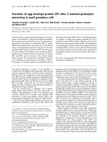
Effects of cocaine on Bax and Bcl-2 protein levels
To determine whether cocaine leads to changes in
Bcl-2 family protein levels in fetal brain, we examined
the Bcl-2 and Bax protein expression. As shown in
Figure 4, Bax protein was minimally detected in con-
trol fetal brain. However, it was dose-dependent in-
creased (% of control) in cocaine 30 mg/kg/day (227.1
± 48.61, P < 0.05) and cocaine 60 mg/kg/day (330.7 ±
31.6, P < 0.05). Bax was minimally present, whereas
Bcl-2 was constitutively expressed in normal fetal rat
brain. As shown in Figure 4, cocaine also significantly
increased Bcl-2 protein levels at the dose of 30
mg/kg/day (145.2 ± 8.16, P < 0.05) and 60 mg/kg/day
(278.8 ± 87.65, P < 0.05). The Bax-to-Bcl-2 ratio in fetal
rat brain was determined at each point by using value
that was normalized to the control protein level within
each group (Fig. 4, bottom panel). The Bax-to-Bcl-2
ratio was significantly higher at the dose of cocaine 30
mg/kg per day than the control group (P < 0.05), but
no difference at the dose of cocaine 60 mg/kg per day.
Int. J. Med. Sci. 2008, 5
299
Figure 4. Effects of maternal cocaine administration on Bax
and Bcl-2 protein expression in fetal rat brain. Cocaine was
administered subcutaneously to the pregnant rats for 7 days as
described in Methods. Immunoblot analysis of Bax and Bcl-2
proteins was performed in fetal rat brain. The top panel shows
the representative immunoblots obtained for Bcl-2 and Bax at
the expected size of 26 kDa and 21 kDa, respectively, and shows
an increase in Bcl-2 and Bax in the brains treated with cocaine.
The middle panel shows quantitative data obtained from five
separate experiments. The bottom panel shows the Bax-to-Bcl-2
ratio in fetal rat brain. Data are expressed as % of control, and
are means ± SEM. *P<0.05 vs. control.
Discussion
We previously showed that maternal cocaine
administration resulted in a decrease in fetal rat body
weight [9]. The present study demonstrated that the
maternal cocaine treatment caused a significant de-
crease in fetal brain weight, as compared with the sa-
line control group. This finding is consistent with the
previous report in pregnant C57BL/6 mice, in which
maternal subcutaneous administration of cocaine from
gestation days 12-18 produced significant decreases in
fetal body and brain weight [27]. The pair-fed studies
demonstrated that maternal undernutrition was not a
likely mediator of the effects caused by cocaine [27, 28].
Moreover, our data indicate that cocaine decreases
fetal brain/body weight ratio, suggesting that cocaine
have higher affinity toxic effects on the fetal brain than
the body. Dow-Edwards [29] reported that fetal brain
had between 26-42% more concentration of cocaine
than fetal plasma after 90 min following either 30 or 60
mg/kg cocaine given via intragastric intubation to
Wistar pregnant rats. It was also reported that cocaine
affinity for brain tissue is similar in the fetus and dam
after subcutaneous injection of cocaine, whereas the
cocaine metabolite benzoylecgonine concentrations in
fetal brain were greater than those observed in mater-
nal brain [30]. Therefore, fetal brain exposure to co-
caine is somewhat prolonged. Our current finding that
cocaine had no effect on the activities of the caspases in
the maternal brain but only in the fetal brain, further
support the idea that these high levels of cocaine or its
active cocaine metabolite may contribute to the pro-
duction of neuronal apoptotic alterations in co-
caine-exposed offspring. Nassogne et al [6] reported
that cocaine selectively affected embryonic neuronal
cells, causing first a dramatic reduction of both num-
ber and length of neurites and then extensive neuronal
death in co-cultures of neurons and glial cell from
mouse embryonic brain. Taken all together, our study
with previous reports demonstrated that cocaine ex-
posure in utero causes severe alterations in the fetal
brain, they could account for the qualitative or quan-
titative defects in neuronal pathways that cause a ma-
jor handicap in brain function following in prenatal
exposure to cocaine.
Although prenatal cocaine use during pregnancy
appears detrimental to the fetus, a causal effect,
mechanism of injury, and the pathway of cocaine in-
duced injury have not well been documented [4]. Pre-
vious studies have reported that cocaine does have
indirect effect on the developing fetus. Cocaine in-
creases circulating catecholamine levels, which may
induce uterine artery vasoconstriction and cause fetal
chronic hypoxia, then, could result in altered fetal so-
matic and fetal brain development [31]. Our finding
that cocaine decreased fetal body weight is consistent
with our previous studies that chronic hypoxia caused
fetal growth restriction [32]. Cocaine induced hypoxia
and increased the susceptibility to hypoxia-induced
brain damage as the outcomes associated with “crack
baby syndrome” represents a common underlying
mechanism [33]. On the other hand, cocaine can exert
direct effects on both the fetal central and peripheral
Int. J. Med. Sci. 2008, 5
300
nervous systems. Studies have demonstrated that
prenatal cocaine exposure has direct long-term effects
on brain structure and function [23-25, 31]. Our current
finding of the asymmetric growth restriction with de-
creased fetal brain-to-body weight ratio further sug-
gests that direct cytotoxic effects of cocaine on the fetal
brain are likely to exist. These data suggests that pre-
natal cocaine exposure has both indirect and direct
effect on the developing fetal brain.
Nassogne et al [22] demonstrated that cocaine
induced injury by apoptosis in vitro cultured cortical
neuronal cells of fetal mice. Whereas, our present
study has demonstrated that cocaine induces apop-
tosis in the fetal brain when it was administrated to the
mother in vivo. In the present study, cocaine-induced
apoptosis in fetal brain was clearly demonstrated by
morphological changes such as cell shrinkage and
rounding, characteristic features of apoptotic death.
Moreover, simultaneous assessment of nuclear chro-
matin morphology further verified that these cells
eventually manifested typical apoptotic condensed
and fragmented nuclei. Similar finding of co-
caine-induced apoptosis has been reported in mice
hepatocytes [14]. In addition, we have confirmed that
the process of apoptosis defined on the basis of cellular
and nuclear chromatin morphology correlates with
apoptosis defined on the basis of internucleosomal
DNA fragmentation assessed by DNA gel electropho-
resis. The discrete ladder of DNA fragments demon-
strated by gel electrophoresis indicates the presence of
DNA cleavage at linker regions producing dou-
ble-strand DNA fragments of integral multiples of
about 200 bp in the cocaine-induced injury fetal brain.
The demonstration of this nucleosomal ladder in the
treatment brain strongly suggests that apoptotic DNA
degradation with internucleosomal digestion by an
endonuclease is involved in the cocaine-induced cell
death in the brain. In present study, DNA was ex-
tracted from the whole fetal brain, so DNA fragmenta-
tion reflected the whole brain region. Future studies
will be needed to study the specific brain region and
cell type undergoing apoptosis in response to cocaine
exposure.
Apoptosis is a process of active cellular
self-destruction that requires the expression of specific
genes [34, 35]. Despite the diversity of signals that can
induce cell death, these pathways share several fea-
tures in their execution. One mechanism, which is
consistently implicated in apoptosis, reflects an or-
chestrated series of biochemical events that is carried
out by a group of cytosolic proteases, termed caspases.
The current finding that activities of caspase-8, cas-
pase-9 and caspase-3 were increased after prenatal
cocaine exposure provides strong evidence that apop-
tosis is activated after cocaine exposure in fetus and
may contribute to secondary cell injury and cell death.
The increased activities of caspae-9 and caspase-3 in-
duced by cocaine suggests that cocaine-induced
apoptosis in the fetal brain was likely mediated by
mitochondria/cytochrome c pathway [36]. On the
other hands, cocaine-induced activation of caspase-8,
which releases two active subunits, p18 and p10, into
the cytosol, activates additional caspases that cleave
other apoptosis-related substrates. Caspase-8 may be
involved in death receptor-mediated apoptosis path-
way [37].
Bcl-2 gene families are identified as apoptosis
regulating genes. Off these genes, bax, bad, bak and
bik promote cell death, whereas bcl-2 and bcl-X
L
in-
hibit apoptosis and promote cell survival [38, 39]. It
has been shown that the Bcl-2 protein physically in-
teracts with several of its homologous proteins, in the
form of heterotypic dimers. The most important inter-
actions are considered to lie in Bcl-2/Bax dimerization.
Thus, we studied the temporal profile of bcl-2 and bax
gene products in terms of protein expression in the
fetal brain after cocaine exposure. The current findings
that cocaine significantly increased the protein levels
of Bcl-2 and Bax in fetal rat brain are consistent to pre-
vious reports that gene transcriptional levels of Bcl-2
and Bax are up-regulated in fetal mouse cerebral wall
[24]. The current result showed that bax gene expres-
sion was markedly induced and dose dependent in-
creased, suggesting that bax was upregulated and
played an important role in the induction of apoptotic
death in the fetal brain after cocaine exposure. How-
ever, in contrast to the aforementioned bcl-2 inhibiting
apoptotic cell death, the present study in fetal rat brain
found that bcl-2 expression was also increased in a
dose dependent manner after cocaine exposure com-
pared to saline control. The increase in anti-apoptotic
Bcl-2 protein in the fetal brain may serve as a com-
pensatory protection of the neural cells upon cocaine
insult. Previous study [40] found that total Bcl-2 pro-
tein is increased in injured brain after traumatic brain
injury. Neurons are resistant to ischemic injury when
Bcl-2 protein is over-expressed in vivo [41-43].
Whether programmed cell death that occurs after brain
injury is maladaptive or beneficial has been addressed
in several studies in animal models of stroke, trauma,
cerebral ischemia and excitotoxicity [44, 45]. These
studies support the hypothesis that Bcl-2 protects
neurons from injury. Thus, Bcl-2 expression could be
an important factor that promotes survival of neurons
injured after cocaine exposure. Although the expres-
sions of Bcl-2 and Bax, both of them, were increased, it
was very interesting that the ratio of Bax/Bcl-2 (pro- to
anti-apoptotic proteins) was also increased after co-
Int. J. Med. Sci. 2008, 5
301
caine exposure in the current study. The findings
support the notion that the relative concentrations of
pro-apoptotic and anti-apoptotic genes may act as a
rheostat for the cell death program [46].
In conclusion, our study has demonstrated that
cocaine induces apoptosis in the fetal brain when it is
administrated to the mother. As reported previously,
our study also demonstrates fetal growth retardation
after cocaine use. Moreover, our data indicate that co-
caine decreases fetal brain/body weight ratio. The
finding of the increased caspases activities re-enforces
the conclusion that cocaine induces apoptosis in the
fetal brain. The current studies also suggest that mul-
tiple mechanisms may be involved in cocaine-induced
apoptosis in the fetal brain. One of the apoptotic
pathways is regulated by specific genes. Of these
genes, Bax is the key gene in upregulation of the co-
caine-induced apoptosis in the fetal brain. However,
Bcl-2 expression could be an important factor that
promotes survival of cocaine-injured neurons. These
findings demonstrate that genes can be orchestrated in
cocaine-induced apoptosis in fetal brain, and provide a
rational for the further development of pharmacol-
ogical and molecular therapies targeting programmed
cell death after cocaine use.
Acknowledgments
The authors thank Dr. Yuhui Xiao for the techni-
cal assistance. This work was supported in part by the
National Institutes of Health grants HL-82779,
HL-83966 (L.Z.) and by the California Tobacco-Related
Disease Research Program Award 14FT-0075 (D.X.).
Conflict of Interest
The authors have declared that no conflict of in-
terest exists.
References
1. Chasnoff IJ. Cocaine, pregnancy, and the growing child. Curr.
Probl. Pediatr. 1992, 22:302-321.
2. Chasnoff IJ, Burns KA, Burns KA. Cocaine use in pregnancy:
Perinatal morbidity and mortality. Neurotoxicol. Teratol. 1987,
9:291-293.
3. Chasnoff IJ, Burns WJ, Schnoll SH, Burns KA. Cocaine use in
pregnancy. N. Engl. J. Med. 1985, 313:666-669.
4. Gingras JL, Weese-Mayer DE, Hume RFJr, O Donnell KL. Co-
caine and development: mechanism of fetal toxicity and neona-
tal consequences of prenatal cocaine exposure. Early Hum. Dev.
1992, 31:1-24.
5. Holzman C, Paneth N. Maternal cocaine use during pregnancy
and perinatal outcomes. Epidemiol. Rev. 1994, 16:315-34.
6. Nassogne MC, Evrard P, Courtoy PJ. Selective neuronal toxicity
of cocaine in embryonic mouse brain cocultures. Proc. Natl.
Acad. Sci. U. S. A. 1995, 92:11029-11033.
7. Li G, Xiao Y, Zhang L. Cocaine induces apoptosis in fetal rat
myocardial cells through the p38 mitogen-activated protein
kinase and mitochondrial/cytochrome c pathways. J. Pharma-
col. Exp. Ther. 2005, 312:112-119.
8. Xiao Y, He J, Gilbert RD, Zhang L. Cocaine induces apoptosis in
fetal myocardial cells through a mitochondria-dependent path-
way. J. Pharmacol. Exp. Ther. 2000, 292:8-14.
9. Xiao Y, Xiao D, He J, Zhang L. Maternal cocaine administration
during pregnancy induces apoptosis in fetal rat heart. J. Car-
diovasc. Pharmacol. 2001, 37:639-648.
10. He J, Xiao Y, Casiano CA, Zhang L. Role of mitochondrial cyto-
chrome c in cocaine-induced apoptosis in coronary artery en-
dothelial cells. J. Pharmacol. Exp. Ther. 2000, 295:896-903.
11. He J, Xiao Y, Zhang L. Cocaine induces apoptosis in human
coronary artery endothelial cells. J. Cardiovasc. Pharmacol. 2000,
35:572-580.
12. He J, Xiao Y, Zhang L. Cocaine-mediated apoptosis in bovine
coronary artery endothelial cells: role of nitric oxide. J. Pharma-
col. Exp. Ther. 2001, 298:180-187.
13. Wu YB, Shen ML, Gu GG, Anderson KM, Ou DW. The effects of
cocaine injections on mouse thymocyte population. Proc. Soc.
Exp. Biol. Med. 1997, 214:173-219.
14. Cascales M, Alvarez A, Gasco P, Fernandez-Simon L, Sanz N,
Bosca L. Cocaine-induced liver injury in mice elicits specific
changes in DNA ploidy and induces programmed death of
hepatocytes. Hepatology 1994, 20:992-1001.
15. Li H, Jiang Y, Rajpurkar A, Dunbar JC, Dhabuwala CB. Cocaine
induced apoptosis in rat testes. J. Urol. 1999, 162:213-216.
16. Hutchins JB, Barger SW. Why neurons die: cell death in the
nervous system. Anat. Rec. 1998, 253:79-90.
17. Johnson EM Jr, Deckwerth TL. Molecular mechanisms of de-
velopmental neuronal death. Annu. Rev. Neurosci.1993,
16:31-46.
18. Wyllie AH, Kerr JF, Currie AR. Cell death: the significance of
apoptosis. Int. Rev. Cytol. 1980, 68:251-306.
19. An SF, Gray F, Scaravilli F. Programmed cell death in brains of
HIV-1-positive pre AIDS patients. Lancet. 1995, 346:911-912.
20. Dickson DW. Apoptosis in the brain. Physiology and pathology.
Am J Pathol. 1995, 146:1040-1044.
21.
Lin
nik MD, Zobrist RH, Hatfield MD. Evidence supporting a
role for programmed cell death in focal cerebral ischemia in rats.
Stroke 1993, 24:2002-2008.
22. Nassogne MC, Louahed J, Evrard P, Courty PJ. Cocaine induces
apoptosis in cortical neurons of fetal mice. J. Neurochem. 1997,
68:2442-2450.
23. Mitchell ES, Snyder-Keller A. c-fos and cleaved caspase-3 ex-
pression after perinatal exposure to ethanol, cocaine, or the
combination of both drugs. Brain Res Dev Brain Res. 2003,
147:107-117.
24. Novikova SI, He F, Bai J, Badan I, Lidow IA, Lidow MS. Co-
caine-induced changes in the expression of apoptosis-related
genes in the fetal mouse cerebral wall. Neurotoxicol Teratol.
2005, 27:3-14.
25. Novikova SI, He F, Bai J, Cutrufello NJ, Lidow MS, Undieh AS.
Maternal cocaine administration in mice alters DNA methyla-
tion and gene expression in hippocampal neurons of neonatal
and prepubertal offspring. PLoS ONE. 2008, 3: e1919.
26. Bae S, Xiao Y, Li G, Casiano CA, Zhang L. Effect of maternal
chronic hypoxic exposure during gestation on apoptosis in fetal
rat heart. Am. J. Physiol. Heart Circ. Physiol. 2003,
285:H983-H990.
27. Middaugh LD, Boggan WO, Bingel SA, Patrick KS, Xu W. A
murine model of prenatal cocaine exposure: effects on the
mother and the fetus. Pharmacol. Biochem. Behav. 1996,
55:565-574.
28. Song J, Guan XW, Ren JQ, He W. Developmental toxicity of
cocaine exposure in mid-pregnancy mice. Acta. Pharmacol. Sin.
2002, 23:1029-1034.
29. Dow-Edwards DL. Fetal and maternal cocaine levels peak rap-
idly following intragastric administration in the rat. J. Subst.
Abuse 1990, 2:427-437.
Int. J. Med. Sci. 2008, 5
302
30. Spear LP, Frambes NA, Kirstein CL. Fetal and maternal brain
and plasma levels of cocaine and benzoylecgonine following
chronic subcutaneous administration of cocaine during gestation
in rats. Psychopharmacology (Berl) 1989, 97:427-431.
31. Slotkin TA. Fetal nicotine or cocaine exposure: which one is
worse? J. Pharmacol. Exp. Ther. 1998, 285:931-945.
32. Xiao D, Ducsay CA, Zhang L. Chronic hypoxia and develop-
mental regulation of cytochrome c expression in rats. J. Soc.
Gynecol. Investig. 2000, 7:279-283.
33. Olsen GD. Potential mechanisms of cocaine-induced develop-
mental neurotoxicity: a minireview. Neurotoxicology 1995,
16:159-167.
34. Kerr JF, Wyllie AH, Currie AR. Apoptosis: a basic biological
phenomenon with wide-ranging implications in tissue kinetics.
Br. J. Cancer 1972, 26:239-257.
35. Oppenheim RW, Prevette D, Tytell M, Homma S. Naturally
occurring and induced neuronal death in the chick embryo in
vivo requires protein and RNA synthesis: evidence for the role
of cell death genes. Dev. Biol. 1990, 138:104-113.
36. Green DR, Reed JC. Mitochondria and apoptosis. Science 1998,
281:1309-1312.
37. Ashkenazi A, Dixit VM. Death receptors: signaling and modula-
tion. Science 1998, 281:1305-1308.
38. Hara A, Hirose Y, Wang A, Yoshimi N, Tanaka T, Mori H. Lo-
calization of Bax and Bcl-2 proteins, regulators of programmed
cell death, in the human central nervous system. Virchows Arch.
1996, 429:249-253.
39. Vaux DL, Cory S, Adams JM. Bcl-2 gene promotes haemopoietic
cell survival and cooperates with c-myc to immortalize pre-B
cells. Nature 1988, 335:440-442.
40. Clark RS, Kochanek PM, Chen M, Watkins SC, Marion DW,
Chen J, Hamilton RL, Loeffert JE, Graham SH. Increases in Bcl-2
and cleavage of caspase-1 and caspase-3 in human brain after
head injury. FASEB. J. 1999, 13:813-821.
41. Lawrence MS, McLaughlin JR, Sun GH, Ho DY, McIntosh L,
Kunis DM, Sapolsky RM, Steinberg GK. Herpes simplex viral
vectors expressing Bcl-2 are neuroprotective when delivered af-
ter a stroke. J. Cereb. Blood Flow Metab. 1997, 17:740-744.
42. Linnik MD, Zahos P, Geschwind MD, Federoff HJ. Expression of
bcl-2 from a defective herpes simplex virus-1 vector limits neu-
ronal death in focal cerebral ischemia. Stroke 1995, 26:670-1674.
43. Martinou JC, Dubois-Dauphin M, Staple JK, Rodriguez I,
Frankowski H, Missotten M, Tschopp J. Overexpression of
BCL-2 in transgenic mice protects neurons from naturally oc-
curring cell death and experimental ischemia. Neuron 1994,
13:1017-1030.
44. Friedlander RM, Gagliardini V, Hara H, Fink KB, Li W, Mac-
Donald G, Fishman MC, Greenberg AH, Moskowitz MA, Yuan J.
Expression of a dominant negative mutant of interleukin-1 beta
converting enzyme in transgenic mice prevents neuronal cell
death induced by trophic factor withdrawal and ischemic brain
injury. J. Exp. Med. 1997, 185:933-940.
45. Hara H, Friedlander RM, Gagliardini V, Ayata C, Fink K, Huang
Z, Shimizu-Sasamata M, Yuan J, Moskowitz MA. Inhibition of
interleukin 1beta converting enzyme family proteases reduces
ischemic and excitotoxic neuronal damage. Proc. Natl. Acad. Sci.
U. S. A. 1997, 94:2007-2012.
46. Adams JM, Cory S. The Bcl-2 protein family: arbiters of cell
survival. Science 1998, 281:1322-1326.