Báo cáo y học: "Soluble Endothelial Selectin in Acute Lung Injury Complicated by Severe Pneumonia" pdf
Bạn đang xem bản rút gọn của tài liệu. Xem và tải ngay bản đầy đủ của tài liệu tại đây (495 KB, 7 trang )
Int. J. Med. Sci. 2011, 8
302
I
I
n
n
t
t
e
e
r
r
n
n
a
a
t
t
i
i
o
o
n
n
a
a
l
l
J
J
o
o
u
u
r
r
n
n
a
a
l
l
o
o
f
f
M
M
e
e
d
d
i
i
c
c
a
a
l
l
S
S
c
c
i
i
e
e
n
n
c
c
e
e
s
s
2011; 8(4):302-308
Research Paper
Soluble Endothelial Selectin in Acute Lung Injury Complicated by Severe
Pneumonia
Daisuke Osaka
1
, Yoko Shibata
1
, Kazunori Kanouchi
2
, Michiko Nishiwaki
1
, Tomomi Kimura
1
, Hiroyuki
Kishi
1
, Shuichi Abe
1
, Sumito Inoue
1
, Yoshikane Tokairin
1
, Akira Igarashi
1
, Keiko Yamauchi
1
, Yasuko Aida
1
,
Takako Nemoto
1
, Keiko Nunomiya
1
, Koji Fukuzaki
1
, and Isao Kubota
1
1. Department of Cardiology, Pulmonology, and Nephrology, Yamagata University School of Medicine, Yamagata, Japan
2. Division of Clinical Laboratory, Yamagata University Hospital, Yamagata, Japan
Corresponding author: Dr. Yoko Shibata, 2-2-2 Iida-Nishi, Yamagata City, Yamagata 990-9585, Japan. Telephone:
+81-23-628-5302, FAX: +81-23-628-5305, Email:
© Ivyspring International Publisher. This is an open-access article distributed under the terms of the Creative Commons License (
licenses/by-nc-nd/3.0/). Reproduction is permitted for personal, noncommercial use, provided that the article is in whole, unmodified, and properly cited.
Received: 2011.03.22; Accepted: 2011.05.02; Published: 2011.05.11
Abstract
Background: Pneumonia is still one of the most frequent causes of death in the elderly.
Complication of acute lung injury (ALI)/acute respiratory distress syndrome (ARDS) by
pneumonia makes patients very ill due to severe respiratory failure. Biomarkers that can
discriminate the presence of complicating ALI/ARDS are required for early detection. The aim
of this research was to investigate whether soluble endothelial selectin (sES) could be a
biomarker for ALI.
Methods: Serum sES levels were measured in 27 pneumonia patients, who were enrolled
between April 2006 and September 2007. Among these patients, six had ALI or a condition
that was clinically comparable to ALI (cALI). All patients who were enrolled were successfully
treated and survived.
Results: Circulating sES levels were elevated in pneumonia patients with ALI/cALI, and sES
levels decreased following treatment of their pneumonia. Univariate and multivariate logistic
regression analyses showed that sES was the only significant factor for identifying complicating
ALI/cALI, independently of C-reactive protein (CRP) and lactate dehydrogenase (LDH). By
receiver operating characteristic (ROC) curve analysis, the cut-off value for sES was 40.1
ng/mL, with a sensitivity of 0.8 and a specificity of 0.8.
Conclusion: sES may be a useful biomarker for discriminating complicating ALI/cALI in pa-
tients with severe pneumonia.
Key words: Pneumonia, acute lung injury, soluble endothelial selectin
INTRODUCTION
Pneumonia is one of the most common infectious
diseases, and is still one of the most frequent causes of
death in the elderly (1). In spite of advances in antibi-
otic therapy, some patients with pneumonia become
severely ill due to delays in receiving adequate
treatment or due to comorbidities such as cancer and
diabetes. In particular, acute lung injury (ALI)/acute
respiratory distress syndrome (ARDS) due to severe
pneumonia results in respiratory failure, prolongs
hospitalization, and sometimes causes death (2,3). The
influx of neutrophils into lung tissue is the initial
hallmark of ALI/ARDS (2,4). The onset and progres-
Int. J. Med. Sci. 2011, 8
303
sion of ALI/ARDS is so acute that it is sometimes
difficult to detect the presence of ALI/ARDS, only by
the use of portable chest X-ray units in the early
phase. Chest computed tomography (CT) is required
for early diagnosis based on the ground glass shadow
that is typical of ALI/ARDS (5,6). However, per-
forming CT scans on all pneumonia patients is costly,
and it is therefore necessary to develop good bi-
omarkers that can discriminate complicating
ALI/ARDS, so that a quick decision can be made on
whether a CT scan should be performed.
Endothelial selectin (ES) is one of the cell adhe-
sion molecules expressed in the vascular endothelium
(7). ES is induced by pro-inflammatory cytokines in
thrombosis (8,9), infectious diseases (10), malignant
tumors (11) and autoimmune diseases (12), leading to
the attachment of leukocytes to endothelial cells and
the accumulation of leukocytes in inflamed tissues. A
part of the extracellular portion of ES is cleaved and
released as a soluble form (13). Circulating levels of
soluble ES (sES) were reported to be elevated in pa-
tients with sepsis and shock (14-16). In addition,
Okajima et al. reported that hypoxia was more prev-
alent in patients with high sES levels and systemic
inflammatory response syndrome (SIRS), compared
to patients with normal sES levels and SIRS (17).
Based on this background information, we hy-
pothesized that measurement of circulating sES levels
may be useful for the discrimination of complicating
ALI/ARDS in severe pneumonia patients. Therefore,
we investigated sES levels in patients with commu-
nity acquired pneumonia, and assessed the associa-
tion between sES levels and complicating ALI.
METHODS
Study Subjects
We measured serum sES levels in 27 patients
who were admitted to Yamagata University Hospital
for the treatment of community acquired pneumonia
between April 2006 and September 2007. The diagno-
sis of pneumonia was based on clinical symptoms
(cough, sputum production, and fever) and chest
X-ray and chest CT findings. Patients with congestive
heart failure were excluded from the analysis, to
avoid misdiagnosis of ALI. In addition, patients with
malignant tumors, thrombotic diseases (deep vein
thrombosis and pulmonary artery thrombosis), or
active systemic inflammatory diseases such as colla-
gen vascular disease, were excluded, because sES
levels are reported to be elevated in these disorders.
The study protocol was approved by the institutional
review board at Yamagata University, and written
informed consent was obtained from all participating
patients.
Assessment of Severity of Pneumonia at Admis-
sion
The age, dehydration, respiratory failure, orien-
tation disturbance and low blood pressure (A-DROP)
scoring system was used to evaluate the severity of
pneumonia (18). This is a 6-point scale (0-5) for as-
sessing the clinical severity of community acquired
pneumonia that was proposed by the Japanese Res-
piratory Society. The A-DROP scoring system assess-
es the following parameters: i) age (male ≥ 70 years,
female ≥ 75 years); ii) dehydration [blood urea nitro-
gen (BUN) ≥ 21 mg/dL]; iii) respiratory failure [(SpO
2
≤ 90% or partial pressure of arterial oxygen (PaO
2
) ≤
60 mm Hg]; iv) orientation disturbance (confusion);
and v) low blood pressure (systolic blood pressure ≤
90 mm Hg).
Diagnosis of ALI and Clinical Status Comparable
to ALI (cALI)
Generally, data on the fraction of inspired oxy-
gen (FiO
2
) is required to accurately diagnose whether
or not patients have ALI (19). However, as all partic-
ipants in this study did not receive mechanical venti-
lation or non-invasive positive pressure ventilation,
the accurate FiO
2
values were not available in this
study. It was previously reported that SpO
2
/FiO
2
(S/F) ratios correlate with PaO
2
/FiO
2
(P/F) ratios,
and S/F ratios of 235 and 315 correlate with P/F ratios
of 200 and 300, respectively, for diagnosing and fol-
lowing up patients with ALI and ARDS (20). Thus,
hypoxic patients were defined as having “ALI/cALI”
using the following criteria: 1) bilateral massive
ground glass shadow on chest X-ray and/or CT scan;
2) no apparent findings suggesting congestive heart
failure, on chest X-ray and ultrasound cardiogram;
and 3) requirement of 5 L/min or more of supple-
mental oxygen therapy using a facial mask, estimated
FiO2 ≥0.35 (21), to maintain SpO
2
>90%, except for
patients with hypoxemia due to their primary chest
disease. By this criterion, subjects with estimated S/F
<257 had a potential to be classified as ALI/cALI
group. The diagnosis of ALI/cALI was made by at
least two pulmonary physicians and a cardiologist,
according to these criteria. All pneumonia patients
received immediate antibiotic therapy. The etiology of
pneumonia was aspiration (ALI/cALI group, n = 9;
non-ALI/cALI group, n = 4), bacterial infection
(ALI/cALI group, n = 11; non-ALI/cALI group, n =
1), and Legionella pneumophilia infection
(non-ALI/cALI group, n = 1). Two patients in the
ALI/cALI group and two in the non-ALI/cALI group
Int. J. Med. Sci. 2011, 8
304
received the neutrophil elastase inhibitor, Sivelestat
(Ono Pharmaceutical, Osaka, Japan) (22), but none of
the patients in this study received glucocorticosteroid
therapy. None of the patients died during the hospital
admission, and all were successfully discharged.
Blood Sampling and Measurement of Soluble
Endothelial Selectin
Repeat blood samples were obtained at intervals
of 3 to 5 days from the time of admission until the
patients were recovered from pneumonia, to measure
sES levels and other biochemical markers. The
time-point of blood sampling was flexibly decided by
each doctor as needed. The median number of blood
sampling was 2 (1 - 3) in non-ALI/cALI group, and 4
(3.75 - 5) in ALI/cALI group [median (inter quartile
range)]. These samples were stored frozen at -20°C
until the measurements were made. sES levels were
measured by latex photometric immunoassay (LPIA)
(Mitsubishi Chemical Medience Corp, Tokyo, Japan)
(17). This assay measures serum sES concentrations
over a linear range of 5.29 to 300 ng/mL (17). It was
reported that the normal range of the plasma sES lev-
els was 4.8 – 29.7 ng/mL (17).
Statistical Analysis
The Mann-Whitney U test for non-parametric
data was used to analyze differences between two
groups. Multiple comparisons were performed by
non-parametric one way analysis of variance (Krus-
kal-Wallis test) followed by the Stu-
dent-Newman-Keuls test. Chi-square tests were used
to evaluate differences in proportions. These compar-
isons and the logistic regression analyses were per-
formed using SigmaPlot version 11 computer soft-
ware (Systat Software, Inc., San Jose, CA, USA) and
JMP version 8 software (SAS Institute Inc., Cary, NC,
USA). Data in the figures are shown as mean ± SD.
Significance was inferred for differences with P < 0.05.
RESULTS
The characteristics of the pneumonia patients
enrolled in this study are shown in Table 1. Age,
gender, and the number of patients requiring sup-
plemental oxygen did not differ significantly between
the non-ALI/cALI and ALI/cALI groups. The length
of hospitalization was significantly greater in the
ALI/cALI group than in the non-ALI/cALI group (P
<0.01). The A-DROP score for severity of community
acquired pneumonia was significantly higher in the
ALI/cALI group, compared with the non-ALI/cALI
group (chi-square test, P <0.05). Among the labora-
tory results on arrival in hospital, only sES and lactate
dehydrogenase (LDH) were significantly higher in the
ALI/cALI group than in the non-ALI/cALI group (P
<0.05). There was a trend for C-reactive protein (CRP)
levels to be higher in the ALI/cALI group than in the
non-ALI/cALI group, but the difference did not reach
statistical significance (P = 0.06). Among patients with
severe pneumonia and a A-DROP score ≥3, there was
a trend for sES levels to be higher in the ALI/cALI
group than in the non-ALI/cALI group, although the
difference was not statistically significant
(non-ALI/cALI, 39.7 ± 23.1 ng/mL; ALI/cALI, 56.0 ±
22.2 ng/mL; P = 0.2).
Table 1. Characteristics of the patients with pneumonia
non-ALI/cALI (n=21)
ALI/cALI (n=6)
Age, years (range)
77.6 (50 - 93)
75.0 (51 - 92)
Male gender, %
44.4
66.7
Hospitalization, days
18.8 ± 15.1
59.3 ± 41.0**
Use of supplemental
oxygen, %
66.6
100
A-DROP score
#
2 / 4 / 9 / 6 / 0 / 0
0 / 0 / 2 / 3 / 1 / 0*
WBC, ×1000/μL
#
11.50± 4.09
12.50± 5.46
CRP, mg/dL
#
11.7 ± 6.3
18.6 ± 11.0
LDH, IU/L
#
195 ± 64
274 ± 104*
BUN, mg/dL
#
18.9 ± 7.7
18.8 ± 10.0
Na, mEq/L
#
137 ± 2.6
134 ± 11.1
Blood glucose,
mg/dL
#
156 ± 52.9
153 ± 44.0
Hematocrit, %
#
37.0 ± 6.28
38.4 ± 5.00
sES, ng/mL
#
33.6 ± 14.8
53.0 ± 17.8*
Data are means ± SD unless indicated otherwise.
#
Data obtained or
evaluated on arrival at the hospital.
* P < 0.05, ** P < 0.01 compared with the non-ALI/cALI group
A-DROP score, a 6-point scale (0-5) for assessing the clinical sever-
ity of community acquired pneumonia, proposed by the Japanese
Respiratory Society. This scoring system assesses the following
parameters: i) age (male ≥ 70 years, female ≥ 75 years); ii) dehydra-
tion (BUN ≥ 21 mg/dL); iii) respiratory failure (SpO
2
≤ 90% or PaO
2
≤ 60 mm Hg); iv) orientation disturbance (confusion); and v) low
blood pressure (systolic blood pressure ≤ 90 mm Hg).
ALI, acute lung injury; BUN, blood urea nitrogen; cALI, clinical
status comparable to ALI; CRP, C-reactive protein; LDH, lactate
dehydrogenase; sES, soluble endothelial selectin; WBC, white blood
cell count
The time courses for sES, CRP and LDH accord-
ing to complicating ALI/cALI are shown in Figure 1.
sES levels were higher in pneumonia patients with
Int. J. Med. Sci. 2011, 8
305
ALI/cALI than in those patients without ALI/cALI.
sES levels decreased after commencement of treat-
ment in the ALI/cALI group (P = 0.017). However, in
the non-ALI/cALI group sES levels did not differ
significantly at each time point (P = 0.075). CRP levels
in pneumonia patients with ALI/cALI tended to be
higher than those in patients without ALI/cALI, alt-
hough the difference was not statistically significant.
CRP levels decreased after the commencement of
treatment in the non-ALI/cALI group (P = 0.008).
However, in the ALI/cALI group, the differences in
CRP levels at each time point did not reach statistical
significance (P = 0.07). LDH levels on day 1, days 3-4,
and days 5-6 were higher in pneumonia patients with
ALI/cALI than in those without ALI/cALI. However,
the differences in LDH levels at each time point were
not statistically significant either in the ALI/cALI
group or in the non-ALI/cALI group (P = 0.444 and P
= 0.527, respectively).
Univariate logistic regression analysis showed
that sES was a significant factor for identifying com-
plicating ALI/cALI (Table 2), whereas age, gender,
white blood cell count (WBC), CRP, LDH, BUN, Na,
blood glucose, and hematocrit were not significant
factors. Furthermore, multiple logistic regression
analysis demonstrated that sES was an independent
factor for identifying the presence of ALI/cALI (Table
3). From analysis of the receiver operating character-
istic (ROC) curve, the cut-off value for sES was 40.1
ng/mL, for discrimination of complicating ALI/cALI
in pneumonia patients, with a sensitivity of 0.8 and a
specificity of 0.8 (Figure 2).
Table 2. Univariate logistic regression analysis for factors
identifying complicating ALI/cALI in patients with pneumo-
nia
Variable
Coefficient
SD
P value
Age, years
0.022
0.038
0.570
Male gender
0.144
0.489
0.769
sES, ng/mL
-0.072
0.036
0.044
WBC, per 1000/μL
-0.059
0.103
0.565
CRP, mg/dL
-0.113
0.067
0.091
LDH, IU/L
-0.012
0.007
0.073
BUN, mg/dL
-0.044
0.049
0.368
Na, mEq/L
0.104
0.08
0.194
Blood glucose, mg/dL
0.0003
0.01
0.977
Hematocrit, %
-0.035
0.078
0.658
ALI, acute lung injury; BUN, blood urea nitrogen; cALI, clinical
status comparable to ALI; CRP, C-reactive protein; LDH, lactate
dehydrogenase; sES, soluble endothelial selectin; WBC, white blood
cell count
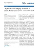
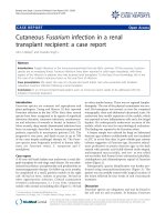
Figure 1. Time courses of soluble endothelial selectin,
C-reactive protein and lactate dehydrogenase after com-
mencement of treatment for pneumonia, according to
complicating acute lung injury. The time courses of
soluble endothelial selectin (sES, A), C-reactive protein
(CRP, B) and lactate dehydrogenase (LDH, C) are shown
according to complicating acute lung injury (ALI)/clinical
status comparable to ALI (cALI). sES levels were higher in
pneumonia patients with ALI/cALI than in those without
ALI/cALI. sES levels decreased after commencement of
treatment in the ALI/cALI group (P = 0.017). However, sES
levels in the non-ALI/cALI group were not significantly
different at each time point (P = 0.075). CRP levels in
Int. J. Med. Sci. 2011, 8
306
pneumonia patients with ALI/cALI tended to be higher than
those in patients without ALI/cALI, although the difference
was not statistically significant. CRP levels decreased after
treatment in the non-ALI/cALI group (P = 0.008). However,
in the ALI/cALI group, the differences in CRP levels at each
time point did not reach statistical significance (P = 0.07).
LDH levels on Day 1, Days 3-4, and Days 5-6 were higher in
pneumonia patients with ALI/cALI than in those without
ALI/cALI. However, the differences in LDH levels at each
time point were not statistically significant either in the
ALI/cALI group or in the non-ALI/cALI group (P = 0.444 and
P = 0.527, respectively). * P < 0.05 compared with Day1; # P
< 0.05 compared with the ALI/cALI group
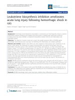
Figure 2. Determination of the soluble endothelial selectin
(sES) cut-off value for discrimination of complicating
ALI/cALI in pneumonia patients. Receiver operating char-
acteristic (ROC) curve analysis was performed to deter-
mine the sES cut-off value for discrimination of complicating
ALI/cALI in pneumonia patients. The area under the curve
(AUC) was 0.875, and the cut-off value was 40.1 ng/mL,
with a sensitivity of 0.8 and a specificity of 0.8.
Table 3. Multiple logistic regression analysis for factors
identifying complicating ALI/cALI in patients with pneumo-
nia
Variable
OR
95% CI
P value
sES, per 1 ng/mL increase
1.099
1.012
1.260
0.021
CRP, per 1 mg/dL increase
1.029
0.829
1.293
0.795
LDH, per 1 IU/L increase
1.017
0.999
1.046
0.052
ALI, acute lung injury; cALI, clinical status comparable to ALI; CI,
confidence interval; CRP, C-reactive protein; LDH, lactate dehy-
drogenase; sES, soluble endothelial selectin; OR, odds ratio
DISCUSSION
This study demonstrated that circulating sES
levels were elevated in pneumonia patients with
ALI/cALI, and that sES levels decreased following
treatment for pneumonia. Although LDH levels were
higher in pneumonia patients with ALI/cALI than in
those without ALI/cALI, the time course of changes
in LDH did not accord with improvement in disease
status. Univariate and multivariate logistic regression
analyses revealed that sES was the only significant
factor for identifying complicating ALI/cALI, inde-
pendently of CRP and LDH. Among patients with
severe pneumonia and an A-DROP score ≥3, sES lev-
els tended to be higher in the ALI/cALI group than in
the non-ALI/cALI group, although the difference was
not statistically significant. Therefore, it may be pos-
sible to predict complicating ALI/cALI in pneumonia
patients from laboratory measurements showing ele-
vated sES levels.
Pneumonia is an infectious disease, in which
pathogenic bacteria or viruses infect the lower respir-
atory tract and proliferate, leading to focal inflamma-
tion of the lungs (4). In patients with severe pneumo-
nia, infiltrating neutrophils produce
pro-inflammatory cytokines and chemokines, result-
ing in the induction of cellular adhesion molecules on
circulating leukocytes and pulmonary endothelial
cells (23,24). Inflamed alveolar cells, in particular al-
veolar macrophages, produce chemoattractant pro-
teins such as interleukin-8 (IL-8), which attract circu-
lating leukocytes into the lung (25). In ALI/ARDS, the
inflamed lesion spreads over the original pneumonic
lung segment, leading to the accumulation of leuko-
cytes in a large pulmonary area. sES is selectively ex-
pressed in vascular endothelial cells, and plays im-
portant roles in the accumulation of leukocytes during
pneumonia (26). Thus, elevation of sES in pneumonia
patients is thought to indicate the presence of major
pulmonary parenchymal inflammation. As the data
from the present study demonstrates, it is possible
that sES becomes a biomarker for ALI/ARDS in pa-
tients with severe pneumonia.
To date, WBC and CRP have been used as bi-
omarkers for the degree of inflammation, with LDH
as a biomarker for tissue damage. We demonstrated
that circulating sES levels were significantly associ-
ated with complicating ALI, whereas WBC, CRP and
LDH were not, indicating the usefulness of sES for
evaluating the presence of ALI in severe pneumonia.
In particular, measurement of sES may be recom-
Int. J. Med. Sci. 2011, 8
307
mended in patients with severe pneumonia and a
high A-DROP score.
The limitations of present study are: 1) since the
study was performed at a single institute, the number
of patients with ALI/cALI was small; 2) patients with
very severe pneumonia, who could not provide writ-
ten informed consent due to severe respiratory failure
comparable to a clinical status of ARDS, were not en-
rolled in this study. All patients, even the patients
with severe pneumonia, were successfully treated and
survived; hence, sES was not measured in very severe
patients in the present study. Therefore, it is necessary
to perform large clinical investigations at multiple
medical institutions to confirm the usefulness of sES
for the discrimination of complicating ALI.
A system for rapid measurement of sES has al-
ready been established (17). In addition, the specific
neutrophil elastase inhibitor, sivelestat, is in clinical
use for SIRS patients (27). Neutrophil elastase induces
IL-8 in alveolar epithelial and bronchial cells through
activation of signaling cascades induced by defor-
mation of cell shape (28-30). Sivelestat has been re-
ported to inhibit the production of cytokines from
lung epithelial cells not only by inhibiting elastolytic
activity but also by modulating signaling cascades
involved in the production of cytokines (31). Early
administration of sivelestat in pneumonia patients
with high sES levels may improve the outcome of
ALI/ARDS treatment by attenuating the influx of
neutrophils into the lung parenchyma.
In conclusion, measurement of sES in patients
with severe pneumonia may be useful for the dis-
crimination of complicating ALI/cALI. The clinical
application of this potentially useful biomarker may
improve the accuracy and rapidity of diagnosis, and
the outcome of treatment in patients with ALI.
Acknowledgements
We thank Taiko Aita and Eiji Tsuchida for their
excellent technical assistance.
Funding
This study was supported by a Grant-in-aid from
the Global COE program of the Japan Society for the
Promotion of Science, and grants-in-aid for Scientific
Research from the Ministry of Education, Culture,
Sports, Science and Technology, Japan (18590835,
18790530, 19590880, and 20590892).
Conflict of Interest
The authors have declared that no conflict of in-
terest exists.
References
1 Murray CJ, Lopez AD. Mortality by cause for eight regions of
the world: Global burden of disease study. Lancet. 1997; 349:
1269-76.
2 Ware LB, Matthay MA. The acute respiratory distress syn-
drome. N Engl J Med. 2000; 342: 1334-49.
3 Matthay MA, Zimmerman GA, Esmon C, et al. Future research
directions in acute lung injury: Summary of a national heart,
lung, and blood institute working group. Am J Respir Crit Care
Med. 2003; 167: 1027-35.
4 Balamayooran G, Batra S, Fessler MB, et al. Mechanisms of
neutrophil accumulation in the lungs against bacteria. Am J
Respir Cell Mol Biol. 2010; 43: 5-16.
5 Endo S, Shibata S, Sato N, et al. A prospective cohort study of
ali/ards in the tohoku district of japan (second report). J
Anesth. 2010; 24: 351-8.
6 Mortelliti MP, Manning HL. Acute respiratory distress syn-
drome. Am Fam Physician. 2002; 65: 1823-30.
7 Albelda SM, Smith CW, Ward PA. Adhesion molecules and
inflammatory injury. FASEB J. 1994; 8: 504-12.
8 Korkmaz S, Ileri M, Hisar I, et al. Increased levels of soluble
adhesion molecules, e-selectin and p-selectin, in patients with
infective endocarditis and embolic events. Eur Heart J. 2001; 22:
874-8.
9 Roldan V, Marin F, Lip GY, et al. Soluble e-selectin in cardio-
vascular disease and its risk factors. A review of the literature.
Thromb Haemost. 2003; 90: 1007-20.
10 Darveau RP, Cunningham MD, Bailey T, et al. Ability of bacte-
ria associated with chronic inflammatory disease to stimulate
e-selectin expression and promote neutrophil adhesion. Infect
Immun. 1995; 63: 1311-7.
11 Benekli M, Gullu IH, Tekuzman G, et al. Circulating intercel-
lular adhesion molecule-1 and e-selectin levels in gastric cancer.
Br J Cancer. 1998; 78: 267-71.
12 McMurray RW. Adhesion molecules in autoimmune disease.
Semin Arthritis Rheum. 1996; 25: 215-33.
13 Gearing AJ, Hemingway I, Pigott R, et al. Soluble forms of
vascular adhesion molecules, e-selectin, ICAM-1, and VCAM-1:
Pathological significance. Ann N Y Acad Sci. 1992; 667: 324-31.
14 Kayal S, Jais JP, Aguini N, et al. Elevated circulating e-selectin,
intercellular adhesion molecule 1, and von willebrand factor in
patients with severe infection. Am J Respir Crit Care Med. 1998;
157: 776-84.
15 Newman W, Beall LD, Carson CW, et al. Soluble e-selectin is
found in supernatants of activated endothelial cells and is ele-
vated in the serum of patients with septic shock. J Immunol.
1993; 150: 644-54.
16 Reinhart K, Bayer O, Brunkhorst F, et al. Markers of endothelial
damage in organ dysfunction and sepsis. Crit Care Med. 2002;
30: S302-12.
17 Okajima K, Harada N, Sakurai G, et al. Rapid assay for plasma
soluble e-selectin predicts the development of acute respiratory
distress syndrome in patients with systemic inflammatory re-
sponse syndrome. Transl Res. 2006; 148: 295-300.
18 Shindo Y, Sato S, Maruyama E, et al. Comparison of severity
scoring systems A-DROP and CURB-65 for communi-
ty-acquired pneumonia. Respirology. 2008; 13: 731-5.
19 Wheeler AP, Bernard GR. Acute lung injury and the acute res-
piratory distress syndrome: A clinical review. Lancet. 2007; 369:
1553-64.
20 Rice TW, Wheeler AP, Bernard GR, et al. Comparison of the
SpO2/FiO2 ratio and the PaO2/FiO2 ratio in patients with
acute lung injury or ARDS. Chest. 2007; 132: 410-7.
21 Kallstrom TJ. Aarc clinical practice guideline: Oxygen therapy
for adults in the acute care facility 2002 revision & update.
Respir Care. 2002; 47: 717-20.
Int. J. Med. Sci. 2011, 8
308
22 Kawabata K, Suzuki M, Sugitani M, et al. Ono-5046, a novel
inhibitor of human neutrophil elastase. Biochem Biophys Res
Commun. 1991; 177: 814-20.
23 Albelda SM. Endothelial and epithelial cell adhesion molecules.
Am J Respir Cell Mol Biol. 1991; 4: 195-203.
24 Standiford TJ, Kunkel SL, Greenberger MJ, et al. Expression and
regulation of chemokines in bacterial pneumonia. J Leukoc Biol.
1996; 59: 24-8.
25 Maus U, Rosseau S, Knies U, et al. Expression of
pro-inflammatory cytokines by flow-sorted alveolar macro-
phages in severe pneumonia. Eur Respir J. 1998; 11: 534-41.
26 Glynn P, Coakley R, Kilgallen I, et al. Neutrophil CD11b and
soluble ICAM-1 and e-selectin in community acquired pneu-
monia. Eur Respir J. 1999; 13: 1380-5.
27 Okayama N, Kakihana Y, Setoguchi D, et al. Clinical effects of a
neutrophil elastase inhibitor, sivelestat, in patients with acute
respiratory distress syndrome. J Anesth. 2006; 20: 6-10.
28 Abe S, Nakamura H, Inoue S, et al. Interleukin-8 gene repres-
sion by clarithromycin is mediated by the activator protein-1
binding site in human bronchial epithelial cells. Am J Respir
Cell Mol Biol. 2000; 22: 51-60.
29 Nakamura H, Yoshimura K, Jaffe HA, et al. Interleukin-8 gene
expression in human bronchial epithelial cells. J Biol Chem.
1991; 266: 19611-7.
30 Shibata Y, Nakamura H, Kato S, et al. Cellular detachment and
deformation induce IL-8 gene expression in human bronchial
epithelial cells. J Immunol. 1996; 156: 772-7.
31 Misumi T, Tanaka T, Mikawa K, et al. Effects of sivelestat, a
new elastase inhibitor, on IL-8 and MCP-1 production from
stimulated human alveolar epithelial type II cells. J Anesth.
2006; 20: 159-65.