Báo cáo sinh học: "The molecular epidemiology of Stenotrophomonas maltophilia bacteraemia in a tertiary referral hospital in the United Arab Emirates 2000–2004" pot
Bạn đang xem bản rút gọn của tài liệu. Xem và tải ngay bản đầy đủ của tài liệu tại đây (234.59 KB, 6 trang )
BioMed Central
Page 1 of 6
(page number not for citation purposes)
Annals of Clinical Microbiology and
Antimicrobials
Open Access
Research
The molecular epidemiology of Stenotrophomonas maltophilia
bacteraemia in a tertiary referral hospital in the United Arab
Emirates 2000–2004
Pauline A Jumaa*
1,2,4
, Agnes Sonnevend
1
, Tibor Pàl
1,2
, Mohammed El Hag
1
,
Ray Amith
1
and Omar Trad
3
Address:
1
Department of Medical Microbiology, Faculty of Medicine and Health Sciences, UAE University, Al Ain, United Arab Emirates,
2
Department of Microbiology, Tawam Hospital, P O Box 15258, Al Ain, United Arab Emirates,
3
Department of Paediatric Oncology, Tawam
Hospital, P O Box 15258, Al Ain, United Arab Emirates and
4
Department of Clinical Microbiology and Infection Control, University Hospital
Birmingham NHS Foundation Trust, Queen Elizabeth Hospital, Edgbaston, Birmingham, B15 2TH, UK
Email: Pauline A Jumaa* - ; Agnes Sonnevend - ; Tibor Pàl - ;
Mohammed El Hag - ; Ray Amith - ; Omar Trad -
* Corresponding author
Abstract
Background: Stenotrophomonas maltophilia is recognised as an important cause of nosocomial
infection, especially in immunocompromised patients, resulting in significant morbidity and
mortality. The treatment of S. maltophilia infection presents a therapeutic challenge. The precise
modes of transmission of S. maltophilia in the hospital environment are not known and such
knowledge is essential to target interventions to prevent spread. There are few published data on
the patterns of nosocomial infection in the United Arab Emirates (UAE). A recent study showed
that S. maltophilia is an established cause of bloodstream infection in Tawam Hospital in the UAE.
Little is known about its epidemiology in the hospital.
Methods: We describe the clinical characteristics of 25 episodes of S. maltophilia bacteraemia
which occurred from 2000–2004. The strains were characterised using pulsed field gel
electrophoresis (PFGE).
Results: All episodes were hospital-acquired and malignancy and central venous catheters were
predisposing factors. Catheter-associated infection comprised 88% infection. Catheter removal
was important for the successful management of catheter-associated infection. The results of PFGE
suggested that there were as many strains as patients. S. maltophilia strains isolated from the same
patient had indistinguishable PFGE profiles.
Conclusion: PFGE is a valid and reproducible typing method for S. maltophilia. The precise sources
and modes of spread of S. maltophilia in the hospital are still not known. Knowledge that person to
person transmission was not a major mode of transmission enabled infection control interventions
for S. maltophilia to be targeted more effectively.
Published: 28 December 2006
Annals of Clinical Microbiology and Antimicrobials 2006, 5:32 doi:10.1186/1476-0711-5-
32
Received: 06 October 2006
Accepted: 28 December 2006
This article is available from: />© 2006 Jumaa et al; licensee BioMed Central Ltd.
This is an Open Access article distributed under the terms of the Creative Commons Attribution License ( />),
which permits unrestricted use, distribution, and reproduction in any medium, provided the original work is properly cited.
Annals of Clinical Microbiology and Antimicrobials 2006, 5:32 />Page 2 of 6
(page number not for citation purposes)
Background
Stenotrophomonas maltophilia is a ubiquitous pathogen and
has been isolated from a wide variety of sources, mainly
water-associated, both inside and outside the hospital
environment [1,2].
S. maltophilia is now recognised as an important cause of
hospital-acquired infection, causing significant morbidity
and mortality in immunocompromised patients [1,3].
Colonisation and infection with S. maltophilia is associ-
ated with mechanical ventilation, the presence of a central
venous catheter (CVC), neutropenia, cytotoxic chemo-
therapy [1]. In most cases, isolation of S. maltophilia from
clinical specimens from non-sterile sites represents colo-
nisation rather than infection and S. maltophilia is consid-
ered a low-virulence pathogen. Bloodstream infection
with S. maltophilia is one of the least subjective diagnoses
to make based on the isolation of S. maltophilia from
blood cultures associated with signs and symptoms of
infection. In contrast, the isolation of S. maltophilia from
respiratory secretions, from which other organisms are
also isolated, may represent colonisation and another
pathogen may be responsible for the clinical signs and
symptoms of pneumonia rather than S. maltophilia. How-
ever, S. maltophilia may cause serious infections such as
bacteraemia, pneumonia, meningitis, skin and soft tissue
infection and there is evidence that its incidence is increas-
ing [1]. Optimizing the antimicrobial treatment of S. mal-
tophilia infection is challenging because S. maltophilia is
inherently resistant to many antimicrobials [3]. Also, rou-
tine antimicrobial susceptibility tests performed using
disc diffusion tests are often unreliable for S. maltophilia
[2,11].
Despite being recognised as a nosocomial pathogen, the
precise modes of transmission of S. maltophilia in the hos-
pital environment are not known. Molecular characterisa-
tion of microorganisms can be used to provide evidence
of epidemiological relationships between strains and is an
important tool in the investigation of the spread of infec-
tious diseases [4]. Such molecular fingerprinting methods
to compare strains can provide useful information about
patterns of infection with S. maltophilia and possible
sources and modes of transmission [5,6]. Pulsed-field gel
electrophoresis (PFGE) has been established as a discrim-
inatory method for typing S. maltophilia strains [5,6].
Recent studies have compared other typing methods such
as ribotyping and ERIC-PCR with PFGE [7,8]. While these
other methods may have advantages over PFGE in terms
of rapidity, PFGE has been shown in studies to be discrim-
inatory [7,8]. PFGE was available in our laboratory.
There are few data on the patterns of nosocomial infection
in the United Arab Emirates (UAE). A literature search
using PubMed did not identify any reports on S. mal-
tophilia from the UAE. However, S. maltophilia is an estab-
lished pathogen in Tawam Hospital, which is a major
tertiary referral hospital in the UAE. In a recent study of
paediatric oncology blood culture isolates in Tawam hos-
pital, 3% of Gram-negative isolates were S. maltophilia [9].
Nothing else is known about the epidemiology of S. mal-
tophilia in the hospital or whether the strains represent a
single clone, suggesting a common source or person to
person spread. We report the clinical characteristics of
bacteraemia in Tawam Hospital, UAE and investigate the
molecular epidemiology of S. maltophilia bacteraemia iso-
lates from the hospital aiming to identify targets to pre-
vent spread.
Methods
Setting
Tawam Hospital is a 400 bed tertiary care and general hos-
pital in Al Ain, UAE and is a major cancer referral centre
for the UAE and the Gulf region. It is also a regional centre
for neurosurgery. Specialist units include intensive care,
renal dialysis, neonatal intensive care, neurosurgery, adult
and paediatric oncology.
Surveillance methods
Clinical and microbiology data were collected from cases
of invasive S. maltophilia infection. Clinical data included:
age; clinical department; underlying disease; whether the
infection was hospital-acquired; origin of bacteraemia;
presence of a central venous catheter; whether the patient
was immunocompromised; antimicrobial therapy; out-
come. Microbiology data included: whether S. maltophilia
was isolated in pure culture; the results of susceptibility
tests using E-tests (ABbiodisk, Solna, Sweden).
Definitions
Hospital-acquired bloodstream infection was defined
according to accepted criteria [10]. Polymicrobial was
defined as more than 1 organism isolated from the same
episode which yielded the S. maltophilia. Outcome was
defined as death within 7 days of S. maltophilia bacterae-
mia.
Microbiology methods
Blood cultures were analysed using the Vital analyzer
(bioMerieux, Marcy-l'Etoile, France) from 2000–2002
and Bactec 9240 (BD Diagnostics, USA) from 2003. Blood
cultures were incubated routinely for 7 days. Positive
blood cultures and other clinical specimens were investi-
gated using routine culture methods. Suspect colonies
were identified as S. maltophilia using the API 20 NE sys-
tem (bioMerieux, Marcy-l'Etoile, France). Strains of S.
maltophilia were saved on nutrient agar slopes wherever
possible. All strains were tested using E-tests (ABbiodisk,
Solna, Sweden) for susceptibility to cotrimoxazole and
meropenem. Meropenem resistance was used to help to
Annals of Clinical Microbiology and Antimicrobials 2006, 5:32 />Page 3 of 6
(page number not for citation purposes)
confirm the identification of S. maltophilia isolates. Only
susceptibility to cotrimoxazole was reported in accord-
ance with available guidelines from the National Com-
mittee for Clinical Laboratory Standards (NCCLS) [11].
Data storage and analysis
Data were stored and analysed in Microsoft Excel.
Molecular typing by Pulsed Field Gel Electrophoresis
(PFGE)
The pulsed field gel electrophoresis technique used was
based on the method published by Denton et al with
modifications [6]. Overnight cultures of S. maltophilia on
Blood Agar Base (Oxoid, Basingstoke, UK) were sus-
pended into SE buffer (25 mM Na-EDTA pH 8.0, 75 mM
NaCl), and the optical density at 600 nm was adjusted to
1.6–1.7. Plugs were prepared by mixing 300 µl of the bac-
terial suspensions with 700 µl 1.0% plug agarose (Sigma-
Aldrich, St. Louis, MO). Cell lysis was carried out in two
steps. First the plugs were incubated for 5 hours at 37°C
in 1 ml lysis buffer (10 mM TRIS-HCl pH 8.0, 100 mM
Na-EDTA pH 8.0, 50 mM NaCl, 0.2% Na-deoxycholate,
1% sarcosyl, 30 µg/ml RNase A, 2 mg/ml lysozyme), than
the lysis buffer was then replaced by 500 µl of proteinase
K buffer (100 mM Na-EDTA pH 8.0, 0.4% Na-deoxycho-
late, 1% sarcosyl) containing 1 mg/ml proteinase K (Gib-
coBRL) and incubated overnight at 56°C. Plugs were
washed 6 times for 20 minutes in TE buffer (10 mM TRIS-
HCl pH 8.0, 1 mM Na-EDTA pH 8.0) than stored in TE
buffer at 4°C.
Prior digestion plugs were placed into 100 µl of restriction
buffer 2 (NEBiolabs, Beverly, USA) for 30 minutes. This
buffer was subsequently replaced with 100 µl fresh restric-
tion buffer 2 containing 30 U Xba I (NEBiolabs, Beverly,
USA) and incubated overnight at 37°C. Electrophoresis
was performed in 1.2% agarose (Sigma-Aldrich, St. Louis,
MO) on a CHEF DRII apparatus (BioRad, Hercules, CA)
with 6 V/cm for 22 hours at 14°C with an initial switch
time of 5 seconds and a final switch time 35 seconds with
linear ramp. Lambda ladder (NEBiolabs, Beverly, USA)
was included at the two side lanes of every gel as molecu-
lar weight marker. The gels were stained with ethidium
bromide, photographed and stored electronically for
analysis. The macrorestriction patterns of the strains were
compared according to Dice similarity index (1% toler-
ance interval) using the GelCompare II software (Applied
Maths, Sint-Martens-Latem, Belgium). A PFGE cluster was
arbitrarily defined as strains showing more than 90% sim-
ilarities in banding patterns.
Results
From 2000–2003, 27 episodes of S. maltophilia bacterae-
mia were identified. In 2 cases, clinical and laboratory
findings suggested that the isolates did not represent true
infection and were contaminants. These were excluded
from the analysis.
The clinical findings are summarised in Table 1. Table 2
shows the patients' details. Table 3 shows the other organ-
isms isolated with S. maltophilia in episodes of polymicro-
bial bacteraemia.
All S. maltophilia isolates were susceptible to cotrimoxa-
zole with minimum inhibitory concentrations (MIC) less
than 0.5 mg/L, and a range of 0.016–0.25 mg/L. All strains
tested were resistant to meropenem with MIC's >32.0 mg/
L.
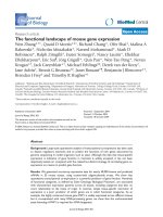
There were 21 strains available for PFGE analysis. Figure 1
shows the PFGE patterns. Of these, 16 distinct patterns,
including three clusters of patterns with over 95% similar-
ity were detected. Two of the clusters, incorporating
strains T46/9 and T49/4 and strains T27/15, T44/6, B6/2,
B6/5 respectively, were isolated from the same patients.
Table 1: Clinical characteristics of Stenotrophomonas maltophilia
bacteraemia Tawam Hospital 2000–2004
Number (n = 25) (%)
Age
Adult 18 (72.0)
Child 7 (28.0)
Hospital Acquired 25 (100)
Clinical Unit
Adult Oncology 11 (44.0)
Paediatric Oncology 5 (20.0)
Intensive Care Unit
+
3 (12.0)
Renal Dialysis Unit 4 (16.0)
Neonatal Intensive Care 1 (4.0)
Paediatric Medical 1 (4.0)
Underlying Disease
Malignancy 19 (76.0)
End stage renal failure 4 (16.0)
Prematurity 1 (4.0)
End stage respiratory failure 1 (4.0)
Immunocompromised 25 (100)
Source
Line-associated 22 (88.0)
Febrile neutropenia 1 (4.0)
Pneumonia 1 (4.0)
Skin soft tissue infection 1 (4.0)
Polymicrobial 9** (36.0)
Treatment of line infection
Line removal 11/12* (91.7)
Antimicrobial therapy
Cotrimoxazole 14/16* (87.5)
Outcome
Death 2
Death attributed to S. maltophilia 0
+
All were adult oncology patients
* Where data were available ** Isolated with 1–3 other organisms
Annals of Clinical Microbiology and Antimicrobials 2006, 5:32 />Page 4 of 6
(page number not for citation purposes)
The latter group was isolated over a period spanning 8
months. The third cluster (V2067 and V3192) were two
strains from two different patients on different wards at
different times. Duplicate strains from the same patient
showed indistinguishable PFGE profiles (data not
shown). Overall, S. maltophilia isolates in our hospital rep-
resented diverse strains and they were unrelated. With the
exception of the three clusters, 2 of which comprised iso-
lates from the same patients, there were as many PFGE
types as patients.
Discussion
The major aim of our study was to try to investigate the
epidemiology of S. maltophilia isolates in our hospital and
thus identify targets to attempt to interrupt its spread in
the hospital environment. While there have been larger,
more detailed studies reported in the medical literature,
this is the first such report from the UAE and therefore we
do not know whether our experience is typical of the UAE.
Although we did not compare our population with
matched controls, the clinical characteristics with S. mal-
tophilia bacteraemia in our hospital in the UAE are broadly
similar to those reported worldwide. All S. maltophilia
bacteraemia episodes were hospital-acquired and malig-
nancy and central venous catheters were major predispos-
ing factors. Line removal seemed important in the
successful management of line-associated infection,
though the numbers investigated were small. Of the 22
cases where the clinical and microbiological features sug-
gested that line infection was likely, in only 12 cases was
it clearly documented in the clinical records that line
removal had taken place.
Table 3: Organisms isolated with Stenotrophomonas maltophilia in
polymicrobial bacteraemia episodes
Organism Number of episodes
Pseudomonas aeruginosa 3
Acinetobacter sp 3
Coagulase-negative staphylococcus 2
Klebsiella sp 2
Enterobacter sp 2
Bacillus sp 1
Kluyvera sp 1
Table 2: Patient details for cases of Stenotrophomonas maltophilia bacteraemia
Patient Gender Age Underlying disease Source Neutropenic Outcome Death attributed to S.
maltophilia
bacteraemia
1 F 9 years ALL Line Y Recovered NA
2 F 5 years Neuroblastoma Line Y Recovered NA
3 M 59 years End Stage Renal Failure Line N Recovered NA
4 M 52 years End Stage Renal Failure Line N Recovered NA
5 F 54 years End Stage renal failure Line N Recovered NA
6 M 15 months Bronchopulmonary dysplasia Line N Recovered NA
7 M 30 years ALL Line Y Died No*
8 M 18 years ALL Cellulitis Y Recovered NA
9 M 25 years T-cell NHL Pneumonia N Died No*
10 M 40 years AML Line N Recovered NA
11 M 56 years AML Line N Recovered NA
12 M 28 years Bowel carcinoma Febrile neutropenia Y Recovered NA
13 M 17 days Prematurity Line-related abscess N Recovered NA
14 F 27 years Osteosarcoma Line Y Recovered NA
15 M 48 years NHL Line N Recovered NA
16 F 46 years Breast carcinoma Line N Recovered NA
17 M 10 years ALL Line N Recovered NA
18 F 22 years ALL Line N Recovered NA
19 F 35 years AML Line N Recovered NA
20 F 6 years ALL Line N Recovered NA
21 F 49 years End Stage Renal Failure Line N Recovered NA
22 M 32 years AML Line Y Recovered NA
23 M 4 years ALL Line N Recovered NA
24 F 18 years ALL Line N Recovered NA
25 M 41 years NHL Line Y Recovered NA
NA = Not applicable; ALL = Acute lymphoblastic leukaemia; AML = Acute myeloid leukaemia; NHL = Non-Hodgkins Lymphoma
* Death attributed to invasive aspergillosis
Annals of Clinical Microbiology and Antimicrobials 2006, 5:32 />Page 5 of 6
(page number not for citation purposes)
We found that S. maltophilia bacteraemia was polymicro-
bial in 9 out of 25 episodes. This characteristic has been
noted some studies, but not in others and the frequency
with which S. maltophilia was isolated from mixed cultures
in earlier studies led to delay in recognising its pathogenic
potential [1,12]. It is not possible to be absolutely sure if
S. maltophilia isolated from polymicrobial episodes repre-
sented a true pathogen, since the other organisms isolated
from polymicrobial episodes in our patients were also
well-recognised pathogens. However, in those cases were
S. maltophilia was the sole isolate, the clinical features sug-
gested that S. maltophilia was behaving as a pathogen.
There were only two deaths in our series of patients and
both of these were attributed to causes other than S. mal-
tophilia bacteraemia. We measured mortality at 7 days and
it is possible that the mortality would have been higher if
we had used 30 day – mortality. Establishing the mortality
of S. maltophilia bacteraemia from the literature is difficult
because different studies have used different mortality
definitions [13,14].
The results of the PFGE suggest that person to person
spread was not the major mode of transmission of S. mal-
tophilia in this hospital. We found repeat that duplicate
isolates of S. maltophilia had indistinguishable PFGE pro-
files, supporting the validity and reproducibility of PFGE
as a fingerprinting method. One patient had 3 separate
episodes of S. maltophilia bacteraemia associated with
portacath infections, spanning 8 months. PFGE of 4
strains from these 3 episodes revealed indistinguishable
profiles, suggesting that the same strain may have been
Pulsed field gel electrophoresis (PFGE) patterns of the Stenotrophomonas maltophilia strainsFigure 1
Pulsed field gel electrophoresis (PFGE) patterns of the Stenotrophomonas maltophilia strains. T 46/9 and T 49/4
are isolates from patient 21 on Table 2. T 27/15, T 44/6, B6/2, B6/5 are isolates from patient 17 on Table 2. V3192 and V2067
represent patients 6 and 9 respectively on Table 2.
Figure 1 PFGE patterns of the Stenotrophomonas maltophilia strains
Annals of Clinical Microbiology and Antimicrobials 2006, 5:32 />Page 6 of 6
(page number not for citation purposes)
acquired from the same unidentified environmental reser-
voir. However, further investigations are necessary to sup-
port this observation.
We did not find the susceptibility tests performed helpful
in distinguishing the strains or in choosing antimicrobial
therapy. Routine susceptibility tests are unreliable for test-
ing S. maltophilia [11]. We recommended cotrimoxazole
for antimicrobial therapy of S. maltophilia infection as it is
considered the antimicrobial of choice for S. maltophilia
infection. All the strains tested were susceptible using the
breakpoint recommended by NCCLS guidelines [11].
In previous studies S. maltophilia has been isolated from a
wide variety of hospital sources [1]. We suggest that S.
maltophilia isolates in this hospital originate from numer-
ous sources in the hospital environment. We were able to
direct our attention to possible environmental sources,
such as improving the compliance with infection control
procedures involved in central venous catheter care rather
than interventions to prevent person to person spread,
such as single room isolation. Further work is necessary to
identify these sources.
Conclusion
Our study highlights that PFGE and other similar typing
schemes are essential to investigate relationships between
isolates and therefore to provide information to identify
targets and strategies to control the spread of nosocomial
pathogens. The sources and precise modes of spread of S.
maltophilia in our hospital are still not known. However,
person to person transmission of S. maltophilia seems a
rare occurrence and knowledge of this is important in ena-
bling scarce infection control resources to be targeted
most appropriately.
Competing interests
The author(s) declare that they have no competing inter-
ests.
Authors' contributions
PJ conceived of the study, participated in its coordination
and design and drafted the manuscript
AS participated in its design and the molecular analysis
TP participated in its coordination and design and molec-
ular analysis
MH Participated in the molecular analysis and the antimi-
crobial susceptibility testing
RA participated in the coordination of the microbiological
and biochemical analysis
OT participated in the collection, analysis and interpreta-
tion of the clinical data
All authors have read and approved the final manuscript
Acknowledgements
This project was supported by a grant from the Faculty of Medicine and
Health Sciences, United Arab Emirates University.
References
1. Denton M, Kerr KG: Microbiological and clinical aspects of
infection associated with Stenotrophomonas maltophilia
infection. Clin Microbiol Rev 1998, 11:57-80.
2. Gilligan PH, Whittier S: Burkholderia Stenotrophomonas, Ralsto-
nia, Brevundimonas, Comomonas and Acidovorax. In Manual of
Clinical Microbiology 7th edition. Edited by: Murray PR, Baron EJ, Pfaller
MA, Tenover FC, Yolken RH. Washington DC: American Society for
Microbiology; 1999:526-538.
3. Gales AC, Jones RN, Forward KR, Linares J, Sader HS, Vernhoef J:
Emerging importance of multidrug-resistant Acinetobacter
species and Stenotrophomonas maltophilia as pathogens in
seriously ill patients: geographic patterns, epidemiological
features and trends in the SENTRY antimicrobial surveil-
lance programme (1997–1999). Clin Infect Dis 2001:S104-S113.
4. Pfaller MA, Acar J, Jones RN, Verhoef J, Turnidge J, Sader HS: Inte-
gration of molecular characterization of microorganisms in
a global antimicrobial resistance surveillance program. Clin
Infect Dis 2001:S156-167.
5. Berg G, Roskot N, Smalla K: Genotypic and phenotypic relation-
ships between clinical and environmental isolates of Steno-
trophomonas maltophilia. J Clin Microbiol 1999, 37:3594-3600.
6. Denton M, Todd N, Kerr K, Hawkey P, Littlewood J: Molecular epi-
demiology of Stenotrophomonas maltophilia isolated from
clinical specimens from cystic fibrosis patients and associ-
ated environmental samples. J Clin Microbiol 1998, 36:1953-1958.
7. Gulmez D, Hascelik G: Stenotrophomonas maltophilia: antimi-
crobial resistance and molecular typing of an emerging path-
ogen in a Turkish University Hospital. Clin Microbiol Infect 2005,
11:880-6.
8. Silbert S, Pfaller MA, Hollis RJ, Barth AL, Sader HS: Evaluation of
three molecular typing techniques for nonfermentative
Gram-negative bacilli. Infect Control Hosp Epidemiol 2004,
25:847-51.
9. Trad O, Jumaa PA, Afify Z: The changing pattern of bloodstream
infection in pediatric oncology patients in the United Arab
Emirates. Pediatr Hematol Oncol 2003, 20:281-289.
10. Garner JS, Jarvis WR, Emori TG, Horan TC, Hughes JM: CDC defi-
nitions for nosocomial infections 1998. Am J Infect Control 1998,
16:128-40.
11. Clinical and Laboratory Standards Institute/NCCLS: Performance
Standards for Antimicrobial Susceptibility Testing: Fifteenth
Informational Supplement. CLSI/NCCLS document M100-
S15. Clinical and Laboratory Standards Institute, Wayne, Pennsylvania,
USA 2005.
12. Jang T-N, Wang FD, Wang LH, Liu CY, Liu LM: Xanthomonas mal-
tophilia bacteremia: an analysis of 32 cases. J Formos Med Assoc
1992, 91:1170-1176.
13. Muder RR, Harris AP, Muller S, Edmond M, Chow JW, Papadakis K,
Wagener MW, Bodey GP, Steckelberg JM: Bacteremia due to
Stenotrophomonas (Xanthomonas) maltophilia: a prospective,
multicenter study of 91 episodes. Clin Infect Dis 1996, 22:508-12.
14. Friedman ND, Korman TM, Fairley CK, Franklin JC, Spelman DW:
Bacteraemia due to Stenotrophomonas maltophilia: an analy-
sis of 45 episodes. J Infect 2002, 45:47-53.