Báo cáo y học: "Mechanisms of Degranulation in Neutrophils" pdf
Bạn đang xem bản rút gọn của tài liệu. Xem và tải ngay bản đầy đủ của tài liệu tại đây (342.47 KB, 11 trang )
98
Neutrophils are highly mobile and short-lived
white blood cells that are densely packed with
secretory granules. They derive from the bone
marrow, where they mature in response to appro-
priate cytokines. Following this, they emigrate
from the bone marrow into the blood and circu-
late to tissues. In healthy individuals, peripheral
blood neutrophils make up the majority of white
blood cells (40–80%). The lungs form the largest
marginated pool of neutrophils in the body. In the
airways, neutrophils fulfill an important sentinel
role in maintaining sterility. As a major effector
cell in innate immunity, neutrophils act as a dou-
ble-edged sword. If neutrophils are absent (eg, in
congenital neutropenia or the more common cyclic
neutropenia), infections result from overgrowth of
bacteria and fungi at sites of injury or exposed
regions of mucosal tissues. At the other extreme,
accumulation and overactivation of neutrophils can
be fatal in disorders such as in septic shock or acute
respiratory distress. The tissue-damaging effects
of neutrophils are completely dependent on the
activation of mediator release.
Mediator release is defined as the secretion
or production of proinflammatory substances that
are derived from intracellular stored granules or
synthesized de novo on stimulation by receptors.
Neutrophils release granule-derived mediators
by degranulation, or exocytosis, of membrane-
bound secretory granules. The neutrophil also
possesses the capacity to release a diverse array
of antimicrobial proteins and enzymes intracel-
lularly into membrane-bound organelles, called
phagosomes, which contain engulfed small
microorganisms. At the same time, neutrophils
release reactive oxygen species and cytokines
outside the cells to kill extracellular bacteria and
recruit additional leukocytes to the region of
infection or inflammation.
Review Article
Mechanisms of Degranulation in Neutrophils
Paige Lacy, PhD
Abstract
Neutrophils are critical inflammatory cells that cause tissue damage in a range of diseases and disor-
ders. Being bone marrow–derived white blood cells, they migrate from the bloodstream to sites of tis-
sue inflammation in response to chemotactic signals and induce inflammation by undergoing receptor-
mediated respiratory burst and degranulation. Degranulation from neutrophils has been implicated as
a major causative factor in pulmonary disorders, including severe asphyxic episodes of asthma. How-
ever, the mechanisms that control neutrophil degranulation are not well understood. Recent observa-
tions indicate that granule release from neutrophils depends on activation of intracellular signalling path-
ways, including -arrestins, the Rho guanosine triphosphatase Rac2, soluble NSF attachment protein
(SNAP) receptors, the src family of tyrosine kinases, and the tyrosine phosphatase MEG2. Some of these
observations suggest that degranulation from neutrophils is selective and depends on nonredundant sig-
nalling pathways. This review focuses on new findings from the literature on the mechanisms that con-
trol the release of granule-derived mediators from neutrophils.
P. Lacy—Pulmonary Research Group, Department of
Medicine, University of Alberta, Edmonton, AB
Correspondence to: Paige Lacy, PhD, 550A HMRC,
Department of Medicine, University of Alberta,
Edmonton, AB T6G 2S2; E-mail
DOI 10.2310/7480.2006.00012
Mechanisms of Degranulation in Neutrophils — Lacy 99
Excessive neutrophil degranulation is a com-
mon feature of many inflammatory disorders,
such as severe asphyxic episodes of asthma, acute
lung injury, rheumatoid arthritis, and septic shock.
1
A recent study by Brinkmann and colleagues
described a novel mechanism by which neutrophils
eliminate bacteria.
2
On activation by a range of
mediators, including interleukin-8 (IL-8),
lipopolysaccharide, and interferon-
␣ with com-
plement 5a,
3
neutrophils were shown to generate
a web of extracellular fibres known as neutrophil
extracellular traps (NETs), composed of deoxyri-
bonucleic acid (DNA), histones, and antimicrobial
granule proteins, which are highly effective at
trapping and killing invasive bacteria. The authors
proposed that NETs amplified the effectiveness of
antimicrobial components by concentrating them
in a fibrous network and reducing their exposure
to host tissues. Although this report fell short on
describing the molecular mechanisms responsible
for NET formation and its association with gran-
ular protein, it opened a new horizon in the field
of neutrophil biology as it relates to mediator
release and bactericidal activity.
Therefore, to attenuate a neutrophilic inflam-
matory response, an effective therapeutic strategy
would be one that is directed at down-regulation
of neutrophil degranulation. Recent findings have
identified a number of important signalling path-
ways in neutrophils that may be useful as targets
for pharmacologic intervention of degranulation.
Granule Types in Neutrophils
Neutrophils contain at least four different types
of granules: (1) primary granules, also known as
azurophilic granules; (2) secondary granules,
also known as specific granules; (3) tertiary gran-
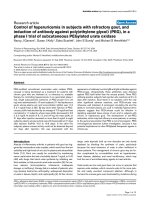
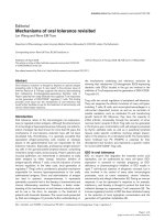
ules; and (4) secretory vesicles (Figure 1). The
Figure 1 Rho guanosine triphos-
phatase and SNAP receptor
(SNARE) signalling pathways
involved in Ca
2+
-dependent neu-
trophil degranulation. Receptor
binding by a chemoattractant
leads to G protein–coupled sig-
nal transduction (G protein–cou-
pled receptor [GPCR]) through
multiple overlapping intracellu-
lar pathways to regulate the
selective release of neutrophil
granules. Some of these path-
ways may be non-redundant, for
example, through G protein–acti-
vated guanine nucleotide
exchange factors (GEFs) to acti-
vate Rac2, which selectively
mobilizes primary granules. ER
= endoplasmic reticulum; fMLP
= F-Met-Lev-Phe; IL = inter-
leukin; InsP3 = inositol 1, 4, 5-
triphosphate; LPTF = lactoperin;
MMP = matrix metalloprotease;
MPO = myeloperoxidase;
VAMP = vesicle-associated
membrane protein.
100 Allergy, Asthma, and Clinical Immunology / Volume 2, Number 3, Fall 2006
primary granules are the main storage site of the
most toxic mediators, including elastase,
myeloperoxidase, cathepsins, and defensins. The
secondary and tertiary granules contain lacto-
ferrin and matrix metalloprotease 9 (also known
as gelatinase B), respectively, among other sub-
stances.
4
The secretory vesicles in human neu-
trophils contain human serum albumin, sug-
gesting that they contain extracellular fluid that
was derived from endocytosis of the plasma
membrane. The secondary and tertiary granules
have overlapping contents but can be discrimi-
nated by their intrinsic buoyant densities when
centrifuged on gradient media.
5
Granules are
prevented from being released until receptors in
the plasma membrane or phagosomal membrane
signal to the cytoplasm to activate their movement
to the cell membrane for secretion of their con-
tents by degranulation. This is an important con-
trol mechanism as the neutrophil is highly
enriched in tissue-destructive proteases.
Degranulation Mechanisms in Neutrophils
When receptor stimulation by a secretagogue
occurs, granules translocate to the phagosomal or
plasma membrane, where they dock and fuse with
the membrane to release their contents. The release
of granule-derived mediators from granulocytes
occurs by tightly controlled receptor-coupled
mechanisms, leading to exocytosis. Exocytosis is
postulated to take place in four discrete steps.
6
The
first step of exocytosis is granule recruitment from
the cytoplasm to target membrane, which is depen-
dent on actin cytoskeleton remodelling and micro-
tubule assembly.
7
This is followed by vesicle teth-
ering and docking, leading to contact of the outer
surface of the lipid bilayer membrane surround-
ing the granule with the inner surface of the tar-
get membrane. Granule priming then follows to
make granules fusion-competent to ensure that
they fuse rapidly, and a reversible fusion pore
structure develops between the granule and the tar-
get membrane. Granule fusion occurs by the expan-
sion of the fusion pore, leading to complete fusion
of the granule with the target membrane to release
granular contents. In the case of exocytosis, this
increases the total surface area of the cell and
exposes the interior membrane surface of the gran-
ule to the exterior.
Translocation and exocytosis of granules in
neutrophils require, as a minimum, increases in
intracellular Ca
2+
, as well as hydrolysis of adeno-
sine triphosphate (ATP) and guanosine triphosphate
(GTP). The target molecules for these effectors are
numerous and include Ca
2+
-binding proteins such
as annexins and calmodulin and GTP-binding
proteins such as G proteins and small monomeric
proteins. ATP is used by ATP-hydrolyzing enzymes
(adenosine triphosphatases) and kinases, which act
by phosphorylating downstream effector mole-
cules. Combined with activation of these effector
molecules is reorganization of the actin cytoskele-
ton, which forms a mesh around the periphery of
the cell as a shield against granule docking and
fusion. The actin cytoskeletal mesh must be dis-
assembled to allow access of granules to the inner
surface of the plasma membrane. It is likely that
the process of granule translocation and exocyto-
sis involves activation and recruitment of many dif-
ferent signalling molecules, only some of which
are beginning to be identified.
Ca
2+
Signalling in Exocytosis
Increases in intracellular Ca
2+
alone are sufficient
to induce the release of many of the granule types
in neutrophils, particularly if the concentration of
Ca
2+
is elevated to sufficiently high levels by the
use of Ca
2+
ionophores such as A23187 or iono-
mycin. A hierarchy of granule release exists in
response to elevating concentrations of Ca
2+
.
8
The
order of release is secretory vesicles > tertiary
granules > secondary granules > primary gran-
ules.
8,9
The release of each type of granule appears
to be regulated by different intracellular signalling
pathways. Many neutrophil receptors activate
increased Ca
2+
levels, including the seven trans-
membrane-spanning G protein–coupled recep-
tors, such as the formyl peptide receptor (that
binds to the bacterial tripeptide f-Met-Leu-Phe) and
chemokine receptors (such as CXCR1). Although
Ca
2+
is a crucial second messenger in the activa-
tion of exocytosis, the specific target molecules for
Ca
2+
in neutrophil degranulation have not yet been
identified (see Figure 1).
Mechanisms of Degranulation in Neutrophils — Lacy 101
Phospholipid Signalling in Degranulation
Numerous studies have indicated a role for phos-
pholipids, particularly polyphosphoinositides, in
the regulation of neutrophil degranulation.
Polyphosphoinositide production, such as phos-
phatidylinositol bisphosphate (PIP
2
), induced by
activation of the hematopoietic cell–specific iso-
form phosphatidylinositol 3-kinase (PI3K)-
␥, has
been shown to be required for granule exocytosis
in permeabilized neutrophil-like cells, HL-60
cells.
10
The intracellular sites of PIP
2
formation in
neutrophils are not known, but it is likely to occur
both at the plasma membrane and on granule
membranes. Regions of PIP
2
enrichment in the
membrane form essential binding sites for many
intracellular signalling molecules, particularly
those that contain pleckstrin homology domains.
Phosphatidylinositol transfer protein has been
shown to be essential for the transport of phos-
phatidylinositol to cellular membranes as a sub-
strate for PI3K activity to generate PIP
2
and is also
capable of restoring exocytotic responses in HL-
60 cells.
10
In addition, a role for phospholipase D
has been indicated in neutrophil degranulation, par-
ticularly for primary and secondary granule release,
as its product, phosphatidic acid, induces the
release of these granules.
11
Thus, membrane lipids
form an essential component of degranulation in
neutrophils.
Role for src Family Kinases
in Neutrophil Degranulation
Protein phosphorylation is a critical event in neu-
trophil activation leading from receptor stimula-
tion to exocytosis. Phosphorylation is carried out
by kinases, which are themselves frequently acti-
vated by phosphorylation by upstream molecules.
This specifically involves the attachment of a
phosphate molecule, donated by intracellular ATP,
to a key site in the effector molecule, leading to
conformational changes that cause activation.
Receptor stimulation through the formyl peptide
receptor by f-Met-Leu-Phe leads to phosphoryla-
tion of a wide range of kinases, which then acti-
vate their respective effector pathways. Kinases can
be discriminated based on their affinity for different
amino acid residues in effector molecules. Thus,
serine/threonine kinases and tyrosine kinases have
been characterized as distinct types of kinases
involved in receptor signalling. Tyrosine kinases
are further differentiated for their intrinsic asso-
ciation with the intracellular domain of receptors
(receptor tyrosine kinases) or as cytosolic enzymes
(nonreceptor tyrosine kinases).
The
src family of nonreceptor tyrosine kinases
has been implicated in the control of exocytosis of
granule products from neutrophils. Three
src fam-
ily members, Hck, Fgr, and Lyn, have been shown
to be expressed in neutrophils and are activated by
f-Met-Leu-Phe receptor stimulation. Interestingly,
different granule populations appear to be associ-
ated with different src kinases. Hck translocates to
the primary granule population following cell acti-
vation
12
whereas Fgr becomes associated with the
secondary granules during exocytosis.
13
The selec-
tive recruitment of src kinases indicates that dif-
ferent signalling pathways exist in neutrophils to
induce the release of each granule population.
Recent studies showed that treatment of human neu-
trophils with the src family inhibitor PP1 led to inhi-
bition of the release of primary granules, secondary
granules, and secretory vesicles in response to f-
Met-Leu-Phe.
14
Neutrophils isolated from
hck
–/–
fgr
–/–
lyn
–/–
triple knockout mice also showed
a deficiency in secondary granule release, although
it was not possible to determine primary granule
release.
14
The deficiency in secondary granule
release correlated with reduced p38 mitogen-acti-
vated protein (MAP) kinase activity, suggesting that
src kinases act upstream of p38 MAP kinase.
Indeed, treatment of neutrophils with the p38 MAP
kinase inhibitor SB203580 led to reduced primary
and secondary granule exocytosis in response to f-
Met-Leu-Phe. Another kinase inhibitor, PD98059,
which blocks extracellular-related kinase (ERK)1/2
activity, did not affect the release of primary and
secondary granules or secretory vesicles. These
findings indicate that
src kinases and p38 MAP
kinase play a role in regulating the release of gran-
ules in response to f-Met-Leu-Phe receptor stim-
ulation in neutrophils and probably act at an early
signalling step proximal to the receptor in this
process (Figure 2).
102 Allergy, Asthma, and Clinical Immunology / Volume 2, Number 3, Fall 2006
-Arrestin Function in Regulating
Exocytosis
The family of scaffolding proteins, -arrestins, may
be required for activating signalling pathways
leading to exocytosis of primary and secondary
granules in neutrophils.
15
-Arrestins are a group
of cytosolic phosphoproteins that were previously
characterized for their role in endocytosis of lig-
and-bound chemokine receptors, particularly
CXCR1, which is the high-affinity receptor for the
neutrophil chemotactic factor IL-8.
-Arrestins act
by uncoupling activated G protein–coupled recep-
tors from their associated heterotrimeric G proteins
and binding directly to the cytoplasmic tail of the
CXCR1 receptor.
15,16
Dominant negative mutants
of
-arrestin were shown to inhibit the release of
granules following transfection of a rat mast cell
line (RBL cells) that serves as a model for neu-
trophil degranulation.
15
Interestingly, -arrestins
also associate with the primary and secondary
granules in IL-8-activated neutrophils, and they do
so by binding to Hck and Fgr, respectively.
15
Thus,
-arrestins act at two sites in the cell during
chemokine activation: one site at the receptor in
the plasma membrane and a second on granule
membranes (see Figure 2).
Requirement for Guanosine
Triphosphatases in Exocytosis
Exocytosis requires binding of GTP to intracellular
effector molecules as the addition of the nonhy-
drolyzable analog GTP
␥S to permeabilized or
patch-clamped neutrophils leads to secretion of
granule-derived mediators.
17
This suggests that
GTP-binding proteins, including guanosine
triphosphatases (GTPases), may be involved in
granule translocation and exocytosis. To date,
over 100 different types of GTPases have been
identified, with heterotrimeric G proteins and
ras-
related monomeric GTPases being two of the
most comprehensively studied families of regu-
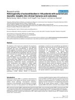
Figure 2 Tyrosine kinases asso-
ciated with chemokine-induced
neutrophil degranulation. Recep-
tor binding leads to direct bind-
ing of the G protein–coupled
receptor (GPCR) by -arrestins,
which also translocate to pri-
mary and secondary granules
along with src family kinases
Hck and Fgr. IL = interleukin;
LTF = ; MAP = mitogen-acti-
vated protein; MMP = matrix
metalloprotease; MPO =
myeloperoxidase.
latory GTPases. Whereas heterotrimeric G proteins
typically bind to the plasma membrane to trans-
duce receptor signals to the cytoplasm, the super-
family of
ras-related GTPases can reside in the
cytoplasm, in actin cytoskeleton, or on mem-
branes in the cell to fulfill a regulatory role in cell
activation.
Ras-related GTPases are important
switches for turning on or off a signalling event.
They are switched on by binding to high-energy
GTP, which is cleaved to form guanosine diphos-
phate to activate the next effector molecule in the
signalling pathway. Binding to GTP induces the
association of many cytosolic GTPases to mem-
brane or cytoskeletal sites within the cell.
Ras-related GTPases can be divided into sev-
eral subfamilies based on their homology at the
amino acid level. One particular group of ras-
related GTPases is the Rho subfamily of GTPases,
which serves a role in regulating actin cytoskele-
tal rearrangement and in the release of reactive oxy-
gen species. Remodelling of the actin cytoskele-
ton is critical for allowing a diverse range of
cellular activities to occur, including cell motility
(chemotaxis), phagocytosis, and exocytosis. The
three prototypical members of the Rho GTPase
subfamily are Rho, Rac, and Cdc42.
18–20
Rac is pre-
sent as three different isoform proteins: Rac1,
Rac2, and Rac3. The functions of Rac1 and Rac2
in superoxide generation and chemotaxis are well
established in neutrophils.
21
Rho GTPases are also
substrates for a number of bacterial toxins, includ-
ing
Clostridium difficile toxin B and Clostridium
sordellii
lethal toxin, which act by glucosylating
Rho GTPases.
22,23
Rac1 and Rac2 possess 92% homology in
their amino acid sequences and differ mainly in the
final 10 amino acids in their carboxyl termini.
Both isoform proteins are expressed in neutrophils,
although human neutrophils express more Rac2
than Rac1.
24
It is because of this high homology
that they serve functionally interchangeable roles
in actin cytoskeletal remodelling and regulation of
the release of reactive oxygen species by activa-
tion of reduced nicotinamide adenine dinucleotide
phosphate (NADPH) oxidase in neutrophils.
25–27
Interestingly, because of sequence variation in a
short carboxyl terminal sequence, Rac2 is the
preferential activator of NADPH oxidase in neu-
trophils.
28
Human neutrophils translocate most of
their Rac protein to intracellular sites of NADPH
oxidase activation following stimulation of res-
piratory burst,
29
suggesting that the neutrophil
oxidase preferentially produces reactive oxygen
species at intracellular sites.
In spite of their high homology, however,
Rac1 and Rac2 are divergent in their functions in
certain types of cellular activities.
30–32
We have
determined that Rac2 serves a crucial and selec-
tive role in degranulation from neutrophils.
32
Gene
deletion of Rac2 led to a profound degranulation
defect in neutrophils, with a complete loss of pri-
mary granule release from murine bone marrow
neutrophils. Release of granule enzymes from
secondary and tertiary granule was normal in
Rac2
–/–
neutrophils, indicating a selective role for
Rac2 in primary granule exocytosis. Rac2
–/–
neu-
trophils express normal or even elevated levels of
Rac1,
28,33,34
further suggesting that Rac2 serves a
unique and distinct role from Rac1 in regulating
translocation and exocytosis of granules. In addi-
tion, although Rac2
–/–
neutrophils showed a loss
of primary granule release, p38 MAP kinase phos-
phorylation was still evident in response to f-Met-
Phe-Leu stimulation. This is in contrast to the
findings of Mocsai and colleagues, who demon-
strated an important role for p38 MAP kinase in
primary granule release by the use of chemical
inhibitors.
14
Rac2
–/–
neutrophils also failed to translocate pri-
mary granules to the cell membrane during f-Met-
Leu-Phe stimulation.
32
Thus, the defect in primary
granule exocytosis in these cells lies in the translo-
cation machinery required to move the granules to
the membrane for docking and fusion. The translo-
cation of granules is likely to require actin cytoskele-
ton remodelling and/or microtubule movements, and
Rac2 has been shown to induce the formation of F-
actin, which is required for chemotaxis.
33
Indeed,
Rac2
–/–
neutrophils did not bind as well as their wild-
type counterparts to adhesion molecules.
33
Identi-
fication of downstream effector molecules of Rac2
that are responsible for regulating actin cytoskele-
tal remodelling and/or microtubule rearrangements
will be important in identifying the pathway(s)
associated with Rac2-mediated primary granule
release (see Figure 1).
Mechanisms of Degranulation in Neutrophils — Lacy 103
104 Allergy, Asthma, and Clinical Immunology / Volume 2, Number 3, Fall 2006
SNARE Molecule Binding in Exocytosis
from Neutrophils
The final step of exocytosis involves the mutual
recognition of secretory granules and target mem-
branes, which is postulated to involve a set of intra-
cellular receptors that guide the docking and fusion
of granules. This led to the formation of the SNAP
receptor (SNARE) paradigm, which states that
secretory vesicles possess membrane-bound recep-
tor molecules that allow their binding by another set
of membrane-bound receptors in target membranes.
35
Studies on yeast and neuronal cells have
yielded significant insights into highly conserved
components of a fusion complex of membrane-
bound proteins proposed to be essential for vesic-
ular docking and fusion in all cell types, known
as SNAREs.
35,36
The prototypical members of this
complex are vesicle-associated membrane pro-
tein (VAMP)-1 (also known as synaptobrevin 1),
syntaxin 1, and synaptosome-associated protein of
25 kD (SNAP-25). The exocytotic SNARE com-
plex consists of a vesicular SNARE VAMP, which
binds to plasma membrane target SNAREs syn-
taxin 1 and SNAP-25. The fusion of membranes
is proposed to depend on cytosolic
N-ethyl-
maleimide-sensitive factor (NSF) and
␣-, -, or ␥-
SNAP (soluble NSF-attachment protein)-medi-
ated disassembly of the SNARE complex.
35
During binding, SNARE molecules form a
coiled-coil structure with four separate
␣-helices
contributed by three different molecules. The
binding region associated with the four
␣-helices
is known as the SNARE motif. The stability of the
bonds within the SNARE structure is such that it
is resistant to treatment with detergents such as
sodium dodecyl sulphate.
37
SNARE molecules are exquisitely sensitive to
cleavage by clostridial neurotoxins containing
zinc endopeptidase activity, in particular, tetanus
toxin (TeNT) and botulinum toxin serotypes
(BoNT/A, B, C, D, E, F, and G).
38
The effects of
these toxins on intracellular SNARE molecules are
likely to be the molecular basis of spastic and
flaccid paralysis induced by tetanus and botu-
linum toxin poisoning, respectively. TeNT and
BoNT holotoxins are only able to enter neuronal
cells since their heavy chain components require
a ganglioside-binding site on the cell surface,
lacking in nonneuronal cells.
38
Other isoforms of
SNAREs have been identified in cells outside the
neuronal system (syntaxin 4 and SNAP-23)
39
whereas VAMP-2 expression is widely distrib-
uted between neuronal and nonneuronal tissues.
40
In addition, VAMP-4,
41
VAMP-5,
42
and the TeNT-
insensitive isoforms VAMP-7 (formerly known as
TeNT-insensitive VAMP or TI-VAMP)
43–46
and
VAMP-8 have been characterized in nonneuronal
tissues.
47–49
Neutrophils have been reported to express
many of the SNARE isoforms so far identified. In
an early report, neutrophils were shown to express
syntaxin 4 and VAMP-2.
50
VAMP-2 was localized
to tertiary granules and CD35
+
secretory vesicles,
and VAMP-2
+
vesicles translocated to the plasma
membrane during Ca
2+
ionophore stimulation. By
reverse transcriptase–polymerase chain reaction,
the messenger ribonucleic acid encoding syntax-
ins 1A, 3, 4, 5, 6, 7, 9, 11, and 16 have been iden-
tified in human neutrophils and a neutrophil-dif-
ferentiated cell line (HL-60).
51
SNAP-23 and
syntaxin 6 appear to be important in regulating neu-
trophil secondary granule exocytosis using anti-
bodies against these molecules in electroperme-
abilized cells stimulated with Ca
2+
and GTP␥S.
52
Finally, the addition of antibodies to VAMP-2 and
syntaxin 4 to electropermeabilized neutrophils
blocked Ca
2+
and GTP␥S-induced exocytosis.
53
Exocytosis in the latter two articles was measured
by flow cytometric analysis of granule markers
CD63 (primary granules) and CD66b (secondary
granules), which are up-regulated on the cell sur-
face during stimulation. It was shown that anti-
VAMP-2 blocked secondary granule CD66b up-
regulation in response to Ca
2+
and GTP␥S whereas
there was no inhibition of CD63
+
primary gran-
ule release with antibody against VAMP-2. In
summary, although VAMP-2 was shown to be
involved in secondary granule exocytosis, there are
no reports describing a VAMP isoform associated
with primary granule exocytosis. This would
appear to be a significant gap in our understand-
ing of the mechanisms of degranulation in these
cells as primary granules are specifically enriched
in bactericidal and cytotoxic mediators, including
elastase and myeloperoxidase.
Mechanisms of Degranulation in Neutrophils — Lacy 105
We recently determined that VAMP-7 is highly
expressed in all neutrophil granule populations and
that it may be an essential component for SNARE-
mediated exocytotic release of primary, secondary,
and tertiary granule release.
54
Inhibition of VAMP-
7 by low concentrations of specific anti-VAMP-7
antibody prevented the release of myeloperoxi-
dase, lactoferrin, and matrix metalloprotease 9 in
streptolysin-O-permeabilized human neutrophils.
These findings indicate that VAMP-7 may play a
promiscuous role in controlling regulated exocytosis
of numerous granule populations. This is compat-
ible with the recent observations that SNARE mol-
ecules are capable of binding multiple cognate and
noncognate partners.
55
Thus, SNARE isoforms are
likely to play a crucial role in the regulation of
granule fusion in neutrophils (see Figure 1).
Other Potential Regulatory Molecules
of Exocytosis in Neutrophils
Recent findings have suggested a role for a pro-
tein tyrosine phosphatase MEG2 in the regulation
of neutrophil degranulation. Neutrophils express
MEG2 in their primary, secondary, and tertiary
granules, which translocates to the phagosomal
membrane on phagocytosis of serum-opsonized
iron beads.
56
MEG2 was recently shown to be a
phosphatase required for dephosphorylation of
NSF, the cytosolic ATPase that is required to cycle
SNARE proteins between bound and unbound
conformations to allow repeated cycles of mem-
brane fusion.
57
This study demonstrated for the first
time that NSF possesses a tyrosine residue that is
phosphorylated and that dephosphorylation trig-
gers the binding of another cytosolic protein,
␣-
SNAP, which is also required for SNARE cycling,
to promote vesicular fusion. Cells expressing a
dephosphorylated form of mutant NSF exhibited
substantial enlargement of their granules, sug-
gesting that the dephosphorylated NSF remained
bound to
␣-SNAP to allow repeated homotypic
granule fusion and enlargement of the granules in
the cells. Transfection of a phosphomimicking
mutant of NSF was shown to inhibit the secretion
of IL-2 from Jurkat T cells.
57
In addition, MEG2
was shown to be activated by polyphosphoinosi-
tides, particularly PIP2,
56
suggesting that MEG2
is directly associated with the membrane fusion
event in granule fusion.
Summary
These recent experimental observations reveal
that a large group of intracellular signalling mol-
ecules exists to regulate translocation of granules
to the cell membrane for docking and fusion to
release their contents. Many of these molecules are
already natural targets for bacterial toxins to inhibit
their function, which highlights their important role
in regulating bactericidal mediator release. It may
be possible to exploit the use of bacterial toxins
as a tool to prevent or modulate neutrophil degran-
ulation. Neutrophil degranulation is an important
event in inflammatory diseases such as asthma and
chronic obstructive pulmonary disease (COPD).
Products of neutrophil degranulation, including the
high-molecular-weight form of matrix metallo-
protease 9 specific to neutrophils, have been shown
to increase in proportion to asthma severity in the
airways of asthmatic patients.
58
Moreover, neu-
trophils and their products are strongly associ-
ated with early pathogenesis of COPD.
59
Further
analysis of the signalling pathways that are specif-
ically activated to induce the release of different
granule populations in neutrophils may create
opportunities for the development of drugs that will
prevent degranulation from neutrophils in airway
diseases and inflammatory disorders.
References
1. Skubitz KM. Neutrophilic leukocytes. In: Lee
GR, Foerster J, Lukens J, et al, editors. Win-
trobe’s clinical hematology. Vol 1. Baltimore:
Williams & Wilkins; 1999. p. 300–50.
2. Brinkmann V, Reichard U, Goosmann C, et al.
Neutrophil extracellular traps kill bacteria. Sci-
ence 2004;303:1532–5.
3. Martinelli S, Urosevic M, Daryadel A, et al.
Induction of genes mediating interferon-depen-
dent extracellular trap formation during neu-
trophil differentiation. J Biol Chem
2004;279:44123–32.
106 Allergy, Asthma, and Clinical Immunology / Volume 2, Number 3, Fall 2006
4. Borregaard N, Cowland JB. Granules of the
human neutrophilic polymorphonuclear leuko-
cyte. Blood 1997;89:3503–21.
5. Kjeldsen L, Sengelov H, Lollike K, et al. Isola-
tion and characterization of gelatinase granules
from human neutrophils. Blood 1994;83:1640–9.
6. Toonen RF, Verhage M. Vesicle trafficking: plea-
sure and pain from SM genes. Trends Cell Biol
2003;13:177–86.
7. Burgoyne RD, Morgan A. Secretory granule exo-
cytosis. Physiol Rev 2003;83:581–632.
8. Sengelov H, Kjeldsen L, Borregaard N. Control
of exocytosis in early neutrophil activation. J
Immunol 1993;150:1535–43.
9. Bentwood BJ, Henson PM. The sequential release
of granule constituents from human neutrophils.
J Immunol 1980;124:855–62.
10. Fensome A, Cunningham E, Prosser S, et al.
ARF and PITP restore GTPgS-stimulated protein
secretion from cytosol-depleted HL60 cells by
promoting PIP2 synthesis. Curr Biol
1996;6:730–8.
11. Kaldi K, Szeberenyi J, Rada BK, et al. Contri-
bution of phospholipase D and a brefeldin A-sen-
sitive ARF to chemoattractant-induced superox-
ide production and secretion of human
neutrophils. J Leukoc Biol 2002;71:695–700.
12. Mohn H, Le Cabec V, Fischer S, Maridonneau-
Parini I. The src-family protein-tyrosine kinase
p59hck is located on the secretory granules in
human neutrophils and translocates towards the
phagosome during cell activation. Biochem J
1995;309(Pt 2):657–65.
13. Gutkind JS, Robbins KC. Translocation of the
FGR protein-tyrosine kinase as a consequence of
neutrophil activation. Proc Natl Acad Sci U S A
1989;86:8783–7.
14. Mocsai A, Jakus Z, Vantus T, et al. Kinase path-
ways in chemoattractant-induced degranulation
of neutrophils: the role of p38 mitogen-activated
protein kinase activated by Src family kinases.
J Immunol 2000;164:4321–31.
15. Barlic J, Andrews JD, Kelvin AA, et al. Regula-
tion of tyrosine kinase activation and granule
release through beta-arrestin by CXCRI. Nat
Immunol 2000;1:227–33.
16. Ferguson SS, Downey WE III, Colapietro AM,
et al. Role of beta-arrestin in mediating agonist-
promoted G protein-coupled receptor internal-
ization. Science 1996;271:363–6.
17. Gomperts BD. GE: a GTP-binding protein medi-
ating exocytosis. Annu Rev Physiol
1990;52:591–606.
18. Wennerberg K, Der CJ. Rho-family GTPases: it’s
not only Rac and Rho (and I like it). J Cell Sci
2004;117:1301–12.
19. Etienne-Manneville S, Hall A. Rho GTPases in
cell biology. Nature 2002;420:629–35.
20. Ridley AJ. Rho family proteins: coordinating
cell responses. Trends Cell Biol 2001;11:471–7.
21. Diebold BA, Bokoch GM. Molecular basis for
Rac2 regulation of phagocyte NADPH oxidase.
Nat Immunol 2001;2:211–5.
22. Just I, Selzer J, Wilm M, Eichel-Streiber C, et al.
Glucosylation of Rho proteins by Clostridium dif-
ficile toxin B. Nature 1995;375:500–3.
23. Popoff MR, Chaves-Olarte E, Lemichez E, et al.
Ras, Rap, and Rac small GTP-binding proteins
are targets for Clostridium sordellii lethal toxin
glucosylation. J Biol Chem 1996;271:10217–24.
24. Heyworth PG, Knaus UG, Xu X, et al. Require-
ment for posttranslational processing of Rac
GTP-binding proteins for activation of human
neutrophil NADPH oxidase. Mol Biol Cell
1993;4:261–9.
25. Werner E. GTPases and reactive oxygen species:
switches for killing and signaling. J Cell Sci
2004;117:143–53.
26. Bokoch GM, Knaus UG. NADPH oxidases: not
just for leukocytes anymore! Trends Biochem Sci
2003;28:502–8.
27. Bokoch GM, Diebold BA. Current molecular
models for NADPH oxidase regulation by Rac
GTPase. Blood 2002;100:2692–6.
28. Li S, Yamauchi A, Marchal CC, et al. Chemoat-
tractant-stimulated Rac activation in wild-type
and Rac2-deficient murine neutrophils: prefer-
ential activation of Rac2 and Rac2 gene dosage
effect on neutrophil functions. J Immunol
2002;169:5043–51.
29. Lacy P, Abdel-Latif D, Steward M, et al. Diver-
gence of mechanisms regulating respiratory burst
in blood and sputum eosinophils and neutrophils
from atopic subjects. J Immunol
2003;170:2670–9.
30. Filippi MD, Harris CE, Meller J, et al. Local-
ization of Rac2 via the C terminus and aspartic
acid 150 specifies superoxide generation, actin
polarity and chemotaxis in neutrophils. Nat
Immunol 2004;5:744–51.
Mechanisms of Degranulation in Neutrophils — Lacy 107
31. Gu Y, Filippi MD, Cancelas JA, et al. Hematopoi-
etic cell regulation by Rac1 and Rac2 guano-
sine triphosphatases. Science 2003;302:445–9.
32. Abdel-Latif D, Steward M, Macdonald DL, et al.
Rac2 is critical for neutrophil primary granule
exocytosis. Blood 2004;104:832–9.
33. Roberts AW, Kim C, Zhen L, et al. Deficiency of
the hematopoietic cell-specific Rho family
GTPase Rac2 is characterized by abnormalities
in neutrophil function and host defense. Immu-
nity 1999;10:183–96.
34. Abdel-Latif D, Steward M, Lacy P. Neutrophil pri-
mary granule release and maximal superoxide gen-
eration depend on Rac2 in a common signalling
pathway. Can J Physiol Pharmacol 2005;83:69–75.
35. Söllner T, Whiteheart SW, Brunner M, et al.
SNAP receptors implicated in vesicle targeting
and fusion. Nature 1993;362:318–24.
36. Sutton RB, Fasshauer D, Jahn R, Brunger AT.
Crystal structure of a SNARE complex involved
in synaptic exocytosis at 2.4 {151} resolution.
Nature 1998;395:347–53.
37. Chen YA, Scales SJ, Patel SM, et al. SNARE
complex formation is triggered by Ca2+ and dri-
ves membrane fusion. Cell 1999;97:165–74.
38. Schiavo G, Matteoli M, Montecucco C. Neuro-
toxins affecting neuroexocytosis. Physiol Rev
2000;80:717–66.
39. Ravichandran V, Chawla A, Roche PA. Identifi-
cation of a novel syntaxin- and synapto-
brevin/VAMP- binding protein, SNAP-23,
expressed in non-neuronal tissues. J Biol Chem
1996;271:13300–3.
40. Rossetto O, Gorza L, Schiavo G, et al.
VAMP/synaptobrevin isoforms 1 and 2 are widely
and differentially expressed in nonneuronal tis-
sues. J Cell Biol 1996;132:167–79.
41. Steegmaier M, Yang B, Yoo JS, et al. Three novel
proteins of the syntaxin/SNAP-25 family. J Biol
Chem 1998;273:34171–9.
42. Zeng Q, Subramaniam VN, Wong SH, et al. A
novel synaptobrevin/VAMP homologous pro-
tein (VAMP5) is increased during in vitro myo-
genesis and present in the plasma membrane.
Mol Biol Cell 1998;9:2423–37.
43. Galli T, Zahraoui A, Vaidyanathan VV, et al. A
novel tetanus neurotoxin-insensitive vesicle-
associated membrane protein in SNARE com-
plexes of the apical plasma membrane of epithe-
lial cells. Mol Biol Cell 1998;9:1437–48.
44. Ward DM, Pevsner J, Scullion MA, et al.
Syntaxin 7 and VAMP-7 are soluble
N-ethylmaleimide-sensitive factor attachment
protein receptors required for late endosome-
lysosome and homotypic lysosome fusion
in alveolar macrophages. Mol Biol Cell
2000;11:2327–33.
45. Hibi T, Hirashima N, Nakanishi M. Rat basophilic
leukemia cells express syntaxin-3 and VAMP-7
in granule membranes. Biochem Biophys Res
Commun 2000;271:36–41.
46. Advani RJ, Yang B, Prekeris R, et al. VAMP-7
mediates vesicular transport from endosomes to
lysosomes. J Cell Biol 1999;146:765–76.
47. Paumet F, Le Mao J, Martin S, et al. Soluble NSF
attachment protein receptors (SNAREs) in RBL-
2H3 mast cells: functional role of syntaxin 4 in
exocytosis and identification of a vesicle-asso-
ciated membrane protein 8-containing secretory
compartment. J Immunol 2000;164:5850–7.
48. Mullock BM, Smith CW, Ihrke G, et al. Syntaxin
7 is localized to late endosome compartments,
associates with VAMP 8, and is required for late
endosome-lysosome fusion. Mol Biol Cell
2000;11:3137–53.
49. Polgar J, Chung SH, Reed GL. Vesicle-associated
membrane protein 3 (VAMP-3) and VAMP-8
are present in human platelets and are required
for granule secretion. Blood 2002;100:1081–3.
50. Brumell JH, Volchuk A, Sengelov H, et al. Sub-
cellular distribution of docking/fusion proteins in
neutrophils, secretory cells with multiple exocytic
compartments. J Immunol 1995;155:5750–9.
51. Martin-Martin B, Nabokina SM, Lazo PA,
Mollinedo F. Co-expression of several human
syntaxin genes in neutrophils and differentiating
HL-60 cells: variant isoforms and detection of
syntaxin 1. J Leukoc Biol 1999;65:397–406.
52. Martin-Martin B, Nabokina SM, Blasi J, et al.
Involvement of SNAP-23 and syntaxin 6 in
human neutrophil exocytosis. Blood
2000;96:2574–83.
53. Mollinedo F, Martin-Martin B, Calafat J, et al.
Role of vesicle-associated membrane protein-
2, through Q-soluble N-ethylmaleimide-sensi-
tive factor attachment protein receptor/R-
soluble N-ethylmaleimide-sensitive factor
attachment protein receptor interaction,
in the exocytosis of specific and tertiary gran-
ules of human neutrophils. J Immunol
2003;170:1034–42.
108 Allergy, Asthma, and Clinical Immunology / Volume 2, Number 3, Fall 2006
54. Logan MP, Lacy P, Odemvyiwa SO, et al. A crit-
ical role for vesicle-associated membrane pro-
tein (VAMP)-7 in exocytosis from human
eosinophils and neutrophils. Allergy.
2006;61:777–84
55. Fasshauer D, Antonin W, Margittai M, et al.
Mixed and non-cognate SNARE complexes.
Characterization of assembly and biophysical
properties. J Biol Chem 1999;274:15440–6.
56. Kruger JM, Fukushima T, Cherepanov V, et al.
Protein-tyrosine phosphatase MEG2 is expressed
by human neutrophils. Localization to the phago-
some and activation by polyphosphoinositides.
J Biol Chem 2002;277:2620–8.
57. Huynh H, Bottini N, Williams S, et al. Control
of vesicle fusion by a tyrosine phosphatase. Nat
Cell Biol 2004;6:831–9.
58. Cundall M, Sun Y, Miranda C, et al. Neutrophil-
derived matrix metalloproteinase-9 is increased
in severe asthma and poorly inhibited by gluco-
corticoids. J Allergy Clin Immunol
2003;112:1064–71.
59. Barnes PJ. Chronic obstructive pulmonary dis-
ease. N Engl J Med 2000;343:269–80.