Báo cáo y học: "Amoxicillin-Induced Eosinophilic Pneumonia with Granulomatous Reaction: Discrepancy between DrugInduced Lymphocyte" pot
Bạn đang xem bản rút gọn của tài liệu. Xem và tải ngay bản đầy đủ của tài liệu tại đây (150.29 KB, 3 trang )
CASE REPORT
Amoxicillin-Induced Eosinophilic Pneumonia with
Granulomatous Reaction: Discrepancy between Drug-
Induced Lymphocyte Stimulation Test Findings and the
Provocation Drug Test
Osamu Matsuno, MD, Ryuichi Takenaka, MD, Masaru Ando, MD, Eishi Miyazaki, MD, and
Tosihide Kumamoto, MD
A 59-year-old man was admitted to the hospital with pulmonary infiltration, fever, erythema, and eosinophilia. Two weeks before
admission, he received amoxicillin, acetaminophen, and shoseiryu-to (a Japanese herbal medicine) for a common cold.
Bronchoalveolar lavage was performed, and an increased number of eosinophils was recovered. Transbronchial biopsy specimens
showed granuloma and interstitial thickening with eosinophils and lymphocytes. Drug-induced eosinophilic pneumonia was
suspected, so all drugs were discontinued. The symptoms and infiltration shadow disappeared. A drug-induced lymphocyte
stimulation test (DLST) was positive for acetaminophen but not for amoxicillin. In contrast to the DLST, a provocation test revealed
that amoxicillin induced the drug allergy. A very striking observation was the coexistence of pulmonary eosinophilia and
granulomatous lung infiltrations. In addition, there was a discrepancy between the DLST and provocation test findings. To our
knowledge, there is no previous report of drug-induced eosinophilic pneumonia with a granulomatous reaction.
Key words: amoxicillin, drug-induced lymphocyte stimulation test, drug-induced pneumonia, granuloma, provocation test
A
llergic reactions to b-lactam drugs, especially peni-
cillin, are commonly reported. The most common b-
lactam-induced drug reactions are maculopapular or
morbilliform and urticarial eruption.
1
Drug-induced
pneumonia, however, is relatively rare.
2–5
There are only
a few reports of the clinical manifestations of penicillin-
induced eosinophilic pneumonia.
6,7
A drug-induced
lymphocyte stimulation test (DLST) measures the pro-
liferation of T cells in response to a drug in vitro. DLST is
rather useful for diagnosing patients with drug eruption.
8
In contrast to drug eruption, the usefulness of a DLST for
identifying the offending drug in drug-induced pneumo-
nia has not been established. Nevertheless, DLST is widely
used in Japan for the diagnosis of drug-induced pneumo-
nia,
5,9
but DLST was not useful for our patient. The drug
allergy was diagnosed by a provocation test. We report the
first case of a granulomatous reaction in the lung
occurring in a patient with eosinophilic pneumonia.
Case Report
A 59-year-old man with fever and dermatologic eruption
was admitted to a local hospital. Two weeks before
admission, he received amoxicillin (750 mg/d, three times
daily), acetaminophen (400 mg/d, as needed), and
shoseiryu-to (a Japanese herbal medicine) (9 g/d, three
times daily) for a common cold at a clinic for 5 days. He
developed erythema on the abdomen 5 days later, and all
drugs were discontinued at that time. Thereafter, the
erythema appeared on his entire body, and high fever
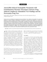
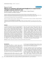
developed 9 days later. Chest computed tomography (CT)
demonstrated diffuse ground-glass opacities, reticular
shadows, and alveolar septal thickening (Figure 1A).
Bronchoalveolar lavage (BAL) was performed and revealed
pulmonary eosinophilia (total cell counts 6.8 3 10
5
/mL,
with eosinophils 83%, lymphocytes 9%, neutrophils 1%,
and alveolar macrophages 7%.
One week after admission (2 weeks after discontinuing
the drug), he was transferred to our hospital for further
Osamu Matsuno, Ryuichi Takenaka, Eishi Miyazaki, and Tosihide
Kumamoto: Division of Respiratory Disease, Department of Brain and
Nerve, Oita University Faculty of Medicine, Yufu-city, Oita, Japan.
Correspondence to: Dr. Osamu Matsuno, Division of Respiratory Disease,
Department of Brain and Nerve, Oita University Faculty of Medicine,
Yufu-city, Oita 879-5593, Japan; e-mail:
DOI 10.2310/7480.2007.00006
70 Allergy, Asthma, and Clinical Immunology, Vol 3, No 2 (Summer), 2007: pp 70–72
evaluation of the disease. His past medical history revealed
neither asthma nor any occupational exposure to toxic
fumes, dust, or animals. Physical examination revealed a
temperature of 38.4uC and blood pressure of 120/70 mm
Hg. Lung auscultation revealed fine crackles in the lower
lung field. There was no heart murmur. Arterial blood
gases were as follows: pH 7.413; HCO
3
23.1 mmol/L;
partial pressure of carbon dioxide 37.1 mm Hg; partial
pressure of oxygen, 81.3 mm Hg; and base excess 21.0
mmol/L. The white blood cell count was 7.9 3 10
3
, with
8.4% eosinophils. His hemoglobin concentration was 12.6
g/dL. C-reactive protein was 7.83 mg/L (normal range 0.0–
0.3 mg/L). Blood chemistry revealed a mild increase in
aspartate aminotransferase and alanine aminotransferase.
The total serum immunoglobulin (Ig)G, IgM, and IgA
levels were 1,470 g/dL, 191 mg/dL, and 320 mg/dL,
respectively, and total serum IgE was 816 IU/mL (normal
range , 170 IU/mL). A cytoplasmic pattern of antineu-
trophil cytoplasmic antibodies with reactivity against
myeloperoxidase was not detected. Angiotensin-converting
enzyme was not elevated. A chest radiograph on admission
indicated that the shadows that were disseminated over
both the mid– and lower lung zones had disappeared.
Chest CT revealed alveolar septal thickening and the
disappearance of ground-glass opacities.
BAL was performed. Sterile saline (150 mL) was
instilled into the right B8 segment in 50 mL aliquots. No
pathogen was identified. The BAL count was 6.04 3 10
5
/
mL, with eosinophils 33.3%, lymphocytes 27.4%, neutro-
phils 0%, and alveolar macrophages 39.3%. The CD4 to
CD8 ratio in the BAL fluid was 0.88. The BAL culture grew
no organism. A transbronchial lung biopsy was performed
and revealed lymphocytic and eosinophilic interstitial
infiltrate with granuloma formation (Figure 1B). There
was no evidence of necrotizing vasculitis. A presumptive
diagnosis of drug-induced pneumonia was made. The
chest radiograph, chest CT scan, and clinical findings
returned to normal 3 weeks after discontinuing the target
drugs without specific treatment.
A DLST for amoxicillin, shoseiryu-to, and acetamino-
phen, expressed as a stimulatory index (over 180% is
considered positive), yielded values of 123%, 101%, and
209%, respectively. These clinical symptoms were more
consistent with penicillin allergy because drug eruption
often occurs in penicillin allergy. Eleven days after
admission to our hospital, a drug provocation test was
performed by oral administration of 3.3% of the daily dose
of the drugs (10% of the single medication dose) with
careful observation. The provocation tests were performed
with the suspected drugs individually. Following provoca-
tion with amoxicillin, but not acetaminophen or sho-
seiryu-to, the dermatologic manifestations (erythema)
reappeared 11 hours later without pulmonary symptoms.
The white blood cell count and eosinophils (%) before the
provocation test were 5.80 3 10
3
/mm
3
and 14%,
respectively, and gradually rose to 8,500 3 10
3
/mm
3
and
23.9%, respectively, 1 week after the provocation. We
therefore concluded that the drug-induced pneumonia was
induced by amoxicillin.
Discussion
The overall prevalence of penicillin-induced pneumonia is
difficult to determine, but it is not a common adverse
reaction compared with penicillin-induced dermatologic
eruptions. There are several reports of penicillin-induced
eosinophilic pneumonia,
2–5,7
but amoxicillin-induced
eosinophilic pneumonia is very rare, with only one
Figure 1. A, Chest computed tomography demonstrated diffuse
ground-glass opacities, reticular shadows, and alveolar septa thicken-
ing. B, Transbronchial lung biopsy specimen revealed lymphocytic and
eosinophilic interstitial infiltrate with granuloma formation (hema-
toxylin and eosin stain, 3100 original magnification).
Matsuno et al, Amoxicillin-Induced Pneumonia 71
reported case in the literature that was diagnosed based on
a DLST.
7
Granulomas of any sort that are identified in the lung
parenchyma raise the spectre of sarcoidosis, mycobacteria,
fungi infection, and drug reaction. Acebutolol, cocaine,
cromolyn sodium, fluoxetine hydrochloride, methotrexate,
nitrofurantoin, procarbazine, pentazocine, sirolimus, and
tripelennamine are the drugs that are most often cited as
producing a granulomatous pneumonia with or without
interstitial infiltration.
10
To our knowledge, there is no
report of penicillin-induced pneumonia associated with a
granulomatous reaction. In addition, other than Churg-
Strauss syndrome, the coexistence of pulmonary eosino-
philia and granulomatous lung infiltration has not been
reported. Our case was not Churg-Strauss syndrome
because there was no evidence of asthma, neuropathy,
systemic organ failure, or vasculitis. This case of drug-
induced pneumonia involved at least two immunologic
mechanisms: delayed hypersensitivity (granuloma forma-
tion) and eosinophilic inflammation. We have no
explanation for their coexistence.
Diagnostic tests are dependent on the type of immune
reaction (antibody or T cell mediated). DLST demon-
strates the specificity of the T-cell reaction to the drug. In
Japan, drug-induced pneumonia and liver injury are
diagnosed on the basis of DLST results,
4,5,7,8
although a
drug provocation test is the most reliable method for
diagnosing a drug allergy.
11
Recent reports, however,
indicate that the DLST is unreliable for the diagnosis of
drug-induced liver injury.
11
In our case, a drug provoca-
tion test supported the diagnosis, but the results were
completely opposite to those of the DLST. Provocation
with 3.3% of the daily amoxicillin dose in the present case
caused the dermatologic manifestations to reappear with-
out the pulmonary symptoms. Wengrower and colleagues
also reported that antibiotic challenges with penicillin
caused a drug-induced skin eruption without reactivation
of the pneumonia.
4
These findings suggest that the
threshold dose for causing drug-induced skin eruptions
is different from that causing drug-induced pneumonia.
In conclusion, we report here the first case of
granuloma formation in the lung occurring in eosinophilic
pneumonia. Caution must be used in interpreting the
DLST for a diagnosis of drug allergy. The clinical course,
exclusion of an alternative cause, and drug readministra-
tion are useful clues to the diagnosis of drug-induced
pneumonia.
References
1. Gruchalla RS. Drug allergy. J Allergy Clin Immunol 2003;111:
S548–59.
2. Anastasiades CN. Severe allergic pneumonitis caused by ampicillin.
J Med Liban 1974;27:679–83.
3. Wengrower D, Tzfoni EE, Drenger B, Leitersdorf E. Erythroderma
and pneumonitis induced by penicillin? Respiration 1986;50:301–3.
4. Kohno S, Yamaguchi K, Yasuoka A, et al. Clinical evaluation of 12
cases of antimicrobial drug-induced pneumonitis. Jpn J Med 1990;
29:248–54.
5. Yonemaru M, Mizuguchi Y, Kasuga I, et al. Hilar and mediastinal
lymphadenopathy with hypersensitivity pneumonitis induced by
penicillin. Chest 1992;102:1907–9.
6. Poe RH, Condemi JJ, Weinstein SS, Schuster RJ. Adult respiratory
distress syndrome related to ampicillin sensitivity. Chest 1980;77:
449–51.
7. Sakito O, Kadota J, Kohno S, et al. Pulmonary infiltration with
eosinophilia and increased serum levels of squamous cell
carcinoma-related antigen and neuron specific enolase. Intern
Med 1994;33:550–3.
8. Pichler WJ, Tilch J. The lymphocyte transformation test in the
diagnosis of drug hypersensitivity. Allergy 2004;59:809–20.
9. Kunichika N, Miyahara N, Kotani K, et al. Pneumonitis induced by
rifampicin. Thorax 2002;57:1000–1.
10. Flieder DB, Travis WD. Pathologic characteristics of drug-induced
lung disease. Clin Chest Med 2004;25:37–45.
11. Mantani N, Kogure T, Tamura J, et al. Lymphocyte transformation
test for medicinal herbs yields false-positive results for first-visit
patients. Clin Diagn Lab Immunol 2003;10:479–80.
72 Allergy, Asthma, and Clinical Immunology, Volume 3, Number 2, 2007