Báo cáo y học: " Chitosan Interferon-c Nanogene Therapy for Lung Disease: Modulation of T-Cell and Dendritic Cell Immune Responses" pps
Bạn đang xem bản rút gọn của tài liệu. Xem và tải ngay bản đầy đủ của tài liệu tại đây (491.79 KB, 11 trang )
ORIGINAL ARTICLE
Chitosan Interferon-c Nanogene Therapy for Lung Disease:
Modulation of T-Cell and Dendritic Cell Immune
Responses
Xiaoyuan Kong, MD, Gary R. Hellermann, PhD, Weidong Zhang, PhD, Prasanna Jena, PhD, Mukesh Kumar, PhD,
Aruna Behera, PhD, Sumita Behera, MSc, Richard Lockey, PhD, and Shyam S. Mohapatra, PhD
The use of chitosan nanoparticles as carriers for expression plasmids represents a major improvement in gene expression
technology. We demonstrated previously that treatment with chitosan interferon-c (IFN-c) plasmid deoxyribonucleic acid (DNA)
nanoparticles (chitosan interferon-c nanogene [CIN]) led to in situ production of IFN-c and a reduction in inflammation and airway
reactivity in mice, but the mechanism underlying the immunomodulatory effects of CIN remains unclear. In this report, the effect of
CIN treatment on the immune responses of CD8
+
T cells and dendritic cells was examined in a BALB/c mouse model of ovalbumin
(OVA)-induced allergic asthma. OT1 mice (OVA-T cell receptor [TCR] transgenic) were also used to test the effects of CIN on OVA-
specific CD8
+
T cells. CIN treatment caused a reduction in IFN-c production in a subpopulation of OVA-specific CD8
+
T cells cultured in
vitro in the presence of OVA. CIN also reduced apoptosis of the CD8
+
T cells. Examination of dendritic cells from lung and lymph
nodes indicated that CIN treatment decreased their antigen-presenting activity, as evident from the reduction in CD80 and CD86
expression. Furthermore, CIN treatment significantly decreased the number of CD11c
+
b
+
dendritic cells in lymph nodes, suggesting
that endogenous IFN-c expression may immunomodulate dendritic cell migration and activation. CIN therapy results in a reduction in
proinflammatory CD8
+
T cells and decreases the number and antigen-presenting activity of dendritic cells.
Key words: allergy, asthma, interferon
T
he past decade has seen tremendous progress in gene
expression technology. Several investigators, includ-
ing us, have used viral vectors for transient gene expression
with some success. The replication-deficient episomal
adenovirus has been the workhorse for gene therapy, but
its toxicity and immunogenicity limit its clinical use.
1–5
Consequently, we have developed and tested a nonviral
platform for gene expression using plasmid deoxyribonu-
cleic acids (pDNAs) that offers ease of preparation and use,
in vivo stability, heat resistance, and the capacity for large
DNA sequences. The plasmids do not integrate into
mammalian genomes or replicate, yet they can persist in
host cells and express the cloned gene for several months.
The drawback of pDNA is its relatively low transfection
efficiency under physiological conditions, especially in
non-dividing or slowly dividing cells, such as epithelial
cells. Some improvement in the transfection efficiency of
pDNA has been made using liposomes or receptor
targeting,
6,7
but the approaches remain largely empirical.
One important development in gene transfer was the
discovery that chitosan (a biocompatible cationic poly-
saccharide derived from crustacean shell chitin) in the
form of nanoparticles (100–200 nm) could be used to
deliver plasmids.
8–12
Chitosan has beneficial immunostim-
ulatory,
13
anticoagulant,
14
wound-healing,
15
and antimi-
crobial properties.
16
It is nontoxic in humans, non-
hemolytic, weakly immunogenic, and slowly biodegradable
and has been widely used in controlled drug delivery.
9,17–21
Xiaoyuan Kong, Gary R. Hellermann, Weidong Zhang, Prasanna Jena,
Mukesh Kumar, Aruna Behera, Sumita Behera, and Shyam Mohapatra:
Division of Allergy and Immunology, Culverhouse Airway Disease
Research and Nanomedicine Center, University of South Florida College
of Medicine, Tampa, FL; and Richard Lockey: James A. Haley VA
Medical Center, Tampa, FL.
These studies were supported by grant 5RO1 HL 071101-02 and VA
Merit Review and Career Scientist Award to S.S.M and the Florida
Biomedical Research Foundation Bankhead-Coley Award and Mabel and
Ellsworth Simmons Professorship to S.S.M., and by the Joy McCann
Culverhouse endowment to the University of South Florida Division of
Allergy and Immunology.
Correspondence to: Shyam S. Mohapatra, PhD, Division of Allergy and
Immunology, Joy McCann Culverhouse Airway Disease Research Center,
University of South Florida College of Medicine, Box MDC-19, 12901
Bruce B. Downs Blvd, Tampa, FL 33612; e-mail: smohapat@
health.usf.edu.
DOI 10.2310/7480.2008.00006
Allergy, Asthma, and Clinical Immunology, Vol 4, No 3 (Fall), 2008: pp 95–105 95
It also has mucoadhesive properties that increase transcel-
lular and paracellular transport across the mucosal
epithelium,
22
which should facilitate gene delivery to
mucosa- and bronchus-associated lymphoid tissue.
Chitosan, therefore, appears to possess all of the attributes
of an ideal gene delivery agent for effective nonviral gene
expression therapy.
Since its discovery, interferon-c (IFN-c) has been
extensively studied for its immunomodulatory and antiviral
activity. Mice lacking the IFN-c receptor exhibit a T-helper
(Th)2-like cytokine profile, implying that IFN-c may be a
key cytokine in asthma.
23
IFN-c provides the stimulatory
signal for interleukin (IL)-12,
23,24
which is a strong inducer
of the Th1 response. IL-12 inhibits Th2 cells by down-
regulating the production of IL-4 and IL-5. The IFN-c-
inducing factor IL-18 also shifts the immune response from a
Th2 to a Th1 state.
25,26
Local administration of aerosolized
IFN-c prevented antigen-induced eosinophil recruitment in
guinea pig trachea.
27
IFN-a,IFN-b,and–c all inhibit
leukotriene C
4
production in murine macrophages,
28
but
IFN-c treatment of guinea pigs induced the release of
prostanoids and nitric oxide, which modify airway smooth
muscle responses through effects on airway epithelium.
29
Patients with allergic asthma exhibit lower than normal
production of IFN-c and IFN-c-dependent IL-12 in whole
blood cultures after stimulation with a mitogen.
30
Because
of the reciprocal regulation of T helper cells, it was
anticipated that increasing IFN-c levels via chitosan pIFN-
c nanoparticles (chitosan interferon-c nanogene [CIN])
would promote a Th1 response by blocking Th2 cytokine
production. IFN-c upregulates the IL-13Ra2 decoy
receptor, leading to diminished IL-13 signaling
31
and
reduced goblet cell hyperplasia and eosinophilia.
32
IFN-c
significantly inhibits release of leukotrienes from periph-
eral blood leukocytes (PBLs) of allergic individuals after
wasp venom immunotherapy
33
and decreases the produc-
tion of sulfidoleukotrienes by human PBLs.
34
IFN-c also
downregulates transforming growth factor b and procolla-
gen I and III and decreases fibrosis in a mouse model of
bleomycin-induced lung injury.
35
Exogenously supplied cytokines, such as IFN-c, IL-12,
and IL-18, have a short half-life in vivo, and systemic
administration at moderate to high doses can cause
substantial adverse effects.
36,37
To overcome these limita-
tions to their therapeutic use, several investigators have used
expression vectors containing the cloned cytokine comple-
mentary deoxyribonucleic acid (cDNA) under the regulation
of a constitutive promoter as a means of boosting in vivo
production of specific cytokines. IFN-c and IL-12 have
proven effective as prophylactics and adjuncts in therapy
against diverse human diseases.
38,39
IL-12 gene transfer and
expression in the mouse airway abrogated airway eosino-
philia and immunoglobulin E (IgE) synthesis,
40
and benefits
from IFN-c, IL-12, and IL-18 gene therapy have been
documented in other animal models.
41–43
However, the
methods used to transduce plasmids in mice are not directly
applicable to humans because of the use of lipofectamine,
which is toxic to humans.
Oromucosal therapy with recombinant IFN reduced
the severity of viral infection,
44
but intranasal administra-
tion of IFN-c pDNA has not been tested. Previous
studies from our laboratory demonstrated that intranasal
administration of IFN-c and IL-12 plasmids inhibited
the induction of IL-5 messenger ribonucleic acid
(mRNA), afforded protection against viral infections,
and significantly decreased airway inflammation and
airway hyperresponsiveness. In this article, we investigated
the mechanism of CIN-mediated immunomodulation
using the mouse OVA-allergic asthma model. IFN-c
treatment reduced cytokine production by a population
of OVA-specific proinflammatory CD8
+
T lymphocytes in
the lung and led to decreased activation of dendritic cells.
Materials and Methods
Animals
All mice were purchased from Jackson Laboratory (Bar
Harbor, ME). BALB/c mice have a predominantly Th2-
type response to allergens, and these were used for most of
the experiments. The transgenic C57BL/6-
TG(TcraTcrb)1100mjb/j mice have CD8
+
T cells specific
for ovalbumin (OVA) amino acids 257 to 264, and these
animals were used in experiments to examine CIN effects
on CD8
+
cells. Female 6- to 8-week-old mice were
maintained in pathogen-free conditions at the University
of South Florida College of Medicine vivarium. All
procedures were reviewed and approved by the
Committee on Animal Research at the University of
South Florida College of Medicine and VA Hospital. A
minimum of four mice were used in each test group, and
experiments were repeated twice.
Preparation of Chitosan IFN-c pDNA Nanoparticles
and Green Fluorescent Protein Test of Expression
Mouse IFN-c cDNA was cloned in the mammalian
expression vector pVAX (Invitrogen, San Diego, CA),
and complexed with chitosan, as described previously.
45
Briefly, plasmids in 25 mM Na
2
SO
4
and chitosan (Vanson,
96 Allergy, Asthma, and Clinical Immunology, Volume 4, Number 3, 2008
Redmond, WA) dissolved in 25 mM Na acetate, pH 5.4, to
a final concentration of 0.02% were separately heated for
10 minutes at 55uC. After heating, the chitosan and DNA
were mixed, vortexed vigorously for 20 to 30 seconds, and
stored at room temperature until use. This treatment
results in nanoparticles of 200 to 300 nm diameter as
measured in transmission electron micrographs and by
dynamic light scattering. The effectiveness of intranasal
chitosan nanoparticle–mediated gene transfer in mice was
tested with a vector that expresses green fluorescent
protein (plasmid-encoding green fluorescent protein
[pEGFP]). Chitosan nanoparticles complexed with
pEGFP were prepared as above, and an amount containing
10 mg of pEGFP was administered intranasally to mice.
After 1, 3, and 7 days, the mice were euthanized and the
lungs were removed and fixed in buffered formalin. Whole
lungs were embedded in paraffin, sectioned, and examined
for GFP by fluorescent microscopy, and an approximate
percentage of GFP-positive cells was estimated.
OVA Sensitization, CIN Treatment, and Preparation
of Lung Sections
Mice were allergen-sensitized by intraperitoneal injection
of 50 mg of OVA adsorbed to 2 mg of alum (Imject, Pierce,
Rockford, IL) followed by an intranasal challenge with 50
mg of OVA after 1 or 2 weeks. To test the effects of
boosting IFN-c production on allergen sensitization, mice
were given an intranasal treatment of 10 mg of CIN,
chitosan with vector, or chitosan alone prior to intraper-
itoneal injection of OVA-alum. This constitutes the
prophylactic CIN regimen. Other groups of mice were
given CIN treatments after OVA sensitization as a
therapeutic regimen. In both prophylactic and therapeutic
treatments, the mice were given a final challenge with OVA
and then euthanized, and the lungs from mice from each
group were perfused in situ with phosphate-buffered saline
(PBS), removed, and fixed in 4% buffered formalin. Lungs
from some mice in each group were left unfixed and
homogenized for lymphocyte isolation (see below). Whole
formalin-fixed lungs were paraffin-embedded, and 15-
micron sections were made. Two sections from each
mouse were dewaxed, rehydrated, and labelled with either
anti-IL-12Rb2–fluorescein isothiocyanate (FITC) (Th1) or
anti-T1/ST2L-rhodamine (Th2). Viewers examining the
stained sections were blinded as to the mouse group from
which the slides were taken. The stained sections were
examined with a Nikon TE300 fluorescence microscope,
and representative areas were photographed.
Isolation of Lung Cells and Flow Cytometry of IFN-c-
Producing, OVA-Specific CD8
+
T Lymphocytes
C57BL/6 mice transgenic for the OVA-specific T cell
receptor (TCR) (amino acids 257–264) and C57BL/6 wild-
type controls were given intranasal CIN treatment (10 mg),
followed by intraperitoneal sensitization with OVA-alum on
day 1. On day 10, the mice were again treated with CIN and
then challenged intranasally with OVA. Twenty-four hours
after the challenge, the mice were euthanized and the lungs
were perfused with PBS and removed. Lungs were weighed,
minced, and homogenized with Teflon homogenizers. The
homogenates were digested with collagenase (50 U/mL) in
the presence of DNase I (200 mg/mL) and passed through a
40-micron cell strainer to prepare single-cell suspensions.
This is a standard method of cell preparation and does not
result in selective loss or enrichment of lymphocytes or
dendritic cells. Viabilities by trypan blue dye exclusion were
. 80%. The cells were cultured for 20 hours with 50 mg/mL
OVA. Cultures were treated with 5 mg/mL brefeldin A for 4
hours prior to harvesting to block secretion of intracellular
cytokines. Class I restricted CD8
+
T cells were surface-
stained with OVA tetramer consisting of the OVA peptide
Ser-Ileu-Ileu-Asn-Phe-Glu-Lys-Leu, bound to four major
histocompatibility complex (MHC)-I molecules conjugated
with a fluorescent phycoerythrin tag (Beckman/Coulter
Immunomics, Fullerton, CA) and intracellularly stained
with FITC-conjugated anti-IFN-c. Cells were counted by
flow cytometry (FACScan, BD Biosciences, San Jose, CA)
with side-scatter/forward-scatter set for lymphocytes and
gating for CD8
+
tetramer. Unstained cells were run as a
control, and non-viable cells were distinguished by staining
with 7-amino-actinomycin D. Tetramers use a mutated
form of MHC-I with low binding affinity to CD8 on non-
OVA-recognizing T cells, so there is no need to run a
tetramer control.
Apoptosis of OVA-Specific CD8
+
T Lymphocytes
from Lung
OVA-sensitized OT-1 mice were given therapeutic CIN
treatment and 24 hours later were challenged intranasally
with OVA. Mice were euthanized 18 hours after OVA
challenge. Lungs were removed, homogenized, digested
with collagenase and DNase, and passed through a cell
strainer to produce single-cell suspensions. The cells were
pelleted by centrifugation at 500g for 5 minutes at 4uC, and
then red blood cells were lysed by resuspending the cell
pellets twice in ice-cold lysis buffer (0.156 M NH
4
Cl,
10 mM KHCO
3
, 0.1 mM ethylenediaminetetraacetic acid,
Kong et al, Chitosan Interferon-c Nanogene Therapy for Lung Disease 97
pH 7.3). Cells were resuspended in Roswell Park Medium I
(RPMI) 1640, plated, and incubated at 37uC for 18 hours.
Non-adherent cells consisting primarily of lymphocytes
were removed by pipetting off the medium. T lymphocytes
were isolated by mouse T-cell enrichment columns (R & D
Systems, Minneapolis, MN) and analyzed for apoptosis
using the TUNEL (terminal deoxynucleotidyl nick end-
labeling) assay (Promega, Madison, WI). The OVA-
specific CD8
+
T-cell population was labelled with phy-
coerythrin-tagged tetramer for OVA peptide conjugated
with MHC-I (Beckman/Coulter Immunomics). Unstained
cells were used as a control. The number of apoptotic
OVA-specific CD8
+
T cells was determined by flow
cytometry (FACScan, BD Biosciences) and expressed as a
percentage of the total number of OVA-specific CD8
+
cells.
Analysis of the Dendritic Cell Population in Lung
Parenchyma
OVA-allergic BALB/c mice were treated therapeutically
with CIN and then challenged with OVA and euthanized
18 hours later. Lungs were removed, and single-cell
suspensions were prepared and plated as described above.
In this case, however, mononuclear cells were isolated
from the cells that had adhered to the dishes after removal
of the lymphocytes. These were further purified using
magnetic beads coated with anti-CD11c (Miltenyi Biotec,
Auburn, CA) with cell recoveries in the range of 3 to 5
million per gram of lung tissue. The CD11c
+
cells were
then seeded into 12-well plates at 5 3 10
5
cells per well and
cultured for 48 hours in the presence of 10 mg of chitosan-
pVAX (vector control) or CIN. Cells were scraped from
the wells, resuspended in PBS + 3% fetal calf serum (FCS),
stained with phycoerythrin meaning (PE)-antiCD11c and
individually with FITC-anti-I-Ad, -CD40, and -CD80.
CD11c
+
cells expressing each of the dendritic cell
activation markers were counted by flow cytometry
(FACScan, BD Biosciences) with appropriate controls.
In another set of experiments, lung mononuclear cells
were isolated as described above but were not cultured. The
cells were stained with FITC-anti-CD11c and rhodamine-
anti-CD11b and counted by flow cytometry (FACScan, BD
Biosciences). The numbers of CD11c
+
b
+
cells were expressed
as a percentage of the total CD11c
+
cells.
Analysis of the Dendritic Cell Population in BAL
Fluid
BALB/c mice were sensitized with OVA, challenged by
intranasal administration of OVA, and 2 months later
given CIN treatment. They were again challenged with
OVA and euthanized 18 hours later. Lungs were lavaged
with 1 mL of PBS in two 0.5 mL aliquots introduced and
withdrawn through the trachea. The BAL fluid was
centrifuged 10 minutes at 300g, and cells were resuspended
in PBS. Buffered paraformaldehyde was added to a final
concentration of 4%, and cells were fixed for 10 minutes at
room temperature. Cells were washed with PBS and
resuspended in PBS with 3% FCS. Aliquots of the cell
suspension were all stained for CD11c and CD11b and
individually for CD40, CD80, CD86, and I-Ad.
Appropriate isotype controls and positive fluorescence
markers for each fluor were also prepared. Cells expressing
each of the four dendritic cell activation markers were
counted by three-colour flow cytometry (FACSCalibur,
BD Biosciences) gated on CD11c
+
and CD11b
+
cells.
Analysis of the Dendritic Cell Population in Lymph
Nodes
BALB/c mice were treated as described above for BAL fluid
isolation, and peribronchial lymph nodes were removed at
euthanasia. Single-cell suspensions were prepared by
maceration of the lymph nodes through 40-micron cell
strainers, and CD11c
+
cells were isolated by magnetic bead
separation. The cells were stained for CD11b and
separately for each of the dendritic cell activation markers
CD40, CD80, CD86, and I-Ad and were counted by flow
cytometry (FACScan, BD Biosciences).
IFN-Inducible Target Gene Array Analysis in
Dendritic Cells
To identify which genes were up- or downregulated by
CIN treatment, CD11c
+
dendritic cells were isolated from
lung cell suspensions of OVA-allergic/-challenged BALB/c
mice (two mice per isolation) using anti-CD11c con-
jugated to magnetic beads (Miltenyi Biotech, Auburn,
CA). Total ribonucleic acid (RNA) was purified from
dendritic cells of mice treated with control vector or CIN,
converted to cDNA using reverse transcriptase, and
labelled with biotinylated deoxyuridine triphosphate
(dUTP). The labelled cDNA was hybridized to a
TranSignal Interferon-inducible Gene Array membrane
(Panomics, Fremont, CA), and signals were detected by
chemiluminescence on x-ray film. The resulting film image
was scanned, and densitometry calculations were done
using the ScionImage (Scion Corporation, www.scioncorp.
com) program to compare the results from untreated mice
98 Allergy, Asthma, and Clinical Immunology, Volume 4, Number 3, 2008
with those from CIN-treated mice. The gene array analysis
was repeated once with a similar expression profile.
Ribonuclease Protection Assay for Cytokine Gene
Expression in Dendritic Cells
BALB/c mice were OVA-sensitized and challenged and given
CIN treatment or chitosan-control vector only. After
treatment, peribronchial lymph nodes were removed and
macerated through 40-micron cell strainers. Single-cell
suspensions were then mixed with anti-CD11c magnetic
beads (Miltenyi Biotec, Auburn, CA), and bound cells were
collected according to the manufacturer’s protocol. Total
RNA was prepared from the CD11c
+
cells, and
32
P-UTP
(Amersham, Piscataway, NJ)-labelled probes were generated
by in vitro transcription of cytokine-specific multiprobe
template sets (BD Pharmingen, San Diego, CA) using T7
RNA polymerase. The labelled probes were purified,
adjusted to 3 3 10
5
cpm/mL, and hybridized to 5 mgof
each RNA. The reactions were subsequently digested with
ribonuclease (Rnase) followed by proteinase K and extracted
with phenol-chloroform. After ethanol precipitation with 4
M ammonium acetate, the protected samples were resus-
pended in loading buffer and separated on a 6% Tris-borate-
EDTA (TBE)-urea gel (Novex Invitrogen, Carlsbad, CA).
The gel was placed on filter paper, dried under a vacuum,
and exposed to Kodak X-OMAT-AR film with intensifying
screen at 280uC.
Determination of Toll-Like Receptor mRNA Levels on
Lymph Node Dendritic Cells
A reverse transcriptase–polymerase chain reaction (RT-
PCR) was carried out on total RNA from CD11c
+
cells
isolated by magnetic bead separation from peribronchial
lymph nodes of OVA-sensitized/OVA-challenged BALB/c
mice treated with vector or CIN. PCR primers specific for
the indicated Toll-like receptors (TLRs) were used along
with b-actin as control, and amplification was performed
for 25, 30, and 35 cycles.
Statistical Analysis
Each test group of mice contained at least four animals,
and experiments were repeated twice. Values for all
measurements are expressed as means 6 SEMs. Groups
were compared by analysis of variance and through the use
of paired Student t-tests. Differences between groups were
considered significant at p , .05.
Results and Discussion
Expression in the Lung of a Chitosan Nanoparticle–
Delivered Plasmid
To determine that pDNAs can be transported in chitosan
nanoparticles and expressed in the lung epithelium,
pEGFP was complexed with chitosan nanoparticles and
administered intranasally to BALB/c mice. At 1, 3, and 7
days thereafter, mice were euthanized, lungs were
removed, and sections were examined by fluorescence
microscopy for GFP expression. By 3 days after pEGFP
nanoparticle administration, roughly half of the lung
epithelial cells appeared to be positive for EGFP (data not
shown), and expression of GFP continued for at least 7
days. We conclude that intranasal delivery of plasmids by
means of chitosan nanoparticles results in a sustained
expression of the encoded protein in the lung and provides
an effective means of supplying therapeutic or prophylactic
levels of an immunomodulatory molecule.
Effects of CIN Therapy on T Lymphocytes
In a previous study, we found that CIN treatment
significantly lowered airway hyperresponsiveness to
methacholine and reduced lung histopathology in a
BALB/c mouse model of OVA-allergic asthma.
46
CIN-
treated mice produced higher levels of IFN-c but less of the
Th2 cytokines IL-4 and IL-5 and had reduced OVA-
specific serum IgE compared with mice given vector alone.
Lung infiltration by eosinophils was significantly reduced
by CIN therapy, and overproduction of mucus was
inhibited within 6 hours of CIN delivery by induced
apoptosis of goblet cells. In this study, we sought to
understand the mechanism of CIN action.
T cells play a critical role in the pathology of asthma
and therefore may be potential targets of agents such as
CIN for ameliorating asthmatic symptoms. CD4
+
T helper
cells can either promote (Th2) or inhibit (Th1) the
inflammation of asthma depending on their phenotype
and the presence or absence of specific cytokines. To
examine the effects of CIN treatment on CD4
+
T
lymphocyte populations in the lung, sections from control
and CIN-treated mice were stained with rhodamine-
labelled anti-T1/ST2L, specific for Th2 cells, or with
FITC-labelled anti-IL-12Rb2 for Th1 cells. Sections were
examined in a blinded fashion by fluorescence microscopy,
and representative photographs were made to show
comparisons of controls with CIN-treated animals. Lungs
from CIN-treated mice showed qualitatively fewer Th2
Kong et al, Chitosan Interferon-c Nanogene Therapy for Lung Disease 99
cells and either the same or slightly more Th1 cells than
control mice (Figure 1A).
Both CD4
+
and CD8
+
T lymphocytes are involved in
the immune response to allergens. To determine if CIN
therapy affects the activity of antigen-specific CD8
+
T cells,
C57BL/6-OT1 mice carrying the transgene for the T-cell
receptor specific for the OVA epitope, amino acids 257 to
264, were given intranasal CIN and then sensitized by
intraperitoneal injection with OVA (day 1). They were
again treated with CIN and then challenged with OVA
intranasally on day 10. Lung lymphocytes were isolated 24
hours later and cultured in the presence of OVA and
brefeldin A for 20 hours. Cell viability was . 80% (trypan
blue dye exclusion test). The OVA-specific CD8
+
T cells
were stained using a tetramer for the 257- to 264-amino
acid OVA peptide, and the IFN-c produced during OVA
exposure was labelled by intracellular cytokine staining.
After appropriate gating, the cells were counted by flow
cytometry. The results (Figure 1B) show that CIN
treatment caused a decrease in the number of IFN-c-
producing, OVA-specific CD8
+
T cells in the lungs of
OVA-challenged mice.
To further assess the fate of OVA-specific CD8
+
T cells,
OVA-allergic C57 mice were treated with CIN and 24
hours later challenged intranasally with OVA. Mice were
euthanized 18 hours after challenge, and the lungs were
used to prepare single-cell suspensions of T cells. The
OVA-specific CD8
+
T-cell population was labelled with
phycoerythrin-tagged tetramer for OVA peptide conju-
gated with MHC-I. CD8
+
cells were analyzed for apoptosis
using the TUNEL assay (Promega) and counted by
FACScan flow cytometry. The percentage of total OVA-
specific CD8
+
T cells that were apoptotic was lower in
CIN-treatedmicecomparedwithuntreatedcontrols
(Figure 1C); therefore, the decrease in IFN-c production
observed in the CD8
+
T cells does not appear to be the
result of destruction of the cells but represents an IFN-c-
mediated downregulation of IFN-c production by a
specific subpopulation of lymphocytes.
These CIN-mediated alterations in T-cell populations
support the hypothesis that CD8
+
T cells are important in
allergen-induced lung pathology and that at least a part of
the protective effect of CIN treatment can be attributed to a
reduction in numbers of a specific CD8
+
T-cell population.
Intranasal administration of antigen to rats was reported to
induce T-cell activation concurrent with a burst of IFN-c
production, whereas subsequent antigen exposure produced
apoptosis and tolerization in a T-cell population.
47
Also,
IFN-c has been reported to induce apoptosis of CD8
+
T
cells.
48,49
Our results in this OVA-allergic asthma model
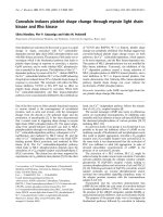
Figure 1. Chitosan interferon-c
nanogene (CIN) modulates T-cell
responses in ovalbumin (OVA)-
allergic mice. A, Localization of T
helper cells in the lungs of C57BL/6
mice using antibody to interleukin-
12Rb (Th1) and T1/ST2L (Th2). B,
Changes in interferon-c (IFN-c) pro-
duction by OVA-specific CD8
+
T cells
from the lung. T-cell receptor OVA-
specific OT1 mice were treated with
CIN and then sensitized with OVA
and challenged 10 days later. T
lymphocytes were isolated from lungs,
cultured with OVA, and stained with
tetramer to label OVA-specific CD8
+
T cells. IFN-c was identified by
intracellular cytokine staining and
counted by flow cytometry after
gating for tetramer-labelled cells. C,
Apoptosis of OVA-specific CD8
+
T
cells from lungs determined by
TUNEL assay and measured by flow
cytometry. The data are based on
cytometry of a minimum of 15,000
cells and were substantiated by a
repeat experiment.
100 Allergy, Asthma, and Clinical Immunology, Volume 4, Number 3, 2008
would suggest that IFN-c may have an autocrine effect,
downregulating its own production in T lymphocytes.
Effects of CIN Therapy on Dendritic Cells
Antigen presentation is a key process in determining the
magnitude of an immune response to an allergen such as
OVA. Since antigen-presenting cells commonly take up
extracellular particles, it is reasonable to suppose that CIN
therapy, which uses chitosan nanoparticles, might involve
cells that interact with or take up the particles and directly
influence the immune response. Dendritic cells are the
predominant antigen-presenting species in regulating T-
cell activation and thus may participate in the protective
effect of CIN therapy.
To determine whether CIN therapy affects the activity
of lung dendritic cells, CD11c
+
mononuclear cells were
isolated from lungs of OVA-allergic mice with or without
CIN treatment and incubated with control nanoparticles
alone or with CIN. Flow cytometry was done on cell
suspensions after labelling for CD11c and for each of the
following markers: I-Ad, CD40, and CD80. The results
showed an increase in CD40 but a decrease in expression
of CD80 and I-Ad in dendritic cells from CIN-treated mice
compared with controls (Figure 2A). CD40 is a cell surface
receptor related to the tumour necrosis factor receptor
superfamily that binds to CD40 ligand, which is expressed
primarily by T cells. Upregulation of CD40 may influence
the interaction of lung dendritic cells with allergen-specific
T lymphocytes.
To further examine the potential effects of CIN on
dendritic cells, ex vivo cultures from BAL fluid and from
thoracic lymph nodes were prepared from mice with or
without CIN treatment and tested for expression of CD40,
the costimulatory molecules CD80 and CD86, and I-Ad
(MHC-II). The results showed that CIN treatment
decreased expression of CD80 and CD86 on dendritic
cells from both BAL fluid and lymph nodes (Figure 2B).
CD40 expression was decreased in lymph node dendritic
cells, whereas the expression of I-Ad was unaffected in
both DC types. These results indicated that dendritic cells
from lung lavage or from peribronchial lymph nodes can
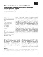
Figure 2. Effect of chitosan inter-
feron-c nanogene (CIN) therapy on
expression of dendritic cell activation
markers. A, In vitro CIN treatment of
CD11c
+
cells from the lungs of
ovalbumin (OVA)-allergic mice.
BALB/c mice were OVA sensitized
and challenged with OVA and sacri-
ficed 18 hours later. Lungs were
removed and CD11c
+
dendritic cells
were isolated as described in Materials
and Methods. Dendritic cells were
cultured with vector alone (control)
or with CIN. Flow cytometry was
performed on cell suspensions after
labelling for CD11c (fluorescein iso-
thiocyanate [FITC]) and for each of
the following markers: I-Ad, CD40,
and CD80 phycoerythrin (PE). Counts
were done using a FACScan gated for
CD11c cells. The figure shows the
percentage of cells that were positive
for CD11c and for each of the
markers. Activation marker expression
in CD11c
+
cells from the bronchoal-
veolar lavage (BAL) fluid and lymph
nodes of OVA-allergic mice treated
with CIN (B). Mice were treated as
described in Materials and Methods,
and CD11c
+
cells were isolated from
BAL fluid and lymph nodes. Cells
were analyzed by flow cytometry for
the expression of CD40, CD80, CD86,
and I-Ad gated to CD11c
+
b
+
. The data
are based on cytometry of a minimum
of 15,000 cells and were substantiated
by a repeat experiment.
Kong et al, Chitosan Interferon-c Nanogene Therapy for Lung Disease 101
be partially inactivated by CIN treatment, and their
capacity to activate T cells may consequently be reduced.
Survival of mature dendritic cells is necessary for
subsequent interaction with T lymphocytes, and CIN may
affect apoptosis of dendritic cells and their activation. To
examine this possibility, OVA-allergic mice were chal-
lenged with OVA and then 2 months later given CIN and
challenged again with OVA. Mononuclear cells were
isolated from lungs as described above, and the relative
numbers of CD11c
+
and CD11b
+
cells were determined by
flow cytometry. CIN treatment decreased the number of
CD11c
+
b
+
cells and increased the number of CD11c
+
b
2
cells (Figure 3). CD11c
+
b
+
cells are considered to be the
most active antigen-presenting cells, so the observation
that CIN therapy caused a significant reduction (fivefold)
in the numbers of CD11c
+
b
+
cells is consistent with the
other data in supporting the idea that CIN treatment
decreases the inflammatory response to an allergen by
inhibiting dendritic cell activation of OVA-specific T cells.
Alteration of Dendritic Cell Gene Expression by CIN
Treatment
Although CD40, CD80, and CD86 are key markers of
activated dendritic cells, many other genes are important
in the allergic immune response and may be up- or
downregulated by CIN treatment. To determine how CIN
affects gene expression, total RNA was isolated from the
lung dendritic cells of control and CIN-treated mice,
converted to cDNA, and labelled as probes, which were
hybridized to a mouse TranSignal Interferon-inducible
Gene Array membrane. The x-ray film images of the
control and CIN arrays were scanned, and spot densities
were analyzed and compared using the ScionImage
program. A two- to threefold increase or decrease in spot
density was considered significant, and an example of
some CIN-upregulated genes is shown in Figure 4A. The
results demonstrate that such arrays can be useful in
detecting significant changes in gene expression and that
novel changes can be identified in lung dendritic cells from
CIN-treated mice compared with controls. To confirm
CIN-mediated changes in expression of selected cytokine
genes, the RNAs were analyzed further by RNase protec-
tion assay. The results show that CIN treatment augments
expression of IL-12 p40, IL-18, IL-1a, and IFN-c in
dendritic cells (Figure 4B). Dendritic cells are activated by
signals generated through recognition of foreign antigens
by their surface TLRs. To examine the possibility that the
observed changes in gene expression were due to CIN-
mediated differential expression of TLR genes, the
expression of a number of TLR genes on peribronchial
lymph node dendritic cells (purified by CD11c magnetic
beads) was assayed by RT-PCR. The results indicated that
CIN treatment did not affect expression of TLR-2, -4, -5,
-6, and -9 at the mRNA level (Figure 5) and, therefore, that
CIN affects the activation of dendritic cells independently
of TLR signalling.
Conclusion
These studies demonstrate that IFN-c delivered via
intranasal CIN treatment reduces the allergic immune
response by means of its effects on CD8
+
T cells and
dendritic cells in mice. The results are consistent with a
mechanism whereby CIN therapy decreases the innate
immune response by altering cytokine production of a
CD8
+
T-cell subpopulation and by decreasing the antigen-
presenting activity of dendritic cells.
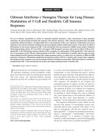
Figure 3. Effect of chitosan interferon-
c nanogene (CIN) therapy on the
dendritic cell population in the lung.
Ovalbumin-allergic/-challenged mice
were treated with or without CIN
therapy as described in Materials and
Methods, and mononuclear cells were
isolated from the lungs. Cells were
stained with fluorescein isothiocyanate
(FITC)–anti-CD11c and rhodamine
(Rhod)-anti-CD11b, and CD11c
+
b
+
cells were counted by flow cytometry.
Values given are the percentage of total
CD11c
+
cells that are also CD11b
+
.The
data are based on cytometry of a
minimum of 15,000 cells and were
substantiated by a repeat experiment.
102 Allergy, Asthma, and Clinical Immunology, Volume 4, Number 3, 2008
Acknowledgement
We would like to thank Sylvia Montalvo for her assistance
in preparation of the manuscript.
References
1. Behera AK, Kumar M, Lockey RF, Mohapatra SS. Adenovirus-
mediated interferon gamma gene therapy for allergic asthma:
involvement of interleukin 12 and STAT4 signaling. Hum Gene
Ther 2002;13:1697–709.
2. Monahan PE, Samulski RJ. AAV vectors: is clinical success on the
horizon? Gene Ther 2000;7:24–30.
3. Senior K. Adeno-associated virus vectors under scrutiny. Lancet
2002;359(9313):1216.
4. Zaiss AK, Liu Q, Bowen GP, et al. Differential activation of innate
immune responses by adenovirus and adeno-associated virus
vectors. J Virol 2002;76:4580–90.
5. Zhao N, Liu DP, Liang CC. Hot topics in adeno-associated virus as
a gene transfer vector. Mol Biotechnol 2001;19:229–37.
6. Cohen AD, Boyer JD, Weiner DB. Modulating the immune
response to genetic immunization. FASEB J 1998;12:1611–26.
7. Donnelly JJ, Ulmer JB, Liu MA. DNA vaccines. Dev Biol Stand
1998;95:43–53.
Figure 4. Chitosan interferon-c nanogene (CIN) treatment induces changes in gene expression in lung dendritic cells. A, Total ribonucleic acid
(RNA) from dendritic cells of ovalbumin (OVA)-allergic/-challenged mice with or without CIN treatment was isolated and assayed with the mouse
TranSignal Interferon-inducible Gene Array Kit, as described in Materials and Methods. The arrays were scanned and the images analyzed using
the ScionImage densitometry program. A two- to threefold increase or decrease in spot density was considered significant, and a selection of genes
upregulated by CIN treatment is shown. B, Ribonuclease protection assay for CIN-induced cytokine gene expression in dendritic cells. Total RNA
was isolated from CD11c
+
lymph node dendritic cells of OVA-allergic/-challenged control and CIN-treated mice and hybridized to probes specific
for interleukin (IL)-12 (p40), IL-1a, IL-18, IL-6, and interferon (IFN)-c. The housekeeping gene, GAPDH, was included as a normalization control.
The data presented are from one typical experiment of two performed. Fcgr1 5 a high-affinity receptor for immunoglobulin G; H1-T23 5
histocompatibility t23 region; H2-Q1 5 histocompatibility 2-Q region; Ifnar2 5 interferon-a receptor 2; Isg15 5 interferon-induced protein, 15
kDa; Krt-17 5 keratin-17; Mt1 5 metallothionein 1; Pml 5 promyelocytic leukemia gene; Stat2 5 signal transducer and activator of transcription
2; Ubc 5 ubiquitin C.
Figure 5. Expression of Toll-like receptors (TLRs) on dendritic cells.
Total ribonucleic acid (RNA) was purified from CD11c
+
cells isolated
by magnetic bead separation from peribronchial lymph nodes of mice
with or without chitosan interferon-c nanogene treatment. RNAs were
reverse-transcribed and used as a template for reverse transcriptase–
polymerase chain reaction (PCR) with primer pairs and PCR
conditions specific for the indicated TLRs. The results of PCRs for
25 cycles are shown with b-actin used as a control (one typical
experiment of two).
Kong et al, Chitosan Interferon-c Nanogene Therapy for Lung Disease 103
8. Erbacher P, Zou S, Bettinger T, et al. Chitosan-based vector/DNA
complexes for gene delivery: biophysical characteristics and
transfection ability. Pharm Res 1998;15:1332–9.
9. Lee KY, Kwon IC, Kim YH, et al. Preparation of chitosan self-
aggregates as a gene delivery system. J Control Release 1998;51:
213–20.
10. Leong KW, Mao HQ, Truong-Le VL, et al. DNA-polycation
nanospheres as non-viral gene delivery vehicles. J Control Release
1998;53:183–93.
11. Richardson SC, Kolbe HV, Duncan R. Potential of low molecular
mass chitosan as a DNA delivery system: biocompatibility, body
distribution and ability to complex and protect DNA. Int J Pharm
1999;178:231–43.
12. Roy K, Mao HQ, Huang SK, Leong KW. Oral gene delivery with
chitosan—DNA nanoparticles generate immunologic protection in
a murine model of peanut allergy. Nat Med 1999;5:387–91.
13. Nishimura K, Nishimura S, Nishi N, et al. Immunological activity
of chitin and its derivatives. Vaccine 1984;2:93–9.
14. Otterlei M, Varum KM, Ryan L, Espevik T. Characterization of
binding and TNF-alpha-inducing ability of chitosans on mono-
cytes: the involvement of CD14. Vaccine 1994;12:825–32.
15. Muzzarelli R, Baldassarre V, Conti F, et al. Biological activity of
chitosan: ultrastructural study. Biomaterials 1988;9:247–52.
16. Pappineau A, Hoover D, Knoor D, Farkas D. Food biotechnol.
New York (NY); Marcel Dekker; 1991;5:45–7.
17. Aspden TJ, Mason JD, Jones NS, et al. Chitosan as a nasal delivery
system: the effect of chitosan solutions on in vitro and in vivo
mucociliary transport rates in human turbinates and volunteers. J
Pharm Sci 1997;86:509–13.
18. Bodmeier R, Chen HG, Paeratakul O. A novel approach to the oral
delivery of micro- or nanoparticles. Pharm Res 1989;6:413–7.
19. Illum L, Farraj NF, Davis SS. Chitosan as a novel nasal delivery
system for peptide drugs. Pharm Res 1994;11:1186–9.
20. Miyazaki S, Nakayama A, Oda M, et al. Chitosan and sodium
alginate based bioadhesive tablets for intraoral drug delivery. Biol
Pharm Bull 1994;17:745–7.
21. Tozaki H, Komoike J, Tada C, et al. Chitosan capsules for colon-
specific drug delivery: improvement of insulin absorption from the
rat colon. J Pharm Sci 1997;86:1016–21.
22. Artursson P, Lindmark T, Davis SS, Illum L. Effect of chitosan on
the permeability of monolayers of intestinal epithelial cells (Caco-
2). Pharm Res 1994;11:1358–61.
23. Coyle AJ, Tsuyuki S, Bertrand C, et al. Mice lacking the IFN-
gamma receptor have impaired ability to resolve a lung
eosinophilic inflammatory response associated with a prolonged
capacity of T cells to exhibit a Th2 cytokine profile. J Immunol
1996;156:2680–5.
24. Romagnani S. Lymphokine production by human T cells in disease
states. Annu Rev Immunol 1994;12:227–57.
25. Wenner CA, Guler ML, Macatonia SE, et al. Roles of IFN-gamma
and IFN-alpha in IL-12-induced T helper cell-1 development. J
Immunol 1996;156:1442–7.
26. Szeto C, Gillespie KM, Mathieson PW. Levamisole induces
interleukin-18 and shifts type 1/type 2 cytokine balance.
Immunology 2000;100:217–24.
27. Gao Y, Chenping Z, Lin XP. [Aerosolized recombinant interferon-
gamma prevent antigen-induced eosinophil recruitment in guinea
pig trachea]. Zhonghua Jie He He Hu Xi Za Zhi 1997;20:287–90.
28. Boraschi D, Censini S, Bartalini M, et al. Interferons inhibit LTC4
production in murine macrophages. J Immunol 1987;138:4341–6.
29. Chen H, Munakata M, Amishima M, et al. Gamma-interferon
modifies guinea pig airway functions in vitro. Eur Respir J 1994;7:
74–80.
30. van der Pouw Kraan TC, Boeije LC, de Groot ER, et al. Reduced
production of IL-12 and IL-12-dependent IFN-gamma release in
patients with allergic asthma. J Immunol 1997;158:5560–5.
31. Daines MO, Hershey GK. A novel mechanism by which interferon-
gamma can regulate interleukin (IL)-13 responses. Evidence for
intracellular stores of IL-13 receptor alpha -2 and their rapid
mobilization by interferon-gamma. J Biol Chem 2002;277:10387–
93.
32. Ford JG, Rennick D, Donaldson DD, et al. IL-13 and IFN-gamma:
interactions in lung inflammation. J Immunol 2001;167:1769–
77.
33. Krasnowska M, Medrala W, Malolepszy J, Krasnowski R. Effect of
recombinant IFN-gamma on IgE-dependent leukotriene genera-
tion by peripheral blood leukocytes in patients with pollinosis and
asthma. Arch Immunol Ther Exp (Warsz) 2000;48:287–92.
34. Pierkes M, Bellinghausen I, Hultsch T, et al. Decreased release of
histamine and sulfidoleukotrienes by human peripheral blood
leukocytes after wasp venom immunotherapy is partially due to
induction of IL-10 and IFN-gamma production of T cells. J Allergy
Clin Immunol 1999;103(2 Pt 1):326–32.
35. Gurujeyalakshmi G, Giri SN. Molecular mechanisms of antifibrotic
effect of interferon gamma in bleomycin-mouse model of lung
fibrosis: downregulation of TGF-beta and procollagen I and III
gene expression. Exp Lung Res 1995;21:791–808.
36. Cohen J. IL-12 deaths: explanation and a puzzle. Science 1995;270:
908.
37. Mohapatra SS. IL-12 possibilities. Science 1995;269:1499.
38. Hogan SP, Foster PS, Tan X, Ramsay AJ. Mucosal IL-12 gene
delivery inhibits allergic airways disease and restores local antiviral
immunity. Eur J Immunol 1998;28:413–23.
39. Murray HW. Current and future clinical applications of
interferon-gamma in host antimicrobial defense. Intensive Care
Med 1996;22 Suppl 4:S456–61.
40. Sur S, Lam J, Bouchard P, et al. Immunomodulatory effects of IL-
12 on allergic lung inflammation depend on timing of doses. J
Immunol 1996;157:4173–80.
41. Dow SW, Schwarze J, Heath TD, et al. Systemic and local
interferon gamma gene delivery to the lungs for treatment of
allergen-induced airway hyperresponsiveness in mice. Hum Gene
Ther 1999;10:1905–14.
42. Li XM, Chopra RK, Chou TY, et al. Mucosal IFN-gamma gene
transfer inhibits pulmonary allergic responses in mice. J Immunol
1996;157:3216–9.
43. Okubo T, Hagiwara E, Ohno S, et al. Administration of an IL-12-
encoding DNA plasmid prevents the development of chronic graft-
versus-host disease (GVHD). J Immunol 1999;162:4013–7.
44. Tovey MG, Maury C. Oromucosal interferon therapy: marked
antiviral and antitumor activity. J Interferon Cytokine Res 1999;19:
145–55.
45. Kumar M, Behera AK, Lockey RF, et al. Intranasal gene transfer by
chitosan-DNA nanospheres protects BALB/c mice against acute
respiratory syncytial virus infection. Hum Gene Ther 2002;13:
1415–25.
104 Allergy, Asthma, and Clinical Immunology, Volume 4, Number 3, 2008
46. Kumar M, Kong X, Behera AK, et al. Chitosan IFN-gamma-pDNA
nanoparticle (CIN) therapy for allergic asthma. Genet Vaccines
Ther 2003;1:3.
47. Laliotou B, Duncan L, Dick AD. Intranasal administration of retinal
antigens induces transient T cell activation and apoptosis within
drainage lymph nodes but not spleen. J Autoimmun 1999;12:145–55.
48. Badovinac VP, Tvinnereim AR, Harty JT. Regulation of antigen-
specific CD8+ T cell homeostasis by perforin and interferon-
gamma. Science 2000;290:1354–58.
49. Lohman BL, Welsh RM. Apoptotic regulation of T cells and
absence of immune deficiency in virus-infected gamma interferon
receptor knockout mice. J Virol 1998;72:7815–21.
Kong et al, Chitosan Interferon-c Nanogene Therapy for Lung Disease 105