Tài liệu Báo cáo Y học: CK2btes gene encodes a testis-specific isoform of the regulatory subunit of casein kinase 2 in Drosophila melanogaster potx
Bạn đang xem bản rút gọn của tài liệu. Xem và tải ngay bản đầy đủ của tài liệu tại đây (279.08 KB, 10 trang )
CK2btes gene encodes a testis-specific isoform of the regulatory
subunit of casein kinase 2 in
Drosophila melanogaster
Alla I. Kalmykova
1
, Yuri Y. Shevelyov
1
, Oksana O. Polesskaya
1,
*, Anna A. Dobritsa
1,
†,
Alexandra G. Evstafieva
2
, Brigitte Boldyreff
3
, Olaf-Georg Issinger
3
and Vladimir A. Gvozdev
1
1
Institute of Molecular Genetics, Russian Academy of Sciences, Moscow, Russia;
2
Belozersky Institute of Physico-Chemical Biology,
Center of Molecular Medicine, Moscow State University, Russia;
3
Department of Biochemistry and Molecular Biology,
University of Southern Denmark, Odense, Denmark
An earlier described CK2btes gene of Drosophila melano-
gaster is shown to encode a male germline specific isoform of
regulator y b subunit of casein kinase 2. Western-analysis
using anti-CK2btes Ig revealed CK2btes protein in
Drosophila tes tes extract. Expression of a CK2btes–
b-galactosidase fusion protein driven by the CK2btes pro-
moter was found in transgenic flies at postmitotic stages of
spermatogenesis. Examination of biochemical characteris-
tics of a recombinant CK2btes protein expressed in
Escherichia coli revealed properties similar to those of CK2b:
(a) CK2btes protein stimulates CK2a catalytic activity
toward synthetic peptide; (b) it inhibits phosphorylation of
calmodulin and mediate s stimulation of CK2 a by polylysine;
(c) it is able to form (CK2btes)
2
dimers,aswellas
(CK2a)
2
(CK2btes)
2
tetramers. Using t he yeast two-hybrid
system and coimmunoprecipitation analysis of protein
extract from Drosophila testes, we demonstrated an associ-
ation between CK2bte s and CK2a. N orthern-analysis has
shown that another regulatory (b¢) subunit found recently in
D. melanogaster genome i s also testis-specific. Thus, we
describe the first example of two tissue-specific regulatory
subunits of CK2 which might serve to provide CK2 sub-
strate recognition during spermatoge nesis.
Keywords: spermatogenesis; casein kinase 2; C K2 b sub unit;
CK2btes; testes.
Protein kinase CK2 is involved in such general cell processes
as cell cycle regulation, transcriptional control, signal
transduction, development and proliferation [1–4]. More
than 160 different proteins serve as substrates for CK2.
Phosphorylation b y CK2 has been found to affect activity
of such Drosophila proteins pivotal for realization of early
developmental program, a s Cut-homeodeomain protein,
Cactus and Antennapedia [5–7]. A CK2 holoenzyme
consists of two a-(ora¢-) and two b subunits. The a subu nit
of CK2 possesses catalytic activity and the regula tory
b subunit was shown to enhance stability of the holoenzyme,
activate CK2a and provide substrate specificity and CK2
targeting in c ells. In spite of CK2b being ubiquitously
represented among eukaryotes, it is far less conserved in
comparison with the catalytic CK2a. This fact might be
explained by a wide spectrum of substrates and partner
proteins interacting with CK2b as a regulatory subunit.
Moreover, other functions, besides being a part o f the CK2
holoenzyme, can be ascribed to the b subunit. For e xample,
it has been demonstrated that the CK2 b subunit is involved
in the regulation of catalytic activity of two other protein
kinases (A-raf a nd Mos kinases [8–10]). The conclusion that
CK2b has a more general functions is supported by the fact
that significant imbalance of its amount in respect to
a subunit is found in tumor cells and some mammalian
tissues such as testicles [11, 12].
Recently it was shown that the CK2 activity as well as the
CK2 protein level are mostly elevated in rat and mouse
testicles [12, 13]. An important role for the CK2 activity in
spermatogenesis was clearly shown by a Ôknock-ou tÕ of the
CK2a¢ gene in mice resulting in a male sterile phenotype
[14]. Spermatogenesis is a complex differentiation process
comprising mitotic and meiotic division s of germline stem
cells followed by sperm morphogenesis. This p rocess is
known to have a lot of common features in Drosophila and
mammals [15]. However, g enetic control and molecular
mechanisms of both Drosophila and mammalian sperma-
togenesis are still poorly understood.
The genomes of most eukaryotes including mammals
carry a single gene encoding bsubunit of CK2. Only
Saccharomyces cerevisiae and Arabidopsis thaliana are
known to have two and three isoforms of CK2b, respect-
ively [16, 17]. Recently, we have described in Drosophila the
SSL gene [18], later renamed CK2btes [19], as a first
candidate on the role of a tissue-specific isoform of the CK2
regulatory subunit. This gene is expressed exclusively in
testes and encodes a protein sharing 45% amino-acid
identity with the ubiquitous Drosophila b subunit. Another
potential Drosophila tissue-specific CK2 regulatory subunit
(b¢) was identified in the yeast two-hybrid screen where
Correspondence to Y. Y. Shevelyov, Department of Molecular
Genetics of Animals, Institute of Molecular Genetics, 123182,
Kurchatov Sq. 2, Moscow, Russia. Fax: + 7 095 1960221;
Tel.: + 7 095 1961909; E-mail:
Abbreviations:CK2,caseinkinase2;CK2b,CK2b subunit; CK2a,
CK2 a subunit; IP, immunoprecipitation; RNAi, RNA interference;
dsRNA, double stranded RNA; X-gal, 5 -bromo-4-chloro-3-indolyl
b-galactopyranoside.
*Presen t address: Molecular Neurobiology Branch, NIDA, NIH, 5500
Nathan Shock Drive, Baltimore, MD, 21224, USA.
Presen t address: Department of Molecular, Cellular and Develop-
mental Biology, Yale University, New Haven, CT 06520, USA.
(Received 25 September 2001, revised 7 De cember 2001, accepted 14
January 2002)
Eur. J. Biochem. 269, 1418–1427 (2002) Ó FEBS 2002
CK2a was used as a bait [20]. In this work we present
compelling evide nce that the CK2btes protein serves as a
tissue-specific isoform of the CK2 regulatory subunit in
Drosophila male germline.
EXPERIMENTAL PROCEDURES
Plasmid constructions
PCRs were performed according to the recommendations
of the manufacturer using GeneAmp XL PCR Kit (Perkin
Elmer, Branchburg, NJ, USA) containing high fidelity
mixture of DNA-polymerases.
CK2btes and CK2a expression constructs: An 850 bp
BamHI–SalI fragment of the CK2btes cDNA #112 (this
cDNA sequence, cloned in the pBlueScript SK- vector,
contains no poly(A) tail and corresponds to nucleotides 72–
840 of the SSL (CK2btes) cDNA #911 sequence deposited
in GeneBank under accession number L42285, see also [18])
was s ubcloned in the pQE 30 expression vector (Stratagene,
La Jolla, CA, USA). The recombinant protein with the
N-terminal His
6
-tag comprises the whole CK2btes ORF
except for the 11 amino acids at the N-terminus.
A 1011-bp fragment of D. melanogaster CK2a gene
comprising the whole ORF region was PCR-amplified from
Drosophila genomic DNA using the f ollowing pair of
primers: 5¢-CAGGATCCATGACACTTCCTAGTGCG
GCTCGC-3¢ and 5¢-CCAAGCTTTTATTGCTGATTAT
TGGGATTCATTTGACCA-3¢ (the gene encoding the
Drosophila CK2 a subunit does not contain introns in the
coding region [21]). The BamHI–HindIII digested PCR
fragment was s ubcloned in the pQE 30 vector.
CK2b¢ probe for Northern-analysis. The161 bp 3¢-f rag-
ment of the C K2b¢ gene was PCR-amplified from Drosophila
genomic DNA using primers 5¢-ATAAGCTTGCTTT
AAAATCCACCCCACG-3¢ and 5¢-TCGGATCCC
AGTGCCCACTTATTCGAAAAG-3¢. HindIII–BamHI
digested PCR product was cloned into the pBlueScript SK-
vector and then recloned by Kpn I–BamHI into the pTZ19R
vector. In vitro transcription was performed for 1 h at 37 °C
in the buffer containing 40 m
M
Tris/HCl, pH 7.5, 60 m
M
MgCl
2
,5 m
M
NaCl, 10 m
M
dithiothreitol, 0.5 m
M
of each of
the ATP, GTP, CTP, 100 ng of the linearized plasmid DNA,
20–100 lCi [a-
32
P]UTP, 2–5 units of T7 RNA polymerase
(Gibco BRL, Life Technologies, CA, USA), 25 U of RNAse
inhibitor (Gibco BRL, Life Technologies, CA, USA).
Constructs for P-element transformation
To make the CK2btes–b-galactosidase fusion construct, a
934-bp fragment of the CK2btes gene including the 161 bp
of promoter region linked to the whole ORF was PCR-
amplified from the DNA of the cosmid clone #9 [18] using
the following pair of primers: 5¢-GACTGCAGTGAAGG
GCATCGAGTCCTCGGG-3¢ and 5¢-GAGGATCCGG
GACATTCCTTAGCCAGGAGGG-3¢.Tomakethe
b-galactosidase expressing construct, a 173-bp PCR frag-
ment of the CK2btes gene including the 161 bp of promoter
region joined with the first 12 bp of the ORF region was
amplified from the DNA of the c osmid clone #9 using the
same direct primer as for the CK2btes–b-galactosidase
fusion construct and the following reverse primer:
5¢-CTGGATCCGGACACGACATGCTCACTCGAA
TAA-3¢.BothPstI–BamHI digested PCR fragments were
clonedinframe,withtheb-galactosidase ORF devoid of the
ATG, into the pCaSpeR-bgal vector [22].
To genera te the CK2btes ÔantisenseÕ co nstruct, the XhoI
fragment of the CK2btes cDNA #421 corresponding to the
12–700 bp region of the sequence of cDNA #911 (one XhoI
site in the cDNA #421 is located in the MCS of BlueScript
SK- vector, and another XhoI site is located in the adaptor
sequence at the opposite side of insert) was cloned into the
modified testis vector kindly provided by H.D. Hoyle
(University of Indiana, Bloomingto n, IN, USA) [23]. This
vector carries the regulatory region of t he b2-tubulin gene
driving testis-specific expression of any gene substituting the
b2-tubulin ORF. The regulatory region had been cloned
upstream of the mini-white gene in the pCaSpeR4 vector.
The m odification of the vector includes the insertion in its
EcoRI cloning site of the MCS polylinker, which may now
be used for cloning with EcoRI, XhoIandKpnI. The
ÔantisenseÕ orientation of the CK2btes cDNA relative to the
b2-tubulin promoter was v erified by restriction digestion.
Constructs for the yeast two-hybrid system assay
To make CK2a AD- and BD- constructs, the whole CK2a
ORF region was amplified from Drosophila genomic DNA
using the following pair of primers: 5¢-CAGAATTCA
TGACACTTCCTAGTGCGGCTCGC-3¢ and 5 ¢-CTG
GATCCTTATTGCTGATTATTGGGATTCATTTGA
CCA-3¢. EcoRI–BamHI digested PCR product was clon ed
as a fusion with t he GAL4 activation domain in the
pGAD424 vector, or as a fusion with the GAL4 DNA-
binding domain in the pGBT9 vector (Clontech, La Jolla,
CA, USA).
To prepare CK2btes AD- and BD- constructs, the whole
CK2btes ORF region was amplified from the cDNA #911
using the following pair of primers: 5¢-CTGGATCCCT
ATGTCGTGTCCCAGGAGCATCGAG-3¢ and 5¢-GTC
TGCAGTTAAAAATTCGGGACATTCCTTAGCCA
GG-3¢. BamHI–PstI digested PCR product was cloned as a
fusion with GAL4bd in the pAS2-1 vector (Clontech, La
Jolla, CA, USA). The CK2btesORFwasexcisedfromthe
pAS2-1 plasmid by joint BamHI and PstI digestion, the PstI
end was blunted by T4 DNA polymerase, and fragment was
cloned in the BamHI–XhoI d igested (the XhoIendwasalso
blunted) pACT2 vector (Clontech, La Jolla, CA, USA) as a
fusion with GAL4ad.
To prepare CK2b AD- and BD- constructs, the whole
CK2b ORF region was amplified from the pEV55Dmb
plasmid DNA (kindly provided by C.V.C. Glover (Uni-
versity of Georgia, Athens, GA, USA), it contains a full size
cDNA of D. melanogaster b subunit [24]) using the follow-
ing p air o f p rimers: 5¢-CAGGATCCCTATGAGCAGC
TCCGAGGAAGTCTCCT-3¢ and 5¢-CTGTCGACTTA
GTTTTTCGCTCGTAGTGGCATTTTAAAATTGGCT
GC-3¢. BamHI–SalI digested PCR fragment was cloned
into BamHI–SalI digested pAS2-1 vector, or into BamHI–
XhoI digested pACT2 vector.
Protein purification, generation of antibodies
Expression and purification of recombinant p roteins from
E. coli using Ni
2+
/nitrilotriacetic acid resin (Qiagen Inc.,
Ó FEBS 2002 Testis-specific isoform of CK2 regulatory subunit (Eur. J. Biochem. 269) 1419
CA, USA) were performed according to the Stratagene
protocols. Drosophila CK2a and CK2btes recombinant
proteins purified under n ondenaturing conditions were used
in the in vitro assays (measurement of CK2a activity, gel
filtration experiments). Human CK2a and Drosophila
CK2btes proteins p urified from E. coli inclusion bodies
under denaturing conditions were used for the g eneration of
antibodies in rabbits. The specificities of isolated antisera
were tested by Western analysis.
RNA isolation and Northern-analysis
Total R NA was isolated by guanidinium thiocyanate
extraction [25] from embryos, pupae, larvae, females, male
carcasses and testes of gt w
a
strain, fractionated by electro-
phoresis in denaturing formaldehyde-agarose g el and
transferred t o a nylon HyBond-N filter (Amersham, Little
Chalfont, UK). Filter prehybridization, hybridization and
washing were performed according to standard protocols
[26]. As a control for the RNA loading, hybridization of the
same filter with the rp49 probe [27] was performed.
CK2 activity test
The equimolar mixture of CK2a and CK2btes proteins
purified under nondenaturing conditions or the CK2a
protein alone were assayed for the CK2 phosphorylation
activity using a synthetic peptide RRRDDDSDDD as a
substrate. The reaction was carried out in the buffer (45 m
M
Tris/HCl, pH 8.0, 5 m
M
MgCl
2
,1 m
M
dithiothreitol, 50 l
M
ATP, 2 lCiÆmL
)1
[c-
32
P]ATP (3000 CiÆmmol
)1
), 200 l
M
peptide) containing different N aCl concentrations (from
0m
M
to 200 m
M
)at37°C for 5 min The reaction aliquots
were loaded onto P81 phosphocellulose paper, washed with
85 m
M
phosphoric acid, and incorporated radioactivity was
measured by the liquid scintillation counter.
For the phosphorylation of calmodulin (kindly provided
by N. B. Gusev, Moscow State University) t he aliquots of
fractions after gel filtration assay containing 50 ng of
CK2a either alone or in combination w ith equimolar amount
of CK2btes protein were used. T he reaction was carried out
in 50 m
M
Tris/HCl, pH 8.0, 10 m
M
MgCl
2
, 150 m
M
NaCl,
20 l
M
ATP, 10 lCiÆmL
)1
[c-
32
P]ATP (3000 CiÆmmol
)1
),
10 l
M
calmodulin at 37 °C for 15 min Polylysine (Sigma,
St Louis, MI, USA) at concentration 100 lgÆmL
)1
was
added where necessary. The reaction was stopped by
cooling in ice, and the samples were subjected to 15%
SDS/PAGE. The gels were dried and autoradiographed.
Assays for detection of protein-protein contacts
in yeast two-hybrid system
Protein–protein interactions were assayed using three
different approaches. For the b-galactosidase filter assay,
single colonies cotransformed with AD- and BD- constructs
were picked and transferred to a Whatman no. 5 paper,
which was further incubated on a fresh plate for 2–3 days.
The filters were frozen in liquid nitrogen, layered over a
second filter prewetted with Z-buffer (16.1 gÆL
)1
Na
2
H-
PO
4
Æ7H
2
O, 5.5 gÆL
)1
NaH
2
PO
4
ÆH
2
O, 0.75 gÆL
)1
KCl,
0.246 g L
)1
MgSO
4
· 7H
2
O), which contained 0.27 mL
2-mercaptoethanol and 1.67 mL 5-bromo-4-chloro-3-indo-
lyl b-galactopyranoside (X-gal; 20 mgÆmL
)1
in dimethyl-
formamide) per 100 mL. Incubation was performed at
30 °C for up to 12 h.
For the liquid assay 5 mL cultures with synthetic
medium were inoculated with single colonies cotransformed
with AD- and BD- c onstructs and were grown until
D
600
1. Each culture (1 mL) was transferred to a
microcentrifuge tube and c entrifuged for 5 s. The yeast
pellet was dissolved in 100 lL of Z buffer and frozen in
liquid nitrogen. After thawing, 700 lL of Z buffer with
mercaptoethanol and 160 lL O-nitrophenyl-b-
D
-galacto-
side ( 4 mgÆmL
)1
in Z buffer) were added and the reaction
was incubated for 1 h at 30 °C. The reaction was stopped
by addition of 400 lL1
M
Na
2
CO
3
. After centrifugation
for 10 min at 13 400 g,theA
420
was measured. b-Galac -
tosidase activity was calculated in Miller units according to
the formula: units ¼ 1000 · A
420
/(culture volume in ml ·
incubation time in min · D
600
).
In addition, the ability of the yeast strain HF7c (carrying
HIS3 reporter gene) being cotransformed with AD- and
BD- constructs to grow on the medium without histidine
was used to verify protein–protein interactions.
Immunoprecipitation
A total of 100 hand-dissected pairs of testes were homo-
genized in the buffer containing 50 m
M
Tris/HCl, pH 8.0,
150 m
M
NaCl, 0.05% Nonidet P40, cocktail of protease
inhibitors. After 3 h of incubation at 4 °C followed by
15 min centrifugation at 4000 g, crude extract was fivefold
diluted with IP buffer (50 m
M
Tris/HCl, pH 8.0, 150 m
M
NaCl, 0.05% NP40, 5 m
M
EDTA, 0.2% BSA, 0.02%
NaN
3
, cocktail of protease inhibitors). Immunoprecipita-
tion was carried out over night at 4 °Cwith1lLofanti-
DmCK2a Ig, kindly provided by C.V.C. Glover. The
complex was precipitated by incubation with protein
A–Sepharose (4 Fast Flow, Pharmacia Biotech, Uppsala,
Sweden) for 1 h at 4 °C. Immunoprecipitate was washed
fourfold with 0.5 mL IP buffer and then fractionated on the
8% SDS/PAGE followed by Western analyses with poly-
clonal anti-( b-galactosidase) Ig (ICN Pharmaceuticals inc.,
Costa Mesa, CA, USA).
Western blot analysis
For the detection of CK2 btes and CK2a proteins in testes
extracts by Western analysis the following antibodies were
used: anti-CK2btes nonpurified serum at a dilution of
1 : 5000; and anti-(Drosophila CK2a) serum, kindly provi-
ded by C.V.C. Glover, at a dilution of 1 : 5000. For
detection of CK2a in gel filtration assay rabbit anti-(human
CK2a) polyclonal IgG was used. Alkaline-phosphatase-
conjugated anti-(rabbit IgG) Ig (Sigma, St Louis, MI, USA)
was used as a secondary reagent.
Samples were resolved by electrophoresis in SDS/PAGE
and blotted onto Hybond-C membrane (Amersham, Little
Chalfont, UK). Blots were developed using the CDP-star
detection system (Tropix, Bedford, MA, USA) according to
the recommendations of the manufacturer.
Gel-filtration experiments
Proteins were passed through the Pharmacia SMART
system chromatographic Superose 6 column in the buffer
1420 A. I. Kalmykova et al. (Eur. J. Biochem. 269) Ó FEBS 2002
(25 m
M
Tris/HCl, pH 8.5, 1
M
NaCl) in the flow rate regime
(40 lLÆmin
)1
).
P-element transformation
Transgenic lines were generated using standard P-element
mediated germline transformation technique [28] with
Df(1)w
67c23(2)
, y strain and the pTURBO transposase
source. Three transformant lines were established for the b-
galactosidase bearing construct, two lines for the CK2btes-
b-galactosidase fusion construct, and one line for the
ÔantisenseÕ CK2btes construct.
Histochemical staining of tissues
For the b-galactosidase staining, testes from adult
Drosophila males, as well as carcasses, were hand-dissected,
fixed in 2% glutaraldehyde in KCl/NaCl/P
i
buffer (8 m
M
Na
2
HPO
4
,137m
M
NaCl, 0 .5 m
M
MgCl
2
,1.6m
M
KH
2
PO
4
,2.7m
M
KCl, pH 8.0) for 30 min, washed twice
in KCl/NaCl/P
i
buffer and stained with 0.25% X-gal at
37 °C for 1.5 h in the buffer containing 150 m
M
NaCl,
10 m
M
NaH
2
PO
4
,pH7.5,1m
M
MgCl
2
,3.1m
M
K
3
[Fe
II
(CN)
6
], 3.1 m
M
K
4
[Fe
III
(CN)
6
].
RESULTS
CK2btes protein is generated in
Drosophila
testes
at postmitotic stages of spermatogenesis
Previously we have revealed the CK2btes transcripts in
Drosophila testes only [18]. To detect the CK2btes protein
in testes, we raised rabbit polyclonal antibodies against a
recombinant CK2 btes protein purified from E. coli.These
antibodies recognize a protein with mobility of approxi-
mately 30 kDa in testes extract, but do not reveal any
specific signal in the corresponding region in the extracts
from males with removed testes and from ovaries
(Fig. 1A). The electrophoretic mobility of the recognized
protein in testes extract is slightly different from that
expected for the protein with the calculated molecular mass
of 25 kDa. The recombinant CK2btes protein purified
from E. coli during electrophoresis also runs slower than
expected. The retardation might be due to the peculiarities
of amino-acids content of the CK2btes protein. A similar
gel retardation was seen in case of ubiquitous Drosophila
CK2b when a 24.8-kDa protein runs as a 28-kDa one [24].
The generated antibodies are expected not to cross react
noticeably with the b and b¢ subunits of CK2 because these
subunits are rather divergent from the CK2btes protein
(45% and 46% of identity, respectively [18,20]). Thus, we
conclude that CK2btes protein is expressed in Drosophila
testes and we are able to detect it using the anti-CK2btes
Ig.
To study spatial e xpression pattern of the CK2btes
protein in the male germline, we generated transgenic flies
expressing either the b-galactosidase protein alone or the
CK2btes–b-galactosidase fusion protein, both being under
the control of the CK2btes promoter (Fig. 1B). Expres-
sion of the reporter genes was monito red by h istochemical
X-gal staining of whole adult testes. Both constructs give
thesameX-galstainingpatternatpremeioticand
postmeiotic stages of spermatogenesis ( Fig. 1C). No
b-galactosidase activity was revealed in the apical part
of a testis where mitotic divisions take place. Other
Drosophila male tissues were not stained also (not shown).
This expression pattern suggests that CK2btes protein is
expressed only at postmitotic stages of m ale germline, and
it re sembles that of other Drosophila male germline
specifically expressed genes, such as b2-tubulin, dhod, Sdic
and others [29–31].
Recombinant CK2btes protein stimulates catalytic
activity of recombinant CK2 a subunit towards
a synthetic peptide substrate
The CK2 holo enzyme is known to be a heterotetramer of
a
2
b
2
structure. The b subunit is catalyticaly inactive by i tself
but it specifically stimulates the phosphorylation activity of
CK2a 5- to 10-fold [1]. To test the CK2btes protein for i ts
ability to stimulate catalytic activity of CK2a, Drosophila
CK2btes and CK2a recombinant proteins were expressed
in E. coli. Proteins purified under nondenaturation condi-
tions were used for the CK2 activity assay. In the reac-
tion buffer without NaCl, the CK2btes protein 2.5-fold
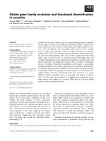
Fig. 1. Expression of the CK2btes protein in test es. (A) Detectio n of
CK2btes protein in Drosophila tissues. Western analysis using poly-
clonal anti-CK2btes Ig: R, recombinant CK2btes protein purified
from E. coli; T, protein extract from 7 pairs of adult testes; C, protein
extract from three male carcasses with removed testes; O, protein
extract from seven pairs of ovaries. Molecular mass markers are shown
to the left. (B) Diagram of microinjected constructs. CK2btes region is
black, b-galactosidase region is gray. (C) X-gal staining (dark region)
of testes from a transgenic fly line carrying the CK2btes–b-galactosi-
dase fusion construct under the control of the CK2btes promoter
region. The arrow marks the tip of the testis, no b-galactosidase
staining of somatic tissues was observed (not shown).
Ó FEBS 2002 Testis-specific isoform of CK2 regulatory subunit (Eur. J. Biochem. 269) 1421
stimulates t he CK2a activity (Fig. 2). Recombinant human
b subunit at the same conditions activates Drosophila
CK2a twofold (not shown). While the CK2a activity is
practically independent of NaCl concentration in the
absence of CK2btes, it is increased 5.5 times under
physiological conditions (150 m
M
NaCl) in the presence
of the equimolar amount of CK2btes (Fig. 2). It was
shown that the regulatory b subunit of D. melanogaster
CK2 purified from baculovirus expression system en -
hanced the activity of catalytic a s ubunit towards synthetic
peptide fivefold [24]. Therefore, CK2btes protein stimulates
CK2a activity in vitro at the optimal NaCl concentration
approximately t o the same extent as the ubiquitous
b subunit.
Recombinant CK2btes protein inhibits the ability
of CK2 a subunit to phosphorylate calmodulin
and this inhibition can be overcome by the polylysine
It was shown that in contrast to the stimulatory effect on
phosphorylation of majority of substrates, b subunit from
Drosophila, as well as from m ammals, suppresses the
calmodulin phosphorylation by the CK2 a subunit [32,
33]. Polybasic compounds such as polylysine and protamine
abolish this inhibition. We asked whether the CK2btes
protein behaves similarly in respect to calmodulin phos-
phorylation by a subunit. As shown in Fig. 3 (lanes 3 and
5), calmodulin is phosphorylated by recombinant Drosophi-
la CK2a, whereas the addition of equimolar amount of
CK2btes results in less efficient incorporation of radio-
activity in this substrate. The addition of polylysine
practically has n o effect on the phosphorylation of
calmodulin by free a subu nit (Fig. 3, lane 4), but drastically
stimulates activity of the equimolar mixture of a with btes
(Fig.3,lane6).Thus,CK2btes protein, such as canonical
b subunit, mediates stimulation of CK2 by polylysine.
Recombinant CK2btes protein forms tetrameric
complexes with CK2 a subunit
in vitro
To elucidate the structure of CK2a–CK2btes complexes in
vitro, recombinant CK2a and CK2btes proteins, purified
under native conditions, were analyzed separately, or in the
equimolar mixture, in gel-filtration experiments. Proteins
eluted from the column were detected by Western blot
analysis using anti-(CK2btes) Ig and polyclonal anti-
(human CK2a) Ig that a lso recognizes the Drosophila
CK2a. Figure 4 shows the results of Western analysis of
fractions 15–20 with a protein marker range from 158 kDa
(fraction 16, IgG) to 17 kDa (fraction 19, myoglobin). It is
seen that CK2btes protein is mainly eluted in the fraction 18
marked with ovalbumin possessing a molecular mass of
44 k Da. The appearance of CK2btesinthisfraction
indicates that most of t he protein molecules are associated
in the (CK2btes)
2
homodimers with a calculated molecular
mass of 50 kDa. When CK2a and CK2btes molecules were
mixed together before passing through the column each
type of subunits was mainly detected in the fraction 17
where protein complexes of a larger size (less than 1 58 k Da,
but more than 44 kDa) were eluted. This elution profile
most likely reflects the proposed (CK2a)
2
(CK2btes)
2
tetr-
amer structu re with the predicted molecular mass o f
130 kDa. The ability to dimerize and to fo rm heterotetra-
metic complexes with the a subunit a re the canonical
features of the regulatory s ubunit of CK2.
CK2btes protein interacts with CK2 a subunit
in yeast two-hybrid system
To examine whether the CK2 a subunit is a ble to interact
with the CK2btes protein in vivo, two-hybrid system
experiements were carried out. This system was designed
to test protein–protein i nteractions in yeast cells. The PCR-
amplified ORF regions of both a subunit and CK2btes
cDNAs were cloned into the two-hybrid system vectors
pACT2 (or pGAD424) and pAS2-1 (or pGBT9) as the
Fig. 2. CK2a phosporylation activity dependence on the NaCl concen-
tration in the presence (open circles) or absence (filled circles) of equi-
molar quantity of CK2btes recombinant protein. The equimolar mixture
of CK2a and CK2btes recombinant proteins purified from E. coli
under nondenaturing condi tions or the CK2a protein alone were as-
sayed for the CK2 phosphorylation a ctivity using a synthetic peptide
RRRDDDSDDD as a s ubstrate. The reaction was carried out in the
buffer containing different NaCl concentrations (from 0 m
M
to
200 m
M
).
Fig. 3. Effect of polylysine on the phosphorylation of calmodulin by
catalytic subunit or by holoenzyme reconstituted from CK2a and
CK2btes proteins. Calmodulin was phosphorylated in the presence
(lanes 2, 4, 6) or absence (lanes 1, 3, 5) of polylysine by either catalytic
subunit alone (lanes 3, 4), or by equimolar mixture of CK2a and
CK2btesproteins(lanes5,6).Lanes1and2,noCK2a was added.
Samples were electrophoresed in 15% SDS/PAGE and autoradio-
graphed. The arrows indicate the position of calmodulin, which runs as
a doub let.
1422 A. I. Kalmykova et al. (Eur. J. Biochem. 269) Ó FEBS 2002
fusions with GAL4-activator (AD), or GAL4-binding (BD)
domains. Besides, the ubiquitous Drosophila CK2 b subunit
was cloned in both AD- and BD-vectors. To assay
interactions, different combination s of AD- and BD-
constructs were cotransformed into SFY526 and HF7c
yeast strains carrying lacZ or HIS3 reporter genes, respect-
ively, under the control of the GAL4-binding sites. When
protein interactions take place, the reporters proteins are
expressed and this expression can be monitored by X-gal
staining or the cell growth on medium without histidine.
The filter and liquid b-galactosidase assays, as well as the
growth on His
–
selection medium were carried out in order
to detect and quantify the strength of an interaction. The
results of these experiments are presented in Table 1. The
pronounced b-galactosidase activity in cells cotransformed
with CK2a(BD) and CK2btes(AD) constructs, as well as
the cell growth on the medium without histidine indicate
that CK2btes protein does interact with the CK2 a subunit
in yeast c ells. Moreover, the strength of such interaction is
nearly the same a s in the case of a/b CK2 interaction (124
vs. 156 Miller units, Table 1). We also observed the nearly
equal ability of different b subu nits to form dimers com-
posed of two b subunits, of two btes subunits and, of the
mixture of b/btes subunits. These interactions are weaker
than interaction of a subunit with btes or b subunit, but
nevertheless, they are quite significant. The two-hybrid
system data give clear evidence that the Drosophila catalytic
CK2 a subunit is able to interact with the CK2btes protein
in yeast cells. It is also seen from the obtained results that
CK2btes protein might compose homodimeric as well as
heterodimeric (with ubiquitous b) structures which are well
known to be the prerequisite for the CK2 h oloenzyme
formation.
CK2btes protein is coimmunoprecipitated
with CK2 a subunit in
Drosophila
testes extracts
To demonstrate the association of CK2btes and CK2a in
vivo in Drosophila testes, coimmunoprecipitation experi-
ments were performed. T he main difficulty in these experi-
ments was the insufficient avidity of polyclonal antibodies
directed against CK2btes protein as well as those directed
against the D. melanogaster CK2 a subunit (the latter were
kindly provided by C.V.C. Glover). This problem was
circumvented by the use of the transgenic flies expressing the
fusion CK2btes–b-galactosidase protein i n testes. Anti-
(CK2a) Ig were used for IP of protein complexes from testes
extracts of two transgenic lines, one of which expressed the
fusion CK2btes–b-galactosidase protein a nd the other, used
as a negative control, expressed b-galactosidase alone (the
structure of transgenic constructs is depicted on Fig. 1B).
The IP c omplexes were bound to protein-A–Sepharose,
washed and separated by SDS/PAGE. The immunostaining
of Western blot was performed by commercially available,
high affinity anti-(b-galactosidase) Ig.
Anti-(b-galactosidase) Ig staining revealed single bands of
different mobility in testes extracts of transgenic flies (Fig. 5,
lanes 1, 3), corresponding to the b-galactosidase or the
CK2btes–b-galactosidase fusion protein, respectively. Lanes
Fig. 4. Analysis of oligomerization status of the CK2a and CK2btes
proteins by gel filtration. The CK2a and CK2btes proteins alone or in
the equimolar mixture were passed through the Pharmacia SMART
system chromatographic Superose 6 column and fractions were ana-
lysed by W estern blotting using anti-CK2a or anti-CK2btes Ig. Posi-
tions of the corresponding protein markers run in parallel are
designated by arrows.
Table 1. CK2 subunits interactions in two-hybrid system. The interactions were determined by growth on His
–
medium (activation of HIS reporter
gene) and quantitative and qualitative assays for b-galactosidase (activation of LacZ reporter gene). BD, pGBT9; BD*, pAS2-1; AD, pGAD424;
AD**, pACT2. Activity values are given as mean values ± standard deviation from two to four different experiments.
Type of interaction Filter b-galactosidase assay Growth on His
–
medium
b-Galactosidase activity
(Miller units)
CK2a(BD): CK2btes(AD**) Blue Yes 124 ± 12
CK2a(BD): CK2b(AD**) Blue Yes 156 ± 7
CK2a(BD): CK2a(AD) White Weak growth 0.05 ± 0.02
CK2a(BD): (AD**) White No 0.06 ± 0.02
CK2b(BD*): CK2btes(AD**) Blue Yes 3.1 ± 0.4
CK2b(BD*): CK2b(AD**) Blue Yes 2.1 ± 0.1
CK2b(BD*): (AD**) White No 0.05 ± 0.05
CK2btes(BD*): CK2btes(AD**) Blue Yes 1.8 ± 0.3
CK2btes(BD*): (AD**) White No 0.05 ± 0.04
Ó FEBS 2002 Testis-specific isoform of CK2 regulatory subunit (Eur. J. Biochem. 269) 1423
2 and 4 show th e results of precipitation. Antibodies against
CK2a precipitate the protein complex containing the
CK2btes–b-galactosidase fusion protein, but not the
b-galactosidase alone. Clearly, the precipitated complex is
formed due to the association betwe en CK2a and CK2btes.
These c omplexes are not the result of nonspecific aggrega-
tion of over-expressed CK2btes protein with a subunit, but
rather they reflect the physiological situ ation, because
Northern-analysis has shown that the CK2btes–b-galac-
tosidase transgene was transc ribed several times less
efficiently than the endogenous CK2btes gene (not shown).
Thus, C K2btes protein is a part of the CK2 holoenzyme in
Drosophila testes.
Drosophila
b¢ subunit of CK2 is also testis-specific
Another Drosophila CK2 regulatory subunit (b¢)was
recently identified in the yeast two-hybrid screen of a
Drosophila embryo cDNA library where CK2a was used as
a bait [20]. However, its profile of expression was not
determined. Using Northern analysis we have examined its
tissue-specific and developmental pattern of transcription
and, to our surprise , f ound the abundant transcript of
b¢ subunit only in testes (Fig. 6). In our experiments no
mRNA in embryos, pupae, larvae, male carcasses and
females w as detected, althougt we c annot exclude the
presence of some minor transcripts in these tissues or
stages of Drosophila development. Consequently, CK2b¢ is
likely to be another testis-specific regulatory subunit in
Drosophila.
DISCUSSION
Our previous studies [18, 19] have shown that the SSL gene,
later renamed CK2btes, is a candidate for being a testis-
specific regulatory subunit of CK2: CK2btes transcripts,
encoding a putative protein with 45% identity to CK2
b subunit, were revealed in Drosophila testes only. The
degree of sequence identity between Drosophila CK2btes
protein and b s ubunit was noticeably lower than among
b subunits from different organisms (chicken, mouse and
human sequences are 100% identical, Drosophila and
human sequences are 88% identical [16]), but still at the
same level as between S. cerevisiae b and b¢ subunits (45%,
[34]). Therefore, it was likely but not strikingly obvious that
CK2btes protein functions as a regulatory subunit of CK2
during Drosophila spermatogenesis. The data of this work
provide direct evidence that the CK2btes gene encodes a
male germline-specific protein possessing typical properties
of the b subunit of CK2. The CK2btes protein is able to
bind the CK2 a sub unit and to stimulate its phosphorylation
activity towards a synthetic peptide in the in vitro experi-
ments. Like the canonical b subunit [32, 33], the CK2btes
protein negatively regulates the CK2 catalytic a ctivity
toward calmodulin and this suppression is overcome by
polylysine. The CK2btes binding with a subunit occurs in
yeast cells as was shown by registration of strong CK2a–
CK2btes interaction in the two-hybrid system experiments.
The CK2btes p rotein forms homodimer molecules in vitro
and in vivo, in yeast cells. It is known [35] that the CK2b
dimerservesasaprecursoroftheformationoftheCK2
holoenzyme tetrameric structure. This (CK2a)
2
(CK2btes)
2
complex has been detected during our gel filtration experi-
ments. Finally, coimmunoprecipitation analysis corrobor-
ates the association between CK2btes and CK2a in
Drosophila testes extracts. Therefore, CK2btes protein is
indeed a testis-specific isoform of the CK2 regulatory
subunit.
The determined crystal structure of human CK2 holo-
enzyme [36] allowed authors t o identify amino-acid residues
in the b subunit participating in the b–b and b–a intersub-
unit contacts. The analysis of conservation of these residues
in the CK2btes sequence has shown that only 19 out of 39
residues contacting between two b subunits, and 12 out of
22 residues contacting between b–a are kept intact in the
btes sequence. This observation underlines the idea that
probably not all contacting residues in the CK2 holoenzyme
are important for the strong subunit interactions. Detailed
analysis is necessary to elucidate amino-acid residues in the
regulatory subunit, which are crucial and sufficient to form
CK2 holoenzyme.
Mutational a nalysis [ 37] o f f unctionally im portant
domains in the b subunit has shown that the acidic region
Fig. 5. Immunoprecipitation of the CK2btes–b-galactosidase fusion
protein from testes extract by anti-CK2a Ig. Protein extracts from testes
of transgenic males expressing b-galactosidase alone (lanes 1) or
CK2btes–b-galactosidase fusion protein (lane 3) were precipitated by
anti-CK2a Ig. The IP complexes were bound to protein-A–Sepharose,
washed, separated by SDS/PAGE followed by Western analysis and
immunostaining with polyclonal anti-(b-galactosidase) Ig (lanes 2 and
4, resp ectively). T, testes extract; IP, immunoprecipitated complexes
bound to protein-A–Se pharose afte r washing. P ositions of CK2btes–
b-galactosidase or b-galactosidase alone are indicated to the right.
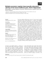
Fig. 6. Testis-specific transciption of CK2b¢ gene. Approximately equal
amounts of total RNA isolated from carcasses (male body remnants
after removal of testes), testes, embryos, larvae, pup ae an d fem ales
wereelectrophoresedin1%formaldehydegel,blottedtoHybond-N
membrane, an d hybridiz ed with either CK2b¢ probe (upper pane l) or
rp49 probe (lower panel). Hybridization signal with the CK2b¢ probe
was detected only in the testes RNA.
1424 A. I. Kalmykova et al. (Eur. J. Biochem. 269) Ó FEBS 2002
(residues 55–64), which is highly conservative among
b subunits from different organisms, is responsible for the
downregulation of catalytic activity of a subunit toward
calmodulin and for the activation by polybasic compounds.
The examination of amino-acid alignment in the acidic
region of three Drosophila CK2 regulatory subunits (Fig. 7)
reveals the lack of two charged residues (Glu57 and Asp60)
in the btes sequence, which might be responsible for less
pronounced CK2btes-mediated effects on the calmodulin
phosphorylation by an asubunit. The b¢ subunit has more
significantly reduced negative charge density in the acidic
region than btes subunit (Fig. 7), but it is still able both to
suppress calmodulin phosph orylation and mediate activa-
tion by polylysine and protamine [20]. Further structural
studies are required to unravel mechanism of this regula-
tion.
In the previous studies it was shown that Drosophila
possesses tandemly repeated Stellate genes e ncoding a
protein with striking sequence similarity to the CK2
b subunit [38]. Moreover, i n the in vitro assay it was
demonstrated that the Stellate protein, although used in
at least 10-fold molar excess, was able t o bind to and
stimulate t he phosphorylation activity of the CK2 a subunit
[39]. The functional homology of the Stellate protein with
the CK2 b subunit rises the f ormal possibility t hat the
Stellate protein may take part in the CK2 regulation.
Nevertheless, all experimental evidences have shown that
the S tellate protein is absent in normal males [38, 39].
Stellate genes are expressed only in testes of the X0 males, or
males lacking the cry locus on the Y-chromosome. In this
case the accumulated Stellate protein forms proteinaceous
crystals in primary spermatocytes of the cry-defic ient males,
thus disturbing the spermatogenesis. Therefore, Stellate
protein c ould not be viewed as an additional testis-specific
isoform of the CK2 r egulatory subunit in normal males.
The CK2 regulatory subunit is ubiquitous among
eukaryotes, but an amino-acid sequence of different b sub-
units is far less conservative as compared to the CK2
a subunits. This fact might be referred to the sup posed
function of b subunit as the regulator of substrate specificity
and targeting of the CK2 holoenzyme in cells. It is suggested
that greater variability and specificity is required for the
realization of these functions. Recent discovery of two
distinct b subunit genes in S. c erevisiae and three genes in
A. thaliana rises a possibility that different b subunits may
serve to provide different substrate specificity or targeting of
the CK2 holoenzyme in cells [16, 17]. Our data extend this
suggestion showing that some b subunits are specialized for
a specific tissue. It seems likely that Drosophila CK2btes and
CK2 b¢ subunit genes were evolutionary adapted for
spermatogenesis.
As was shown earlier [12], quantity of the b subunit
reaches its maximum in the testicles of mammals, as
compared to other tissues. On the other hand, in Drosophila
the ubiquitous CK2 b subunit gene is poorly expressed in
testes [18]. Therefore, the supposed requirement of massive
b subunit production during spermatogenesis is resolved by
different ways in Drosophila and in mammals: while
mammals utilitize the upregulation of expression of a single
b subunit gene, the fruit fly has generated in evolution two
specialized genes f or this purpose.
Despite the accumulation of large amount of information
about it, CK2 remains an enigmatic enzyme. Its un-
doubtedly crucial role in signalling is based only on a
variety of indirect observations, rather than on clear
evidence of cause-and-effect relations. Xu et al.[14]have
shown that the CK2 activity was essential for the spermato-
genesis in mammals. The gene Ôknock-outÕ of the CK2
a¢ subunit in mice resulted in male sterility without any
other physiological defects. In S . cerevisiae , the deletions of
both CK2 a and a¢ subunit genes appeared to be lethal [40],
whereas, the disruption of CK2b,orCK2b¢, or both
resulted in no phenotype or morphology alterations except
the elevated sensitivity to salt concentration in the medium
[34]. Thus, the question concerning vital functions of the
CK2 b subunits in higher eukaryotes is still open.
We tried to address this issue on the model of spermato-
genesis in Drosophila by making a Ôknock-downÕ of the
CK2btes gene by means of the RNAi mechanism. This
approach has been applied recently in Drosophila for
disruption of gene function as an alternative to the classical
mutational analysis [41]. To use such an approach, we
generated transgenic flies transcribing in testes the ÔantisenseÕ
CK2btes RNA under the control of the b2-tubulin promo-
ter. We hoped that this RNA would anneal in vivo to the
CK2btes mRNA t hus forming dsRNA, a nd that this would
lead to the CK2btes mRNA degradation. In fact, we
observed a detectable decrease in the CK2btes mRNA and
protein level (2–4 times lower) in testes of transgenic males
when the Drosophila stock w as maintained at 28 °C, while
no effect on amount of RNA and protein was observed at
18 °C (not shown). The example of temperature sensitivity
of the RNAi effect was already described in Drosophila [42]
but the molecular mechanism underlying it is unclear.
Nevertheless, we were able to Ôknock-downÕ to some extent
the CK2btes gene in Drosophila testes, althought it should
be mentioned that this effect was rather unreproducible.
These unreproducible variations in the degree of the
CK2btes protein drop down did not allow us to make any
conclusions concerning the influ ence of the CK2btes
decrease on the Drosophila male fertility.
Recent evidence for the existence of a Ôfree Õ fraction of the
CK2 b subunit in mouse testicles [12] implicates a new role
for the b subunit in spermatogenesis, a part from the
regulation of CK2 catalytic activity. It is known that the
CK2 b subunit might specifically, but with lower strength,
interact with some partner proteins, other t han CK2
a subunit. These interactive partners are represented, for
example, by A-raf and Mo s kinases [8–10]. If this is the case
in Drosophila, the achieved decrease of the CK2btes p rotein
level in testes of transgenic males might be insufficient to
affect the CK2 activity, a s it could be compensated by the
initial m olar excess of total pool of b subunits over the CK2
a subunit. Taking into account the CK2btes ability to form
Fig. 7. Alignment of acidic region 55–64 in three Drosophila CK2
regulatory subunits. The GenBank accession numbers for t he se quen-
ces shown are the following: D. melanogaster CK2b (M16535),
D. melanogaster CK2b¢ (U51209), D. melanogaster CK2btes
(L49382). Dashes indicate gaps introduced to improve the alignment.
Ó FEBS 2002 Testis-specific isoform of CK2 regulatory subunit (Eur. J. Biochem. 269) 1425
heterodimers with other b subunits shown in ou r yeast two-
hybrid system experiments, it is reasonable to suppose a
possibility of a replacement of one b subunit by another i n
the case of a deficiency of any of the subunits, i.e. a so called
ÔbypassÕ mechanism may operate in order to maintain
appropriate levels and targeting of CK2 activity in testes. In
accordance with this hypothesis are our results showing that
two to fourfold downregulation of the CK2btes gene in
transgenic males does not lead to the noticeable decrease of
total CK2 activity in testes (not shown). Drosophila CK2b-
related genes expressed in testes undoubtedly require further
investigation a s a system for understanding how evolution
of structural properties i s responsible for subtle functional
differences between related genes.
ACKNOWLEDGEMENTS
We are grateful to Dr C.V.C. Glover for providing us with the
pEV55Dmß plasmid a nd the a nti-DmCK2a antiserum, and to Dr H .D.
Hoyle for providing the testis vector. We wou ld like to thank P rof. N.B.
Gusev for providing calmodulin and for fruitful advice. We thank B.
Guerra for the help with gel filtration experiments and M. Silicheva for
technical assistance. This work was supported by the Russian Founda-
tion for Bas ic Researches Grants # 00 -15-97896 and # 03-04-48420, as
well as by a FEBS short-term fellowship to A. I. K. and by an EMBO
short-term fellowship (ASTF 9160) to Y. Y. S.
REFERENCES
1. Allende, J.E. & Allende, C.C. (1995) Protein kinase CK2: an
enzyme with multiple substrates and a puzzling regulation.
FASEB J. 9, 313–323.
2. Pinna, L.A. & Meggio, F. (1997) Protein kinase CK2 (Ôcasein
kinase-2Õ) and its implication in cell division and proliferation.
Prog.CellCycleRes.3, 77–97.
3. Glover, C.V.C. (1998) On the physiological role of casein kinase II
in Saccharomyces cerevisiae. Prog. Nucleic Acid Res. Mol. Biol. 57,
95–133.
4. Guerra, B. & Issinger, O G. (1999) Protein kinase CK2 and its
role in cellular proliferation, development a nd pathology. Elec-
trophoresis 20, 391–408.
5. Coqueret, O., Martin, N., Berube, G., Rabbat, M., Litchfield,
D.W., Nepveu, A. (1998) DNA binding by Cut homeodomain
proteins is down -regulated by casein kinase 2. J. Biol. Chem. 273,
2561–2566.
6. Liu, Z.P., Galindo, R.L., Wasserman, S.A. (1997) A role for CKII
phosphorylation of the cactus PEST domain in dorsoventral
patterning of the Drosophila embryo. Genes Dev. 11, 3413–3422.
7. Jaffe, L., Ryoo, H D., Mann, R.S. (1997) A role for phos-
phorylation by casein kinase 2 in modulating Antennapedia
activity in Drosophila. Genes Dev. 11, 1327–1340.
8. Boldyreff, B. & Issinger, O G. (1997) A-Raf kinase is a new
interacting partner of protein kinase CK2 b subunit. FEBS Lett.
403, 197–199.
9. Hagemann,C.,Kalmes,A.,Wixler,V.,Wixler,L.,Schuster,T.,
Rapp, U.R. (1997) The regulatory subunit of protein kinase CK2
is a specific A-Raf activator. FEBS Lett. 403, 200–202.
10. Chen, M. & Cooper, J.A. (1997) The b-subunit of CKII negatively
regulates Xenopus oocyte matu ration. Proc. Natl Acad. Sci. USA
94, 9136–9140.
11. Lu
¨
scher, B. & Litchfield, D. (1994) Biosynthesis of casein kinase II
in lymphoid cell lines. Eur. J. Biochem. 220, 521–526.
12. Guerra, B., Siemer, S., Boldyreff, B., Issinger, O G. (1999) Protein
kinase CK2: evidence for a protein kinase CK2b subunit fraction,
devoid of the catalytic CK2a subunit, in mouse brain and testicles.
FEBS L ett. 462, 353–357.
13. Diaz-Nido, J., Mizuno, K., Nawa, H., Marshak, D.R. (1994)
Regulation of protein kinase CK2 isoform expression during rat
brain d evelopm ent. Cell. Mol. Biol. Res. 40, 581–585.
14. Xu, X., Toselli, P.A., R ussel, L.D., Seldin, D.C. (1999) Globo-
zoospermia in mice lacking the casein kinase II a catalytic subunit.
Nat. Genet. 23, 118–121.
15. Fuller, M.T. (1998) Genetic control of cell proliferation and dif-
ferentiation in Drosophila spermatogenesis. Semin. Cell. Devel.
Biol. 9, 433–444.
16. Reed, J.C., Bidwai, A.P., Glover, C.V.C. (1994) Cloning and
disruption of CKB2, the gene encodin g the 32-kDa regulatory
b-subun it of Saccharomyces cerevisiae casein kinase II. J. Biol.
Chem. 269, 18192–18200.
17. Sugano, S., Andronis, C., Green, R.M., Wang, Z Y., Tobin, E.M.
(1998) Protein kinase CK2 interacts with and phosphorylates the
Arabidopsis circadian clock-associated 1 protein. Proc. Natl Acad.
Sci. US A 95, 11020–11025.
18. Kalmykova, A.I., Shevelyov, Y.Y., Dobritsa, A.A., Gvozdev,
V.A. (1997) Acquisition and amplification of a testis expressed
autosomal gene, SSL,bytheDrosophila Y chro mosome. Proc.
Natl Aca d. Sci. USA 94, 6297–6302.
19. Kalmykova, A.I., Dobritsa, A.A., Gvozdev, V.A. (1997) The Su
(Ste) repeat in the Y chromosome and bCK2tes gene encode
predicted isoforms of regulatory b-subunit of protein kinase CK2
in Drosophila melanogaster. FEBS Lett. 416, 164–166.
20. Bidwai, A.P., Zhao, W., Glover, C.V.C. (1999) A gene located at
56F1-2 in Drosophila melanogaster encodes a novel metazoan
b-like subunit of casein kinase II. Mol. Cell. Biol. Res. Commun. 1,
21–28.
21. Saxena, A., Padmanabha, R., Glover, C.V.C. (1987) Isolation and
sequencing of cDNA clones encoding a and b subunits of Dro-
sophila melanogaster casein kinase II. Mol. Cell. Biol. 7, 3409–
3417.
22. Thummel, C.S., Boulet, A.M., Lipshitz, H.D. (1988) Vectors for
Drosophila P-element-mediated transformation and tissue culture
transfection. Gene 74, 445–456.
23. Hoyle, H.D. & Raff, E.C. (1990) Two Drosophila b tubulin iso-
forms are not functionally equivalent. J. C ell Biol. 111, 1009–1026.
24. Birnbaum, M.J., Jianguo, W., O’Reilly, D.R., Rivera-Marrero,
C.A., Hanna, D.E., Miller, L.K., Glover, C.V.C. (1992) Expres-
sion and purification of the a and b subunits of Drosophila casein
kinase II using a baculovirus vector. Prot.Exp.Purif.3, 142–150.
25. Chomczynsky, P. & Sacchi, N. (1987) Single-step method of RNA
isolation by acid guanidinium th iocyanate- phenol-ch loroform
extraction. Anal. Biochem. 162, 156–159.
26. Sambrook, J., Fritsch, E.F., M aniatis, T. (1989) Molecular Clo-
ning. A Laboratory Manual. Cold Spring Harbor Laboratory
Press, Cold Spring Harbor, New York, USA.
27. O’Connel, P.O. & Rosbash, M. (1984) Sequence, structure and
codon preference of the Drosophila ribosomal protein 49 gene.
Nucleic Acids Res. 12, 5495–5513.
28. Spradling, A.C. (1986) P-element-mediated transformation. In
Drosophila, a Practical Approach (Roberts, D.B., ed.), 1 78–197.
IRL P re ss, Oxford, UK.
29. Michiels, F., Gasch, A., Kaltschmidt, B., Renkawitz-Pohl, R.
(1989) A 14 bp promoter element directs the testis specificity of the
Drosophila b2 tubulin gene. EMBO J. 8, 1559–1565.
30. Yang, J., Porte r, L., Rawls, J. (1995) Expression of the
dihydroorotate dehydrogenase gene, dhod, during spermato-
genesis i n Drosophila melanogaster. Mol. Gen. Genet. 246, 3 34–341.
31. Nurminsky, D.I., Nurminskaya, M.V., De Aguiar, D., Hartl, D.L.
(1998) Selective sweep of a newly evolved sperm-specific gene in
Drosophila. Nature 396, 572–575.
32. Bidwai, A.P., Reed, J.C., Glover, C.V. (1993) Phosphorylation
of calmodulin by the catalytic subunit of casein kinase II is
inhibited by the regulatory subunit. Arch. Biochem. Bioph ys. 300,
265–270.
1426 A. I. Kalmykova et al. (Eur. J. Biochem. 269) Ó FEBS 2002
33. Meggio, F., Boldyreff, B., Marin, O., Marchiori, F., Perich, J.W.,
Issinger, O G., Pinna, L.A. (1992) The effect of polylysine on
casein-kinase-2 activity is influenced by both the structure of the
protein/peptide substrates and the subunit composition of the
enzyme. Eur. J. Biochem. 205, 939–945.
34. Bidwai, A.P., Reed, J.C., Glover, C.V. (1995) Cloning and dis-
ruptionofCKB1,thegeneencodingthe38-kDab subunit of
Saccharomyces cerevisiae casein kinase II (CKII). Deletion of
CKII regulatory subunits elicits a salt-sensitive phenotype. J. Biol.
Chem. 270, 10395–10404.
35. Gietz, R.D., Graham, K.C., Litchfield , D.W. (1995) Interaction s
between the subunits of casein kinase II. J. Biol. Chem. 270,
13017–13021.
36. Niefind, K., Guerra, B., Ermakowa, I., Issinger, O G. (2001)
Crystal structure of human protein kinase CK2: insights into basic
properties of the CK2 holoenzyme. EMBO J. 20, 5320–5331.
37. Meggio, F., Boldyreff, B., Issinger, O G., Pinna, L.A. (1994)
Casein kinase 2 down-regulation and activation by polybasic
peptides are mediated by acidic residues in the 55–64 region of the
b-su bunit. A study with calmod ulin as phosphorylatable sub-
strate. Biochemistry 33, 4336–4342.
38. Livak, K.J. (1990) Detailed structure of the Drosophila mela-
nogaster Stellate genesandtheirtranscripts.Genetics 124, 303–
316.
39. Bozzetti, M.P., Massari, S., Finelli, P., Meggio, F., Pinna, L.A.,
Boldyreff,B.,Issinger,O G.,Palumbo,G.,Ciriaco,C.,Bonac-
corsi, S., Pimpinelli, S. (1995) The Ste locus, a component of the
parasitic cry-Ste system o f Drosophila melanogaster, encodes a
protein that forms crystals in primary spermatocytes and mimics
properties of the b subunitofcaseinkinase2.Proc. Natl Acad. Sci.
USA 92, 6067–6071.
40. Padmanabha, R., Chen-Wu, J.L., Hanna, D.E., Glover, C.V.
(1990) Isolation, sequencing, and disruption of the yeast CKA2
gene: c asein kinase II is essential for viability in Saccharomyces
cerevisiae. Mol. Cell. Biol. 10, 4089–4099.
41. Misquitta, L. & Paterson, B .M. (1999) Targeted disruption of gene
function in Drosophila by RNA interference (RNA-i): a role for
nautilus in embryonic somatic muscle formation. Proc. Natl Acad.
Sci. USA 96, 1451–1456.
42. Fortier, E. & Belote, J.M. (2000) Temperature-dependent gene
silencing by an expressed inverted repeat in Drosophila. Genesis 26,
240–244.
Ó FEBS 2002 Testis-specific isoform of CK2 regulatory subunit (Eur. J. Biochem. 269) 1427