Báo cáo y học: "Abnormalities of B cell phenotype, immunoglobulin gene expression and the emergence of autoimmunity in Sjögren’s syndrome" pdf
Bạn đang xem bản rút gọn của tài liệu. Xem và tải ngay bản đầy đủ của tài liệu tại đây (287.21 KB, 12 trang )
360
BCA-1 = B-cell-attracting chemokine 1; CDR = complementarity-determining region; GC = germinal center; IFN = interferon; IgV = Ig variable
region; IL = interleukin; LT = lymphotoxin; NHL = non-Hodgkin lymphoma; pSS = primary Sjögren’s syndrome; RA = rheumatoid arthritis; RAG =
recombination activating gene; SDF-1 = stromal cell-derived factor 1; SLE = systemic lupus erythematosus; TGF = transforming growth factor;
Th = T helper; TNF = tumor necrosis factor.
Arthritis Research Vol 4 No 6 Dörner and Lipsky
Introduction
Primary Sjögren’s syndrome (pSS) represents an idio-
pathic inflammatory exocrinopathy characterized by both
organ-specific autoimmunity, preferentially affecting the
salivary and/or lacrimal glands, and systemic manifesta-
tions. The characteristic hallmarks of pSS are focal
lymphocytic infiltrates and subsequent destruction of the
lacrimal and salivary glands, resulting in keratoconjunctivitis
sicca and xerostomia. In addition, there is a broad variety
of accompanying clinical and laboratory manifestations,
emphasizing that pSS is a systemic disorder [1]. In most
patients, these laboratory parameters include hyper-
gammaglobulinemia, circulating immune complexes and
autoantibodies, such as those against the Ro-SSA and/or
La-SSB autoantigens and rheumatoid factors. The typical
production of autoantibodies and polyclonal hypergamma-
globulinemia indicates that abnormalities of humoral
immunity are significant in pSS and have been included in
the classification criteria [2]. However, the factors driving
autoimmunity and leading to the differentiation of auto-
reactive lymphocytes into autoantibody-producing plasma
cells remain largely unknown, although several epitope
mapping studies have suggested that autoimmunity in
pSS is driven by autoantigens.
The glandular infiltration in pSS is composed mainly of
CD4
+
T lymphocytes [3] but usually also contains a sub-
stantial number of B cells and plasma cells [4,5]. The
degree of glandular destruction and symptoms of dryness
do not seem to be directly related to the number of
infiltrating lymphocytes. Indeed, the mechanism of glandular
damage remains incompletely delineated, although a role
for CD4
+
T cells has been proposed, either directly or
through the action of secreted cytokines.
Primary Sjögren’s syndrome (pSS) is an autoimmune disorder characterized by specific pathologic
features and the production of typical autoantibodies. In addition, characteristic changes in the
distribution of peripheral B cell subsets and differences in use of immunoglobulin variable-region genes
are also features of pSS. Comparison of B cells from the blood and parotid gland of patients with pSS
with those of normal donors suggests that there is a depletion of memory B cells from the peripheral
blood and an accumulation or retention of these antigen-experienced B cells in the parotids. Because
disordered selection leads to considerable differences in the B cell repertoire in these patients, the
delineation of its nature should provide important further clues to the pathogenesis of this autoimmune
inflammatory disorder.
Keywords: autoimmunity, B cells, IgV gene usage, lymphocytes, Sjögren’s syndrome
Review
Abnormalities of B cell phenotype, immunoglobulin gene
expression and the emergence of autoimmunity in Sjögren’s
syndrome
Thomas Dörner
1
and Peter E Lipsky
2
1
Department of Medicine, Rheumatology and Clinical Immunology, University Hospital Charité, Berlin, Germany
2
National Institute of Arthritis and Musculoskeletal and Skin Diseases, National Institutes of Health, Bethesda, Maryland, USA
Corresponding author: Thomas Dörner (e-mail: )
Received: 22 July 2002 Revisions received: 5 September 2002 Accepted: 16 September 2002 Published: 25 September 2002
Arthritis Res 2002, 4:360-371 (DOI 10.1186/ar603)
© 2002 BioMed Central Ltd (
Print ISSN 1465-9905; Online ISSN 1465-9913)
Abstract
361
Available online />Although local autoantibody production in the glands has
been suspected and autoantibodies have been found in
the saliva [6], the pathogenetic role of the glandular B
lymphocytic infiltrates remains largely unknown. In this
regard, it is notable that autoantibodies to the M3 type of
the muscarinergic acetylcholine receptor might function to
inhibit salivary flow (reviewed in [7]) in a manner compara-
ble to the antibody-mediated blockage of nicotinergic
receptors in patients with myasthenia gravis. Cell clusters
resembling germinal center (GC)-like structures have been
reported in the focal lymphocytic sialadenitis of the minor
(labial) salivary glands in patients with pSS [4], although it
is not known whether they function completely as lymphoid
GCs. Because similar ectopic GC-like reactions have also
been observed in the synovium in rheumatoid arthritis (RA)
[8,9] as well as in a variety of other diseases (such as
spondylarthopathies, myasthenia gravis and thyroiditis), it
has been proposed that such potentially functional B cell
aggregates can be induced in several extrafollicular tissues
in autoimmune disorders [4,8–10]. The formation of these
aggregates seems to be clearly dependent on the interac-
tion of chemokines and their receptors [10–12].
Cytokines and chemokines in pSS
In patients with pSS the salivary and lacrimal glands are
the main target organs for the immune system and, at least
in part, subsequent autoimmune-mediated tissue damage.
Local production of cytokines by mononuclear cells and
also epithelial cells might contribute to the immune-
mediated destruction of exocrine glands in pSS [13]. pSS
has therefore also been called ‘autoimmune epithelitis’
[14], emphasizing that epithelial cells are thought to be
important in the immunopathogenesis.
Cytokines
Several cytokines have been demonstrated in inflamed
tissue by using reverse-transcriptase-mediated poly-
merase chain reaction technologies as well as in animal
models. Cytokines, such as tumor necrosis factor (TNF),
lymphotoxin α (LTα) and interleukin (IL)-1β have been
found to influence the destruction of the acinar structure in
human salivary gland cell clones [15]. The most prominent
cytokines detected in affected salivary glands of patients
with pSS are IL-1, IL-6, IL-10, transforming growth factor
(TGF)-β, interferon (IFN)-γ and TNF. Fox et al. [16] found
that salivary gland CD4
+
T cells produced over 40-fold
more IL-2, IFN-γ and IL-10 than peripheral-blood CD4
+
T cells from patients with SS or from controls. Moreover,
salivary gland epithelial cells produced 40-fold more IL-1α,
IL-6 and TNF mRNA than epithelial cells from individuals
with histologically normal salivary glands.
It has therefore been suggested that T helper type 1 (Th1)
cytokines, such as IFN-γ and IL-2, as well as IL-10, IL-6
and TGF-β, might be important in the induction and/or
maintenance of pSS [10], whereas Th2 cytokines,
detected in some cases in association with a striking B
cell accumulation in the labial salivary glands, might be
involved in the progression of the disease. Furthermore, it
has been suggested that TGF-β, an important immunoreg-
ulatory cytokine whose absence can lead to systemic
autoimmune disease [17], might be deficient in SS. In this
regard, reduced levels of TGF-β have been found in SS
glands with intense lymphocytic infiltrates [18]. Further-
more, mice that fail to express TGF-β develop an
exocrinopathy resembling SS [17]. Although several
studies have analyzed cytokines that seem to be involved
in the pathogenesis of SS, reports on cytokine poly-
morphisms are very limited and do not allow any firm
conclusion. It should be noted that the interaction
between epithelial cells and infiltrating T cells has been
characterized in detail, but the cytokines involved in local
B cell activation remain largely unknown.
Chemokines
The infiltration of lymphocytes into glandular aggregates
apparently has a crucial role in the tissue pathology of SS.
This process seems to be tightly regulated at least in part
by chemokines and the local expression of their receptors.
Chemokines and the expression of chemokine receptors
by the inflamed tissue as well as by lymphocytes are there-
fore likely to be of importance in the evolution of tissue
pathology in pSS. Mice deficient in lymphoid-homing
chemokine receptors CXCR4 and CXCR5 lack normal
lymphoid organs [19–21], indicating that these receptors
as well as the chemokines CXCL12 (stromal cell-derived
factor 1; SDF-1) and CXCL13 (B-cell-attracting
chemokine 1; BCA-1) are important for lymphoid organo-
genesis. In addition, studies in CXCL13 transgenic mice
found that this chemokine, together with TNF and LTβ, is
crucial in lymphoid organogenesis, whereas the LTβ
knockout mouse lacks the formation of GC structures. A
recent review reported that CXCL13 (BCA-1), CCL19
(ELC), CCL21 (SLC) and CXCL12 (SDF-1) all contribute
to lymphoid homing and to the persistence of chronic
inflammation in pSS [11]. Moreover, studies on RA
demonstrated that CXCL12 [22] and CXCL13 [12] are
involved in the formation of GC in the rheumatoid syn-
ovium. Thus, recent studies have shed light on factors
involved in directing lymphocytes into inflamed tissue and
maintaining inflammation in SS, whereas the etiologic
factors of SS remain to be delineated.
Of potential importance is the fact that enhanced levels of
B lymphocyte stimulator (BLyS; also known as B cell acti-
vating factor belonging to the TNF family [BAFF] or trans-
membrane activator and CAML interactor [TACI]) have
been demonstrated in patients with SS [23], levels that
are higher than previously identified in systemic lupus
erythematosus (SLE) [24]. In addition, expression of BLyS
has been found to be markedly enhanced in the inflamed
salivary glands [23], indicating that activation of B cells
362
Arthritis Research Vol 4 No 6 Dörner and Lipsky
might take place in the parotids. In this regard, it is notable
that BLyS transgenic mice develop SS with age after the
development of manifestations of lupus.
General aspects of Ig variable region (IgV)
gene usage
Autoantibodies are features of most systemic autoimmune
diseases, including pSS. The production and persistence
of autoantibodies in autoimmune conditions is considered
to occur because of immune dysregulation with a resultant
break in tolerance, regardless of whether these autoanti-
bodies are pathogenic [25]. Despite intensive work on the
characterization of autoantigens, the cellular basis of their
production and the strong association of certain autoanti-
bodies with particular MHC class II alleles, and despite
increasing knowledge about tissue B cell immunology and
the role of cytokines and chemokines, little is known about
the usage of IgV genes in autoimmune conditions and
about the respective autoantibodies.
The observation that certain autoantibodies are frequently
encoded by a limited number of IgV gene segments sug-
gested that biases in the development of the B cell recep-
tor repertoire might have a role in the tendency of specific
individuals to develop these autoantibodies. Whether the
usage of these specific gene segments is different in
normals and patients with systemic autoimmune disorders
remains controversial. Recent approaches have made it
possible to address this issue and to estimate the differen-
tial impact of molecular and selective influences in shaping
the IgV gene repertoire in normals and patients with
autoimmune diseases, including those with pSS.
Previous analyses of autoimmune B cells focused almost
exclusively on productive V gene rearrangements and
therefore could not discern the impact of molecular and
selective influences. Analysis of the nonproductive reper-
toire is especially important to assess the immediate
impact of molecular processes, such as recombination
and somatic hypermutation [26–32], because the non-
productive rearrangements do not encode an expressed
Ig molecule and are therefore not influenced by selection.
In contrast, the distribution of B cells and their productive
IgV gene rearrangements can be influenced by a variety of
selective events during development and subsequent anti-
genic stimulation because of the nature of the expressed
heavy and/or light chain and the cognate (auto)antigen.
Recent analysis of the nonproductive V gene repertoire in
patients with pSS and SLE has documented minimal
abnormalities in the nonproductive V
H
, Vκ and Vλ gene
repertoires [33–41]. Although only a few patients with
pSS and with SLE have been analyzed with this approach
to assess the overall repertoire, the data indicate that IgV
gene usage in the nonproductive repertoire is not signifi-
cantly different from normal, suggesting that the basic
process of IgV gene recombination is largely normal in
patients with these autoimmune diseases. This does not
rule out the possibility that abnormalities in the usage of
specific V
H
or V
L
genes might contribute to autoimmunity.
However, it is apparent that there is unlikely to be a gener-
alized abnormality in V(D)J recombination in these auto-
immune diseases. A study by de Wildt et al. [42]
confirmed on the mRNA level that the IgV gene usage in
patients with SLE or mixed connective-tissue disease
(MCTD) was comparable to normal. Moreover, the large
number of V
H
genes that encode specific autoantibodies
make it unlikely that an abnormality of V gene usage
underlies autoimmunity in most patients.
Although there is no conclusive evidence for recombination
biases predisposing to systemic autoimmunity in SLE and
SS, there are some exceptions. In some circumstances,
specific autoantibodies express marked biases in IgV gene
usage. One example is the almost exclusive usage of V
H
4-
34 in cold agglutinin disease [43]. In addition, the 16/6 idio-
type expressed by some anti-DNA antibodies is encoded by
V
H
3-23 and the 9G4 epitope encoded by V
H
4-34 is over-
represented in anti-DNA antibodies in SLE. In these
instances, autoantibodies preferentially employ specific V
H
genes for the variable regions. Despite the apparent
increase in the use of these specific genes, the generalized
higher frequency of somatic hypermutation, abnormal pat-
terns of targeted mutations toward specific DNA motifs,
indications of increased receptor editing and differences in
the entire IgV gene repertoire of peripheral B cells from
patients with SLE [34,35,37,44] indicate that broad abnor-
malities in B cell selection are characteristic of SLE. B cell
repertoire abnormalities are therefore not restricted to
clones of autoreactive B cells in this autoimmune disease.
A recent study [45] demonstrated that V
H
4-34 is clearly
negatively selected among post-GC B cells in normals, as
already shown at the single-cell level for peripheral B cells
[27] and post-switch tonsilar plasma cells [46]. By con-
trast, patients with SLE do not negatively select these
cells appropriately in their GC, allowing their expansion
after encountering antigen and receiving help from T cells.
An enhanced frequency of V
H
4-34 has previously been
observed in peripheral plasma cells of a patient with active
SLE [33]. This contrasts markedly with the normal post-
switch plasma cell repertoire, in which V
H
4-34 is strictly
excluded [46]. This finding is consistent with the conclu-
sion that disturbances in selection have a key role in SLE.
Notably, different autoimmune diseases, such as pSS and
SLE, seem to have characteristic abnormalities of particu-
lar censoring mechanisms.
IgV gene usage in patients with pSS is
preferentially shaped by disordered selection
Distinct abnormalities in the B cell repertoire have been
identified in pSS. Recent studies in three patients with
363
Available online />pSS found that major abnormalities were related mainly to
influences of selection, because the usage of V
H
and V
L
gene segments in the nonproductive repertoire was
largely normal. However, significant abnormalities were
found in the productive repertoires, especially by affecting
V
L
distribution [36,37]. Notably, four Vλ genes (2A2, 2B2,
2C and 7A) represented 56% of all functional Vλ joints
[37]. In the productive Vκ repertoire, three Vκ genes (L12,
O12/O2 and B3) comprised 43% of all amplified VκJκ
joints [36]. It is of interest that VκA27, a gene frequently
employed by autoantibodies, rheumatoid factor and
lymphomas in SS, was found less frequently in the periph-
eral nonproductive repertoire of the patients with pSS
than in normal controls (8% versus 14%; P < 0.05). By
contrast, this gene was found at an increased frequency in
the parotid gland (29%) compared with the blood (8%;
P < 0.05) in the one patient examined [39]. Moreover, 2 of
15 VκA27–Jκ5 rearrangements found in the parotids were
clonally related [38]. Furthermore, 11 clonally unrelated
VκA27–Jκ2 rearrangements representing 34% of all pro-
ductive Jκ2 using rearrangements were found in the
parotid. Accumulation or local expansion of B cells
expressing VκA27–Jκ2 rearrangements in the parotid
glands seems to be a characteristic of pSS.
Receptor editing is a mechanism by which B cells are able
to escape deletion by revising their autoreactive receptors.
To the best of our knowledge, there are no reports
addressing the role of editing in SS, whereas several
studies did so for SLE. Formerly it was thought that
expression of the IgB cell receptor (BCR) extinguished
subsequent Ig rearrangements by downregulating the
expression of recombination activating gene (RAG) 1 and
RAG2 enzymes in the bone marrow. However, recent
studies provide evidence that immature B cells outside the
bone marrow [9,47,48] retain RAG activity and can there-
fore replace their receptors by secondary Ig gene recom-
bination (receptor editing/revision). This is noted with
increased frequency in secondary lymphoid organs
[9,46,47,49] and in the fetus [50]. The extent to which the
presence of recombination enzymes is correlated with
actual editing is uncertain.
There is a controversy over whether defects in receptor
editing or secondary rearrangements are involved in
shaping the B cell repertoire in autoimmunity. The possibil-
ity that deficiencies in central or peripheral receptor
editing could have a role in generating autoimmunity has
been suggested [51]. In addition, analysis of autoreactive
hybridomas [52] generated from patients with SLE
demonstrated an overusage of J-proximal Vκ1 genes and
a preferential use of J elements proximal to Vκ, suggesting
that receptor editing in SLE might be defective, because
skewing towards the usage of Jκ, distal Vκ genes and
Jκ5-expressing V gene products [9,34,53] has been taken
as an indication of active receptor editing. Because recep-
tor editing at the V
L
loci is thought to have a major role in
rescuing autoreactive B cells from deletion [52], defects in
receptor editing could have a role in the etiology of SLE
[25,34,35,49–52].
Recent studies in patients with RA [9,54,55] provided evi-
dence that receptor editing/revision might also be more
active in the synovium of these patients than in normals. In
contrast with these patients with RA, patients with pSS
seem to have decreased receptor editing/revision, as
identified by an enhanced usage of V-proximal J
L
seg-
ments. It is possible that this reflects a defect or infrequent
usage of receptor editing in pSS [36,37,39]. In this
regard, a recent analysis of six monoclonal antibodies with
rheumatoid factor activity obtained from the peripheral
blood of patients with pSS showed that all used Vλ-proxi-
mal Jλ2/3 gene segments [56], which is consistent with
the conclusion that receptor editing/revision might be
defective in pSS. The role of abnormalities in this mecha-
nism in permitting the emergence of autoimmunity remains
to be fully delineated.
Influences of selection by direct comparison
of the B cell receptor repertoire in the
parotids versus blood in pSS
As already mentioned, recent studies [36–41] addressed
the question of whether there are differences in the IgV
chain gene repertoire of CD19
+
B cells by comparing two
immune compartments, the peripheral blood and the
inflamed parotid gland, a target tissue in pSS. Although
only one patient was analyzed, the data obtained provide
new insights into this disease.
The underlying assumption of this study was that the
peripheral circulating B cell repertoire reflects a complex
group of cells expressing IgV genes that might have been
influenced by a variety of immune compartments, whereas
the IgV genes of B cells infiltrating the parotid might
provide a more skewed population owing to the local
selection and/or (antigen-dependent) proliferation. Alter-
natively, inflammation might have induced the migration of
polyclonal B cells into the affected tissue [40,56–58].
Whereas clonal B cell expansions in the target tissues of
SS patients are well established [4,5] and early studies
examining anti-idiotypes have suggested that B cell infiltra-
tions in pSS represent a highly selected population [59], a
molecular analysis of 37 Ig heavy chain rearrangements
from labial salivary gland biopsies in pSS [60] has shown
a rather polyclonal pattern of IgV gene usage, comparable
to that of circulating B cells from normals. Thus, the lack of
direct comparison between the B cell repertoire in the
blood and that in the parotids has prevented the drawing
of firm conclusions about the extent to which selective
pressures influence the repertoire in autoimmune diseases
with characteristic extrafollicular germinal centers, such as
pSS.
364
IgV gene usage
With the exception of the V
H
7 family, a single gene family
that is known to be related to an insertion/deletion poly-
morphism of the human V
H
locus [61], members of all V
H
families were found in the patient’s peripheral blood as
well as in the parotid gland. Notably, however, there were
specific differences in the V
L
gene repertoire when blood
and parotid were compared [38]. Strong selective influ-
ences were detected in the parotid gland of the patient in
that B cells with rearranged VκA27, VκA19 and Vλ2E as
well as Vλ1C were markedly enriched. Furthermore, there
was evidence of clonal expansion of VκA27–Jκ5 and
VκA19–Jκ2 rearrangements in the patient’s parotid gland
as well as of Vλ1C–Jλ3 rearrangements in both blood and
parotid gland. An increase in VκA27 preferentially
rearranged to Jκ2 but not clonally related in the gland is
noteworthy because there was a significantly lower
frequency of VκA27 in the periphery of patients with pSS
compared with normals. These data are consistent with
the conclusion that there is clonal expansion within the
salivary gland B cells as well as the selection of cells
expressing particular light chains. In addition, a polyclonal
population of B cells was present.
Positive selection of particular V
L
chain genes by foreign
antigens or autoantigens present in the gland seems to
shape the productive V
L
chain repertoire in the inflamed
tissue. This is in contrast to the V
H
repertoire of the patient
analyzed, which was similar in the peripheral blood and in
the parotid gland. These results suggest an important role
for V
L
chain gene usage in the immune activation of B
cells within the parotid gland of the pSS patient studied. A
restriction of the V
L
chain repertoire has been described
after vaccination. As an example, antibodies against
Haemophilus influenzae (Hib) B that develop as part of a
T
H
2 response have been identified as being frequently
encoded by VκA2, O8/O18, L11, A17 and A27 [62].
Moreover, Vλ genes of the Vλ2 and Vλ7 family were found
in the Hib-antibody V
L
gene repertoire [62]. In addition,
VκA27 and Vλ2C, 2E, 2A2 or 10A were also shown to
encode antibodies against Streptococcus pneumoniae
[63]. Interestingly, VκA27 and Vλ2E, which were fre-
quently found in the parotid gland of this patient, with
VκA27 expanded clonally, have also been shown to
encode antibodies against rabies virus [64]. Thus, micro-
bial antigens, including bacterial and viral epitopes that
could be involved in the pathogenesis of pSS, might also
be involved in the selective processes shaping the V
L
gene repertoires of B cells accumulated in the parotid
gland of this patient with SS.
In contrast, it is possible that autoantigens might be
involved in the accumulation of parotid gland B cells in this
patient. In this regard, VκA27 was frequently used by
rheumatoid factors in patients with RA [65]. Rheumatoid
factor is typically present in the sera of patients with pSS
and was also detected in the saliva or in salivary gland biop-
sies [66] of these patients. In this regard, Martin et al. [66]
described two salivary-gland lymphomas that developed in
patients with pSS from rheumatoid-factor-specific B cells.
Moreover, VκA27 has been reported to be frequently
employed by lymphomas developing in the salivary gland of
patients with pSS [67]. Despite the presence of clonally
expanded B cells expressing VκA27, the patient studied did
not develop lymphoma during a follow-up period of 3 years
after the examination, indicating that additional factors or
further persistence of the chronic B cell proliferation are
essential for the development of lymphoma.
Analysis of mutations
In the peripheral-blood B cells of patients with pSS, less
than a third (28.3%) of the CD19
+
B cells expressed
somatically mutated productive V
H
rearrangements [39].
The frequency of mutations was lower than that previously
reported for circulating CD19
+
B cells of normals (1.4
versus 2.6%; P < 0.001) [26,28]. Although a direct com-
parison of B cell subsets was not performed at the begin-
ning of these analyses, decreased levels of memory
CD27
+
B cells in patients with pSS probably account for
the difference in mutations [39,40]. By contrast, the vast
majority (about 80%) of the parotid B cells used mutated
V
H
rearrangements, and both the nonproductive and pro-
ductive glandular rearrangements exhibited significantly
increased mutational frequencies compared with the
blood counterpart. Because mutated IgV genes are char-
acteristic of memory B cells [68], this finding indicates an
accumulation of memory-type B cells in the inflamed
parotid gland.
The mutational frequency and the percentage of mutated
light-chain genes were also greater in the productive V
L
chain rearrangements of B cells from the parotid gland
than in cells from the peripheral blood, but the V
L
rearrangements accumulated a large number of silent
mutations. Interestingly, productively rearranged Vλ genes
from the parotid gland (3.32%) exhibited a significantly
greater mutational frequency than the Vκ gene rearrange-
ments (2.35%; P < 0.001) [38].
Because GC-like structures have previously been
described in the parotid gland [3,4], this site might be able
to act as a secondary lymphoid organ facilitating somatic
hypermutation and selection of antigen-specific B cells.
Antigen-driven germinal-center reactions might proceed
within ectopic lymphoid follicles in the parotid gland, giving
rise to highly mutated antigen-specific B cells. However, the
migration of highly mutated antigen-specific B cells from the
patient’s blood to the parotid gland could also contribute to
the observed differences in the mutational frequencies.
The analysis of the replacement/silent (R/S) ratio and the
mutational ‘hot spots’ of productive V
L
chain rearrange-
Arthritis Research Vol 4 No 6 Dörner and Lipsky
365
ments of peripheral and parotid gland B cells revealed no
major abnormalities when compared with normal donors,
indicating intact selective mechanisms with selection
against R mutations in the frame work regions that might
cause structural constraints of the Ig molecule. In the pro-
ductive V
L
chain repertoire of B cells from the parotid
gland, we found the frequency of S mutations to be
increased, which was consistent with a reduced R/S ratio
in the complementarity-determining regions (CDRs)
[38,39]. This is in accordance with the observations of
other studies (Gellrich et al. [60], Stott et al. [4] and
Miklos et al. [69]). Stott et al. [4] described a decreased
R/S ratio in the CDRs of V
H
and V
L
gene rearrangements
of B cells obtained by minor salivary-gland biopsies from
two patients with SS. Detailed analyses of the frequency
of the distribution of mutations revealed that mutations in
nonproductive V
L
rearrangements of B cells from the
parotid gland were less targeted towards the highly
mutable RGYW [R(purine)/G/Y(pyrimidine)/W(A or T)]
motifs. However, these targeted mutations of RGYW in V
L
gene rearrangements were highly selected in B cells from
the parotid gland. Although no firm conclusion can be
drawn, it is possible that these targeted mutations are
generated in the parotids of the patient, with retention of
particular mutated V
L
rearrangements.
CDR3 analysis of IgV gene rearrangements
Remarkably, the productive glandular V
H
rearrangements,
but not V
L
rearrangements, were found to exhibit a signifi-
cantly shorter CDR3 region than their peripheral produc-
tive counterparts [38,39]. To a considerable extent, this
was accounted for by a less frequent usage of the J
H
6
segment in the glandular rearrangements when compared
with the patient’s peripheral repertoire as well as with that
reported previously for normals [27,28]. It is noteworthy
that the J
H
6 segment encodes the longest CDR3 compo-
nent (29 nucleotides) of all J
H
genes and can thereby con-
tribute to rearrangements with longer CDR3 regions. This
confirms conclusions that J
H
6 is positively selected in the
expressed preimmune repertoire [70] but negatively
selected in the mutated repertoire. Moreover, selective
influences on productive rearrangements seem to favor
shorter CDR3 regions [27,28]. The finding of V
H
sequences with shorter CDR3 regions is in accordance
with the conclusion that memory B cells that accumulate
in the parotids are recruited by antigen. The CDR3 region
of V
H
rearrangements might be more important in reacting
to parotid antigens than other regions of the V
H
molecule.
Evidence of clonal expansions
The major salivary glands are known to be the site of pref-
erential B cell expansions and in some cases of lympho-
proliferation in pSS. B cells from the parotid gland have
been identified as a distinct population showing preferen-
tial expansion and somatic mutation of particular V
L
chain
rearrangements, such as VκA27–Jκ5, VκA19–Jκ2 and
Vλ1C–Jλ2/3, in comparison with peripheral B cells [38].
Clonal expansion was not associated with any evidence of
intraclonal diversification. Thus, this glandular expansion
might be derived from proliferation of the very small subset
of proliferating mantle-zone (founder) B cells or from an
early state of dark-zone germinal center cells [71].
However, it is uncertain whether these cells are able to
leave the germinal center.
Accumulation of memory-type B cells in the inflamed
parotid gland supports the conclusion that there is an
enhanced influx/homing of particular memory-type B cells
into the inflamed gland, rather than a proliferation of a few
founder B cells entering the parotid GC structures in
patients with pSS, despite evidence of clonally expanded
B cells in the tissue.
Analysis of B cell subsets in Sjögren’s
syndrome allows differentiation from SLE
Several groups, including our own, have performed
studies on the distribution of peripheral B cell subsets in
systemic autoimmune diseases, such as SLE and pSS
[33,40,57,72]. In this regard, the identification of CD27 as
a marker of memory B cells [33,68,73,74] made it possi-
ble to characterize peripheral naive (IgM
+
/CD27
–
) and
memory (CD27
+
) B cells. Interaction of CD27 with its
ligand on T cells, CD70, serves as a pathway of differenti-
ation of B cells into plasma cells [73,75,76]. Recently,
homotypic interaction of CD27 and CD70 expressed by B
cells only [77] was also reported to be sufficient for B cell
differentiation, raising the possibility that B cells might be
able to regulate themselves by CD27–CD70 interactions.
In another recent study [78] it was shown that CD27
–
B
cells can be differentiated into IgG-producing or IgE-pro-
ducing plasma cells in vitro. It needs emphasis that class
switching, but not somatic hypermutation, could be
induced, although the recently discovered activation-
induced deaminase (AID) [79] has been found to be
expressed in these cells.
On the basis of the available data on the distribution of B
cell subsets in SLE versus pSS, there is increasing evi-
dence that diseases associated with immunologic activity
can be characterized by unique features of B cell distribu-
tion [33,72,80–83]. Whereas patients with active SLE
[33] revealed increased circulating CD27
+
memory B
cells, reduced naive CD27
–
B cells and markedly
increased CD27
high
plasma cells that seemed to be
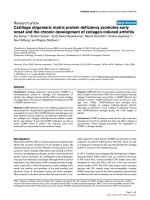
related to lupus disease activity, analysis in pSS [40]
showed a clear predominance of CD27
–
naive B cells
(Fig. 1) that also lacked expression of CD5 compared with
normal donors as well as patients with SLE (P < 0.001)
and a significant decrease in the frequency of memory
CD27
+
B cells that were predominantly CD5
+
[40]. The
reduced frequency of CD27
+
B cells in pSS was signifi-
cant when compared with either normal controls
Available online />366
Arthritis Research Vol 4 No 6 Dörner and Lipsky
(P < 0.05) or patients with SLE (P < 0.0002). Another
recent study [57] characterized peripheral B cells in 11
patients with pSS as well as patients with RA and normal
controls. These investigators also found a predominance
of naive B cells that were CD27
–
and a reduced frequency
of memory B cells in patients with pSS.
The difference between pSS and SLE (Fig. 2) is noteworthy
because of the many common clinical and serologic similari-
ties (hyperimmunoglobulinemia, positive anti-Ro and anti-La
autoantibodies, rheumatoid factor) between patients with
pSS and those with SLE. Another difference between
patients with pSS and with SLE was the normal peripheral
B cell count in the former, whereas patients with SLE exhib-
ited significant decreases in peripheral B cell frequencies
[33,40]. In one study [57], patients with RA also manifested
an increased frequency of CD27
+
memory B cells and a
normal frequency of CD27
–
naive B cells. The pattern of B
cell subpopulations in pSS, RA and SLE defined by CD27
expression therefore seemed to be unique.
Previous data have shown that the frequency of CD27
+
B
cells reflects the accumulation of antigen experience of an
individual that is, at least in part, related to age [73,82].
Cord blood and blood from hyper-IgM patients normally
do not contain CD27
+
B cells [73]. Because of the usually
more advanced age of patients with pSS than of those
with SLE, the actual differences identified between the
Figure 1
Analysis of the distribution of peripheral CD19
+
B cell subsets demonstrates that patients with primary Sjögren’s syndrome (pSS) have reduced
frequencies of CD27
+
memory B cells in the peripheral blood compared with normal donors. In addition, patients with pSS with secondary non-
Hodgkin lymphoma exhibited an increase in CD27
+
B cells in the blood.
CD19
CD27
pSS patient #13 pSS patient #5Healthy Control
pSS patient with NHL
59.1%
40.1%
0.8%
94.3%
7.7%
0.9%
91.3%
5.5%
0.3%
34.7%
40.1%
35.2%
Figure 2
Schematic distribution of B cell subsets in peripheral blood of normals compared with patients with systemic lupus erythematosus and Sjögren’s
syndrome.
Sjögren
Sjögren
‘s
’s
syndrome
syndrome
Naïve
B cells 80%
Naïve
B cells ≥80%
Memory
B cells
Memory
B cells
Plasmablasts
CD27 Expression
Normals
Normals
Naïve
B cells 60%
Naïve
B cells 60%
Memory
B cells
Memory
B cells
Plasmablasts
CD27 Expression
Systemic
Systemic
Lupus
Lupus
Naïve
B cell
s
Naïve
B cells
Memory
B cells
Memory
B cells
Plasmablasts
CD27 Expression
367
Available online />SLE and pSS groups might have been underestimated. In
contrast, the peripheral status of B cell distributions looks
very similar in patients with pSS and in those with HIV with
predominantly naive B cells [40,80]. Because CD4
+
T cells are depleted in HIV but not in pSS, one interpreta-
tion of these observations may be that T cell dependent
priming of B cells might be less in patients with pSS than
in normals and patients with SLE.
Remarkably, the CD27
–
B cells could be further subdi-
vided by the mutational status of their productive V
H
rearrangements into a majority of naive cells (35 of 39 with
no mutations; mutational frequency less than 0.1%) and a
minority of memory-type cells (4 of 39 with mutations;
mutational frequency 4.6%), whereas all but one of the
CD27
+
B cells (31 of 32) analyzed expressed mutated IgV
genes [40]. Currently, the finding of the small population
of CD27
–
B cells expressing mutated V
H
rearrangements
remains unclear. It is noteworthy that this population had a
significantly lower mutational frequency than CD27
+
B cells (4.6% versus 7.8%; P = 0.0009). Possible expla-
nations might be transient or low-level CD27 expression,
shedding of CD27, or stimulation that results in mutations
but fails to upregulate CD27. Notably, however, there are
no striking differences in the frequency of this population
between SS patients and normals [40], and, very recently,
this population has also been detected in other normal
and abnormal conditions by studies on single cells. Impor-
tantly, the regulation of CD27 and its association with the
acquisition of IgV
H
mutations seems to be normal in
patients with pSS.
Previous molecular analysis documented that CD27 can
be taken as a reliable marker for memory B cells in healthy
normals [68,74] as well as in patients with SLE [33]. The
analysis of a patient with SLE revealed a mutational fre-
quency of 0.4% in the CD27
–
B cells and 6.1% in the
CD27
+
B cells. Overall, there was no major difference in
the frequency of mutations in V
H
rearrangements of
CD27
–
and CD27
+
B cells, respectively, obtained from
patients with pSS or SLE and from normals, which is con-
sistent with the conclusion that expression of CD27 indi-
cates previous antigen contact by the respective B cell.
Several studies have identified an enhancement of CD5-
expressing B cells in the periphery of patients with pSS
[84–87], although to the best of our knowledge no study
has analyzed in detail the proportions of CD5
+
B cells
among naive and memory B cells. In contrast, a few
reports did not identify an enhanced frequency of CD5
+
B
cells in pSS [88]. Earlier studies [89] found enhanced fre-
quencies of CD5
+
B cells in about half of patients with
pSS, as well as in about half of patients with RA and
about a quarter of patients with SLE and normals. By con-
trast, there was an increase only of CD5
–
/CD27
–
naive B
cells in patients with pSS [40], whereas there was no sig-
nificant increase in the overall CD5
+
B cell population.
However, a subgroup of seven patients with pSS with the
highest frequencies of naive B cells (86–94%) also had
larger numbers of CD5
+
/CD27
–
naive B cells
(14.2–37.2%). In the patients analyzed, there were no
clinical features that distinguished these seven patients
with enhanced CD5
+
B cells from the remainder. These
data indicate that the previously known enhancement in
CD5
+
B cells in SS stems preferentially from an increase
in the immature B cell pool. It should be noted that B cells
infiltrating the parotids frequently express CD5, further
supporting the hypothesis of homing and activation of
specific B cells in the glands.
B cell malignancies and Sjögren’s syndrome
In contrast to the focal sialadenitis of the minor (labial) sali-
vary glands, the lymphocytic lesions of the major salivary
glands often contain secondary lymph follicles. B cells
have been shown to infiltrate the glandular duct epithelium
and thereby to contribute to the characteristic pattern of
chronic lymphocytic inflammation called myoepithelial
sialadenitis (MESA) or benign lymphoepithelial lesion [90].
These lesions are thought to form the substrate for the
development of extranodal non-Hodgkin lymphomas
(NHLs) [91,92]. In this context, it is well known that
patients with pSS have an increased risk of developing
such lymphomas compared with normals. Extranodal lym-
phomas in pSS are almost exclusively of B cell origin and
are frequently identified in the major salivary glands.
Recently, the suggested linkage between autoimmunity,
autoantibody-producing cells and lymphoma [66,93,94]
has been emphasized by the demonstration of two cases
of parotid gland lymphomas in pSS producing mono-
specific rheumatoid factors [66].
A remarkably biased usage of individual V
H
segments (in
particular the V
H
1-69/DP-10 and V
H
3-07/DP-54 segments)
has been shown in both benign and malignant clonal B cell
expansions in the salivary glands of patients with pSS,
exhibiting some evidence for (auto)antigen selection, for
example by rheumatoid factor activity [4,58,66,95,96].
Moreover, a previous anti-idiotypic study has suggested that
B cells expressing V
H
1-69/DP10 cross-reactive idiotypes
G6, G8 and H1 are increased in infiltrates in the minor sali-
vary glands of patients with pSS [65].
Support for a role of B cell activation in the development
of lymphoma comes from phenotypic analyses of periph-
eral B cells in patients with pSS that demonstrated an
enhanced frequency of CD27
+
memory B cells in their
peripheral blood, contrasting with patients with pSS but
no lymphoma.
Because the expression of CD27 as well as its ligand,
CD70, is strictly regulated on normal lymphocytes, it is
striking that neoplastic B cells at different stages of B cell
368
differentiation strongly express CD27 [97,98]. Notably,
this included B cell malignancies with a putative origin
from antigen-inexperienced B cells, such as mantle-zone
lymphomas [98]. In addition, a recent study reported that
7 of 10 high-grade lymphomas from HIV-positive patients
and 6 of 10 HIV-negative patients with different lym-
phomas expressed CD27 [81]. The extent to which these
findings indicate a loss of regulation of CD27 expression
by the malignant cells and the nature of these abnormali-
ties remain unknown. Potential explanations for the differ-
ent expression of CD27 by lymphoma might be alterations
in the circulation or stimulation of these cells as well as a
loss of normal regulatory activity. Importantly, co-expres-
sion of both CD27 and CD70 by several tumors indicate
that this receptor–ligand pair promote autocrine growth
regulation of these lymphomas [98].
It is notable that exceptionally high frequencies of CD27
+
(including CD27
high
) B cells were seen in two patients
with pSS and secondary NHL, in contrast with all other
patients with pSS (Fig. 1). Although in both cases these
lymphomas were putatively derived from later stages of B
cell differentiation (immunocytoma and plasmocytoid lym-
phoma), CD27 expression has been shown in almost all
types of B cell NHL potentially associated with pSS
[97,98]. Thus, the observations suggest that significantly
enhanced expression of CD27 might serve as an early
indicator of the development of an NHL in pSS, a disease
with a well-known increased risk for secondary NHL but
for which there are no reliable early laboratory parameters.
A recent study of a patient with cold agglutinin disease
subsequently developing NHL demonstrated that almost
all peripheral B cells were CD27
+
, although not all B cells
belonged to the lymphoma [99]. Whether the B cells
expressing CD27 in the patients with pSS without lym-
phoma differ from the cells found in patients with NHL or
merely reflect a higher overall activation of B cells in these
patients needs to be further examined.
Conclusions
Characteristic disturbances of peripheral B cell homeo-
stasis with depletion of memory B cells in the peripheral
blood, and evidence for the accumulation and retention of
these antigen-experienced B cells in the parotids, together
with new findings of the role of chemokines and chemokine
receptors, permitted new insight into the immunopathogen-
esis of pSS. Although most the current data indicate that
there is no major molecular abnormality in generating the
IgV heavy and light chain repertoire in patients with pSS,
influences of disordered selection apparently lead to
remarkable differences in V gene usage by B cells in these
patients. Most notably, selective influences after encounter-
ing (auto)antigen lead to preferential changes in V
L
gene
usage and the length of the CDR3 of V
H
rearrangements in
patients with pSS. One possible explanation is that fine
tuning of the antigen-binding pocket is preferentially active
on the V
H
CDR3 and IgV
L
chains. Overall, concentration
and maintenance of B cell activation in the salivary glands
of patients with pSS leads to a significant depletion of
memory B cells in the peripheral blood, probably resulting
in autoantibody production and potential malignant trans-
formation of B lymphocytes in the glands. It will be impor-
tant to identify factors directing the migration and
accumulation of B lymphocytes in order to interrupt the
apparent immunopathology in patients with SS.
Acknowledgements
This work was supported by Deutsche Forschungsgemeinschaft
Grants Sonderforschungsbereich 421/TP C7, Do 491/4-1, 4-3 and 5-
1, and by National Institutes of Health Grant AI 31229.
References
1. Fox RI, Stern M, Michelson P: Update in Sjögren syndrome.
Curr Opin Rheumatol 2000, 12:391-398.
2. Vitali C, Bombardieri S, Jonsson R, Moutsopoulos HM, Alexander
EL, Carsons SE, Daniels TE, Fox PC, Fox RI, Kassan SS, Pillemer
SR, Talal N, Weisman MH: European Study Group on Classifi-
cation Criteria for Sjögren’s Syndrome. Assessment of the
European classification criteria for Sjögren’s syndrome in a
series of clinically defined cases: results of a prospective
multicentre study. The European Study Group on Diagnostic
Criteria for Sjögren’s Syndrome. Ann Rheum Dis 1996, 55:
116-121.
3. Xanthou G, Tapinos NI, Polihronis M, Nezis IP, Margaritis LH,
Moutsopoulos HM: CD4 cytotoxic and dendritic cells in the
immunopathologic lesion of Sjögren’s syndrome. Clin Exp
Immunol 1999, 118:154.
4. Stott DI, Hiepe F, Hummel M, Steinhauser G, Berek C: Antigen-
driven clonal proliferation of B cells within the target tissue of
an autoimmune disease. The salivary glands of patients with
Sjögren’s syndrome. J Clin Invest 1998, 1102:938-946.
5. Bodeutsch C, deWilde PC, Kater L, van den Hoogen FH, Hene
RJ, van Houwelingen JC, van de Putte LB, Vooijs GP: Monotypic
plasma cells in labial salivary glands of patients with Sjö-
gren’s syndrome: prognosticator for systemic lymphoprolifer-
ative disease. J Clin Pathol 1993, 46:123-128.
6. Horsfall AC, Venables PJ, Allard SA, Maini RN: Co-existent anti-
La antibodies and rheumatoid factors bear distinct idiotypic
markers. Scand J Rheumatol 1988 (Suppl), 75:84-88.
7. Fox RI, Konttinen Y, Fisher A: Use of muscarinic agonists in the
treatment of Sjögren’s syndrome. Clin Immunol 2001, 101:
249-263.
8. Schröder AE, Greiner A, Seyfert C, Berek C: Differentiation of B
cells in the nonlymphoid tissue of the synovial membrane of
patients with rheumatoid arthritis. Proc Natl Acad Sci USA
1996, 93:221-225.
9. Meffre E, Davis E, Schiff C, Cunningham-Rundles C, Ivashkiv LB,
Staudt LM, Young JW, Nussenzweig MC: Circulating human B
cells that express surrogate light chains and edited receptors.
Nat Immunol 2000, 1:207-213.
10. Amft N, Bowman SJ: Chemokines and cell trafficking in Sjö-
gren’s syndrome. Scand J Immunol 2001, 54:62-69.
11. Amft N, Curnow SJ, Scheel-Toellner D, Devadas A, Oates J,
Crocker J, Hamburger J, Ainsworth J, Mathews J, Salmon M,
Bowman SJ, Buckley CD: Ectopic expression of the B cell-
attracting chemokine BCA-1 (CXCL13) on endothelial cells
and within lymphoid follicles contributes to the establishment
of germinal center-like structures in Sjögren’s syndrome.
Arthritis Rheum 2001, 44:2633-2641.
12. Shi K, Hayashida K, Kaneko M, Hashimoto J, Tomita T, Lipsky PE,
Yoshikawa H, Ochi T: Lymphoid chemokine B cell-attracting
chemokine-1 (CXCL13) is expressed in germinal center of
ectopic lymphoid follicles within the synovium of chronic
arthritis patients. J Immunol 2001, 166:650-655.
13. Halse A, Tengner P, Wahren-Herlenius M, Haga H, Jonsson R:
Increased frequency of cells secreting interleukin-6 and inter-
leukin-10 in peripheral blood of patients with primary Sjö-
grens syndrome. Scand J Immunol 1999, 49:533-528.
Arthritis Research Vol 4 No 6 Dörner and Lipsky
369
Available online />14. Moutsopoulos HM, Kordossis T: Sjögren’s syndrome revisited:
autoimmune epithelitis. Br J Rheumatol 1996, 35:204-206.
15. Taga K, Cherney B, Tosato G: IL-10 inhibits apoptotic cell
death in human T cells starved of IL-2. Int Immunol 1993, 5:
1599-1608.
16. Fox R, Kang HI, Ando D, Abrams J, Pisa E: Cytokine mRNA
expression in salivary gland biopsies of Sjögrens syndrome. J
Immunol 1994, 152:5532-5539.
17. Ngo VN, Korner H, Gunn MD, Schmidt KN, Riminton DS, Cooper
MD, Browning JL, Sedgwick JD, Cyster JG: Lymphotoxin-
ααββ
and
tumor necrosis factor are required for stromal cell expression
of homing chemokines in B and T cell areas of the spleen. J
Exp Med 1999, 189:403-412.
18. Gunn MD, Kyuwa S, Tam C, Kakiuchi T, Matsuzawa A, Williams
LT, Nakano H: Mice lacking expression of secondary lym-
phoid organ chemokine have defects in lymphocyte homing
and dendritic cell localization. J Exp Med 1999, 189:451-
460.
19. Cyster JG: Chemokines and cell migration in secondary lym-
phoid organs. Science 1999, 286:2098-2102.
20. Forster R, Mattis AE, Kremmer E, Wolf E, Brem G, Lipp M: A
putative chemokines receptor, BLR1, directs B cell migration
to defined lymphoid organs and specific anatomic compart-
ments of the spleen. Cell 1996, 87:1037-1047.
21. Luther SA, Lopez T, Bai W, Hanahan D, Cyster JG: BCA-1
expression in pancreatic islets causes B cell recruitment and
lymphotoxin-dependent lymphoid neogenesis. Immunity 2000,
12:471-481.
22. Nanki T, Hayashida K, El-Gabalawy HS, Suson S, Shi K, Girschick
HJ, Yavuz S, Lipsky PE: Stromal cell-derived factor-1-CXC
chemokine receptor 4 interactions play a central role in CD4+
T cell accumulation in rheumatoid arthritis synovium. J
Immunol 2000, 165:6590-6598.
23. Groom J, Kalled SL, Cutler AH, Olson C, Woodcock SA, Schnei-
der P, Tschopp J, Cachero TG, Batten M, Wheway J, Mauri D,
Cavill D, Gordon TP, Mackay CR, Mackay F: Association of
BAFF/BLyS overexpression and altered B cell differentiation
with Sjögren’s syndrome. J Clin Invest 2002, 109:59-68.
24. Zhang J, Roschke V, Baker KP, Wang Z, Alarcon GS, Fessler BJ,
Bastian H, Kimberly RP, Zhou T: Cutting edge: a role for B lym-
phocyte stimulator in systemic lupus erythematosus. J
Immunol 2001, 166:6-10.
25. Dörner T, Lipsky PE: Immunoglobulin variable-region gene
usage in systemic autoimmune diseases. Arthritis Rheum
2001, 44:2715-2727.
26. Brezinschek HP, Foster SJ, Dörner T, Brezinschek RI, Lipsky PE:
Pairing of variable heavy and variable kappa chains in individ-
ual naive and memory B cells. J Immunol 1998, 160:4762-
4767.
27. Brezinschek HP, Brezinschek RI, Lipsky PE: Analysis of the
heavy chain repertoire of human peripheral blood B cells
using single-cell polymerase chain reaction. J Immunol 1995,
155:190-202.
28. Brezinschek HP, Foster SJ, Brezinschek RI, Dörner T, Domiati-
Saad R, Lipsky PE: Analysis of the human VH gene repertoire.
Differential effects of selection and somatic hypermutation on
peripheral CD5+/IgM+ and CD5-/IgM+ B cells. J Clin Invest
1997, 99:2488-2501.
29. Farner NL, Dörner T, Lipsky PE: Molecular mechanisms and
selection influence the generation of the human V
λλ
J
λλ
reper-
toire. J Immunol 1999, 162:2137-2145.
30. Foster SJ, Brezinschek HP, Brezinschek RI, Lipsky PE: Molecular
mechanisms and selective influences that shape the kappa
gene repertoire of IgM+ B cells. J Clin Invest 1997, 99:1614-
1627.
31. Dörner T, Brezinschek HP, Brezinschek RI, Foster SJ, Domiati-
Saad R, Lipsky PE: Analysis of the frequency and pattern of
somatic mutations within non-productively rearranged human
VH genes. J Immunol. 1997, 158:2779-2789.
32. Dörner T, Brezinschek HP, Foster SJ, Brezinschek RI, Farner NL,
Lipsky PE: Comparable impact of mutational and selective
influences in shaping the expressed repertoire of peripheral
IgM+/CD5- and IgM+/CD5+ B cells. Eur J Immunol 1998; 28:
657-668.
33. Odendahl M, Jacobi A, Hansen A, Feist E, Hiepe F, Burmester
GR, Lipsky PE, Radbruch A, Dörner T: Disturbed peripheral B
lymphocyte homeostasis. J Immunol 2000, 165:5970-5979.
34. Dörner T, Foster SJ, Farner NL, Lipsky PE: Immunoglobulin
kappa chain receptor editing in systemic lupus erythemato-
sus. J Clin Invest 1998, 102:688-694.
35. Dörner T, Farner NL, Lipsky PE: Immunoglobulin lambda and
heavy chain gene usage in early untreated systemic lupus
erythematosus suggests intensive B cell stimulation. J
Immunol 1999, 163:1027-1036.
36. Heimbächer C, Hansen A, Pruss A, Jacobi A, Reiter K, Lipsky PE,
Dörner T: Immunoglobulin V
κκ
light chain analysis in patients
with Sjögren’s syndrome. Arthritis Rheum 2001, 44:626-637.
37. Kaschner S, Hansen A, Jacobi A, Reiter K, Monson NL, Odendahl
M, Burmester GR, Lipsky PE, Dörner T: Immunoglobulin V
λλ
light
chain gene usage in patients with Sjögren’s syndrome. Arthri-
tis Rheum 2001, 44:2620-2632.
38. Jacobi AM, Hansen A, Kaufmann O, Burmester GR, Lipsky PE,
Dörner T: Analysis of immunoglobulin light chain rearrange-
ments in the salivary gland and blood of a patient with Sjö-
gren’s syndrome. Arthritis Res 2002, 4:R4.
39. Hansen A, Jacobi AM, Burmester GR, Lipsky PE, Dörner T: Com-
parison of heavy chain rearrangements in the blood and
parotid gland of a patient with Sjögren’s syndrome. Scand J
Immunol 2002, in press.
40. Hansen A, Odendahl M, Reiter K, Jacobi AM, Feist E, Scholze J,
Burmester GR, Lipsky PE, Dörner T: Evidence for the migration
and accumulation of memory B cells in the salivary glands of
patients with Sjögren’s syndrome. Arthritis Rheum 2002, 46:
2160-2171.
41. Dörner T, Kaschner S, Hansen A, Pruss A, Lipsky PE: Perturba-
tions in the impact of mutational activity on V
λλ
genes in sys-
temic lupus erythematosus. Arthritis Res 2001, 3:368-374.
42. de Wildt RM, Hoet RM, van Venrooij WJ, Tomlinson IM, Winter G:
Analysis of heavy and light chain pairings indicates that
receptor editing shapes the human antibody repertoire. Mol
Biol 1999, 285:895-901.
43. Silberstein LE, Jefferies LC, Goldman J, Friedman D, Moore JS,
Nowell PC, Roelcke D, Pruzanski W, Roudier J, Silverman GJ:
Variable region gene analysis of pathologic human autoanti-
bodies to the related i and I red blood cell antigens. Blood
1991, 78:2372-2386.
44. Dörner T, Heimbacher C, Farner NL, Lipsky PE: Enhanced muta-
tional activity of V
κκ
gene rearrangements in systemic lupus
erythematosus. Clin Immunol 1999, 92:188-196.
45. Pugh-Bernard AE, Silverman GJ, Cappione AJ, Villano ME, Ryan
DH, Insel RA, Sanz I: Regulation of inherently autoreactive
VH4-34 B cells in the maintenance of human B cell tolerance.
J Clin Invest 2001, 108:1061-1070.
46. Yavuz S, Grammer AC, Yavuz AS, Nanki T, Lipsky PE: Compara-
tive characteristics of mu chain and alpha chain transcripts
expressed by individual tonsil plasma cells. Mol Immunol
2001, 38:19-34.
47. Kelsoe G: Life and death in germinal centers (Redux). Immu-
nity 1996, 4:107-111.
48. Nemazee D, Weigert M: Revising B cell receptors. J Exp Med
2000, 191:1813-1817.
49. Girschick HJ, Grammer AC, Nanki T, Mayo M, Lipsky PE: RAG1
and RAG2 expression by B cell subsets from human tonsil
and peripheral blood. J Immunol 2001, 166:377-386.
50. Lee J, Monson NL, Lipsky PE: The V
λλ
J
λλ
repertoire in human
fetal spleen: evidence for positive selection and extensive
receptor editing. J Immunol 2000, 165:6322-6333.
51. Radic MZ, Zouali M: Receptor editing, immune diversification
and self-tolerance. Immunity 1996, 5:505-511.
52. Bensimon C, Chastagner P, Zouali M: Human lupus anti-DNA
autoantibodies undergo essentially primary V
κκ
gene
rearrangements. EMBO J 1994, 13:2951-2962.
53. Chen C, Luning-Prak E, Weigert M: Editing disease-associated
autoantibodies. Immunity 1997, 6:97-105.
54. Itoh K, Meffre E, Albesiano E, Farber A, Dines D, Stein P, Asnis
SE, Furie RA, Jain RI, Chiorazzi N: Immunoglobulin heavy chain
variable region gene replacement as a mechanism for recep-
tor revision in rheumatoid arthritis synovial tissue B lympho-
cytes. J Exp Med 2000, 192:1151-1164.
55. Zhang Z, Wu X, Limbaugh BH, Bridges SL Jr: Expression of
recombination-activating genes and terminal deoxynu-
cleotidyl transferase and secondary rearrangement of
immunoglobulin kappa light chains in rheumatoid arthritis
synovial tissue. Arthritis Rheum 2001, 44:2275-2284.
370
Arthritis Research Vol 4 No 6 Dörner and Lipsky
56. Elagib KE, Borretzen M, Thompson KM, Natvig JB: Light chain
variable (VL) sequences of rheumatoid factors (RF) in
patients with primary Sjögren’s syndrome (pSS): moderate
contribution of somatic hypermutation. Scand J Immunol
1999, 50:492-498.
57. Bohnhorst J, Bjorgan MB, Thoen JE, Natvig JB, Thompson KM:
Bm1–bm5 classification of peripheral blood B cells reveals
circulating germinal center founder cells in healthy individuals
and disturbance in the B cell subpopulations in patients with
primary Sjögren’s syndrome. J Immunol 2001, 167:3610-3618.
58. Bahler DW, Swerdlow SH: Clonal salivary gland infiltrates
associated with myoepithelial sialadenitis (Sjögren’s syn-
drome) begin as nonmalignant antigen-selected expansion.
Blood 1998, 91:1864-1872.
59. Kipps TJ, Tomhave E, Chen PP, Fox RI: Molecular characteriza-
tion of a major autoantibody-associated cross-reactive idio-
type in Sjögren’s syndrome. J Immunol 1989, 142:4261-4268.
60. Gellrich S, Rutz S, Borkowski A, Golembowski S, Gromnica-Ihle
E, Sterry W, Jahn S: Analysis of V(H)-D-J(H) gene transcripts in
B cells infiltrating the salivary glands and lymph node tissues
of patients with Sjögren’s syndrome. Arthritis Rheum 1999, 42:
240-247.
61. Tomlinson IM, Cook GP, Walter G, Carter NP, Riethman H,
Buluwela L, Rabbitts TH, Winter G: A complete map of the
human immunoglobulin VH locus. Ann NY Acad Sci 1995,
764:43-46.
62. Insel RA, Adderson EE, Carroll WL: The repertoire of human
antibody to the Haemophilus influenzae type b capsular poly-
saccharide. Int Rev Immunol 1992, 9:25-43.
63. Sun Y, Park MK, Kim J, Diamond B, Solomon A, Nahm MH:
Repertoire of human antibodies against the polysaccharide
capsule of Streptococcus pneumoniae serotype 6B. Infect
Immun 1999, 67:1172-1179.
64. Ikematsu W, Kobarg J, Ikematsu H, Ichiyoshi Y, Casali P: Clonal
analysis of a human antibody response. III. Nucleotide
sequences of monoclonal IgM, IgG, and IgA to rabies virus
reveal restricted V
κκ
gene utilization, junctional V
κκ
J
κκ
and V
λλ
J
λλ
diversity, and somatic hypermutation. J Immunol 1998, 161:
2895-2905.
65. Deacon EM, Matthews JB, Potts AJ, Hamburger J, Mageed RA,
Jefferis R: Expression of rheumatoid factor associated cross-
reactive idiotypes by glandular B cells in Sjögren’s syndrome.
Clin Exp Immunol 1991, 83:280-285.
66. Martin T, Weber JC, Levallois H, Labouret N, Soley A, Koenig S,
Korganow AS, Pasquali JC: Salivary gland lymphomas in
patients with Sjögren’s syndrome may frequently develop from
rheumatoid factor B cells. Arthritis Rheum 2000, 43:908-916.
67. Bahler DW, Miklos JA, Swerdlow SH: Ongoing Ig gene hyper-
mutation in salivary gland mucosa-associated lymphoid
tissue-type lymphomas. Blood 1997, 89:3335-3344.
68. Klein U, Rajewsky K, Küppers R: Human immunoglobulin
(Ig)M+IgD+ peripheral blood B cells expressing the CD27 cell
surface antigen carry somatically mutated variable region
genes: CD27 as a general marker for somatically mutated
(memory) B cells. J Exp Med 1998, 188:1679-1689.
69. Miklos JA, Swerdlow SH, Bahler DW: Salivary gland mucosa
associated lymphoid tissue lymphoma immunoglobulin VH
genes show frequent use of V1-69 with distinctive CDR3 fea-
tures. Blood 2000, 95:3878-3884.
70. Rosner K, Winter DB, Tarone RE, Skovgaard GL, Bohr VA,
Gearhart PJ: Third complementarity-determining region of
mutated VH immunoglobulin genes contains shorter V, D, J, P,
and N components than non-mutated genes. Immunology
2001, 103:179-187.
71. Küppers R, Zhao M, Hansmann ML, Rajewsky K: Tracing B cell
development in human germinal centres by molecular analy-
sis of single cells picked from histological sections. EMBO J
1993, 12:4955-4967.
72. Arce E, Jackson DG, Gill MA, Bennett LB, Banchereau J, Pascual
V: Increased frequency of pre-germinal center B cells and
plasma cell precursors in the blood of children with systemic
lupus erythematosus. J Immunol 2001, 167:2361-2369.
73. Agematsu K, Nagumo H, Yang FC, Nakazawa T, Fukushima K, Ito
S, Sugita K, Mori T, Kobata T, Morimoto C: B cell subpopula-
tions separated by CD27 and crucial collaboration of CD27+
B cells and helper T cells in immunoglobulin production. Eur J
Immunol 1997, 27:2073-2079.
74. Tangye SG, Liu YJ, Aversa G, Phillips JH, de Vries JE: Identifi-
cation of functional human splenic memory B cells by
expression of CD148 and CD27. J Exp Med 1998, 188:1691-
1703.
75. Agematsu K, Nagumo H, Oguchi Y, Nakazawa T, Fukushima K,
Yasui K, Ito S, Kobata T, Morimoto C, Komiyama A: Generation of
plasma cells from peripheral blood memory B cells: synergis-
tic effect of interleukin-10 and CD27/CD70 interaction. Blood
1998, 91:173-180.
76. Nagumo H, Agematsu K, Shinozaki K, Hokibara S, Ito S, Takamoto
M, Nikaido T, Yasui K, Uehara Y, Yachie A: CD27/CD70 interac-
tion augments IgE secretion by promoting the differentiation
of memory B cells into plasma cells. J Immunol 1998, 161:
6496-6502.
77. Shinozaki K, Yasui K, Agematsu K: Direct B/B-cell interactions
in immunoglobulin synthesis. Clin Exp Immunol 2001, 124:
386-391.
78. Nagumo H, Agematsu K, Kobayashi N, Shinozaki K, Hokibara S,
Nagase H, Takamoto M, Yasui K, Sugane K, Komiyama A: The
different process of class switching and somatic hypermuta-
tion; a novel analysis by CD27
-
naive B cells. Blood 2002, 99:
567-575.
79. Muramatsu M, Kinoshita K, Fagarasan S, Yamada S, Shinkai Y,
Honjo T: Class switch recombination and hypermutation
require activation-induced cytidine deaminase (AID), a poten-
tial RNA editing enzyme. Cell 2000, 102:553-563.
80. Moir S, Malaspina A, Ogwaro KM, Donoghue ET, Hallahan CW,
Ehler LA, Liu S, Adelsberger J, Lapointe R, Hwu P: HIV-1 induces
phenotypic and functional perturbations of B cells in chroni-
cally infected individuals. Proc Natl Acad Sci USA 2001, 98:
10362-10367.
81. Widney D, Gundapp G, Said JW, van der Meijden M, Bonavida B,
Demidem A, Trevisan C, Taylor J, Detels R, Martinez-Maza O:
Aberrant expression of CD27 and soluble CD27 (sCD27) in
HIV infection and in AIDS-associated lymphoma. Clin Immunol
1999, 93:114-123.
82. Agematsu K, Nagumo H, Shinozaki K, Hokibara S, Yasui K,
Terada K, Kawamura N, Toba T, Nonoyama S, Ochs HD:
Absence of IgD-CD27
+
memory B cell population in X-linked
hyper-IgM syndrome. J Clin Invest 1998, 102:853-860.
83. Denz A, Eibel H, Illges H, Kienzle G, Schlesier M, Peter HH:
Impaired up-regulation of CD86 in B cells of ‘type A’ common
variable immunodeficiency patients. Eur J Immunol 2000, 30:
1069-1077.
84. Ebo D, DeClerck LS, Bridts CH, Stevens WJ: Expression of CD5
and CD23 on B cells of patients with rheumatoid arthritis, sys-
temic lupus erythematosus and Sjögren’s syndrome. Rela-
tionship with disease activity and treatment. In Vivo 1994, 8:
577-580.
85. Zeher M, Suranyi P, Nagy G, Szegedi G: B cells expressing CD5
in minor labial salivary glands of patients with primary Sjö-
gren’s syndrome [comment]. Arthritis Rheum 1990, 33:453.
86. Brennan F, Plater-Zyberk C, Maini RN, Feldmann M: Coordinate
expansion of ‘fetal type’ lymphocytes (TCR
γγ∆∆
+T and CD5+B)
in rheumatoid arthritis and primary Sjögren’s syndrome. Clin
Exp Immunol 1989, 77:175-178.
87. Plater-Zyberk C, Brennan FM, Feldmann M, Maini RN: ‘Fetal-type’
B and T lymphocytes in rheumatoid arthritis and primary Sjö-
gren’s syndrome. J Autoimmun 1989, Suppl. 2:233-241.
88. Foster HE, Calvert JE, Kelly CA, Griffiths ID: Levels of CD5+ B
cells are not increased in probands or relatives in a family
study of primary Sjögren’s syndrome. Autoimmunity 1992,
12:207-214.
89. Dauphinee M, Tovar Z, Talal N: B cells expressing CD5 are
increased in Sjögren’s syndrome. Arthritis Rheum 1988, 31:
642-647.
90. DiGiuseppe JA, Corio RL, Westra WH: Lymphoid infiltrates of
the salivary glands: pathology, biology, and clinical signifi-
cance. Curr Opin Oncol 1996; 8:232-237.
91. De Vita S, Boiocchi M, Sorrentino D, Carbone A, Avellini C: Char-
acterization of prelymphomatous stages of B cell lymphopro-
liferation in Sjögren’s syndrome. Arthritis Rheum 1997, 40:
318-331.
92. Kassan SS, Thomas TL, Moutsoloulos HM, Hoover R, Kimberly
RP, Budman DR, Costa J, Decker JL, Cused TM: Increased risk
of lymphoma in sicca syndrome. Ann Intern Med 1978, 89:
888-892.
371
Available online />93. Chen PP, Olsen NJ, Yang PM, Soto-Gil RW, Olee T, Siminovitch
KA, Carson DA: From human autoantibodies to the fetal anti-
body repertoire to B cell malignancy: it’s a small world after
all. Intern Rev Immunol 1990, 5:239-251.
94. Mackay IR, Rose NR: Autoimmunity and lymphoma: tribula-
tions of B cells. Nat Immunol 2001, 2:793-795.
95. Bahler DW, Miklos JA, Swerdlow SH: Ongoing Ig gene hyper-
mutation in salivary gland mucosa-associated lymphoid
tissue type lymphomas. Blood 1997, 89:3335-3344.
96. Miklos JA, Swerdlow SH, Bahler DW: Salivary gland mucosa-
associated lymphoid tissue lymphoma immunoglobulin VH
genes show frequent use of V1-69 with distinctive CDR3 fea-
tures. Blood 2000, 95:3878-3884.
97. van Oers MH, Pals ST, Evers LM, van der Schoot CE, Koopman
G, Bonfrer JM, Hintzen RQ, von dem Borne AE, van Lier RA:
Expression and release of CD27 in human B-cell malignan-
cies. Blood 1993, 82:3430-3406.
98. Lens SM, Tesselaar K, van Oers MH, van Lier RA: Control of lym-
phocyte function through CD27–CD70 interactions. Semin
Immunol 1998, 10:491-499.
99. Ruzickova S, Pruss A, Odendahl M, Wolbart K, Burmester GR,
Dörner T, Hansen A: Chronic lymphocytic leukemia preceeded
by cold agglutinin disease: intraclonal Ig light chain diversity
in VH4-34 expressing single B cells. Blood 2002, in press.
Correspondence
Thomas Dörner, MD, Department of Medicine, Rheumatology and Clini-
cal Immunology Charite, Schumannstraße 20/21, 10098 Berlin,
Germany. Tel: +49 30 4505 13017; fax: 49 30 4505 13917; e-mail: