Báo cáo y học: "CD44 in rheumatoid arthritis" pot
Bạn đang xem bản rút gọn của tài liệu. Xem và tải ngay bản đầy đủ của tài liệu tại đây (219.92 KB, 11 trang )
105
CD44s = standard CD44; CD44v = CD44 variant; CIA = collagen-induced arthritis; DTH = delayed type hypersensitivity; ECM = extracellular
matrix; FGF-2 = fibroblast growth factor-2; GAG = glycosaminoglycan; HA = hyaluronic acid; ICAM-1 = intercellular adhesion molecule-1; IL =
interleukin; LFA-1 = lymphocyte function-associated antigen-1; mAb = monoclonal antibody; OA = osteoarthritis; RA = rheumatoid arthritis; TNF =
tumor necrosis factor; VCAM-1 = vascular cell adhesion molecule-1; VEGF = vascular endothelial growth factor; VLA-4 = very late antigen-4.
Available online />Introduction
Inflammation, a local accumulation of fluid, plasma pro-
teins and leukocytes (mostly neutrophils, macrophages
and lymphocytes) initiated by physical injury, infection or
an immune response, is normally a self-limiting episode.
The inflammatory response is fostered by the upregulation
of adhesion molecules on the surface of the inflammatory
cells and endothelium, the activation of cell-surface and
tissue enzymes, the delivery of chemoattractants, type I
cytokines, growth factors and oxygen-derived free radi-
cals, and by an intensive process of angiogenesis and
continuous transendothelial migration of leukocytes from
the blood vessels into the extravascular tissue. The ulti-
mate outcome of an acute inflammatory response to infec-
tion is the eradication of the pathogenic microorganism,
with minimal environmental damage. In contrast, the
chronic version of this activity, promoted by persistent
infection or an autoimmune reaction, is consistently being
increased, like a rolling snowball, provoking irreversibly
destructive consequences.
To initiate and maintain their biological functions, both acute
and chronic inflammations exploit virtually similar mecha-
nisms, namely similar adhesion molecules, enzymes, type I
cytokines, chemoattractants, growth factors and oxygen
radicals. Constant targeting of elements associated with
chronic inflammation can therefore cause damage to the
defense mechanism against pathogenic microorganisms.
Review
CD44 in rheumatoid arthritis
David Naor and Shlomo Nedvetzki
The Lautenberg Center for General and Tumor Immunology, Hebrew University-Hadassah Medical School, P.O. Box 12272, Jerusalem 91120,
Israel
Corresponding author: David Naor (e-mail: )
Received: 6 Jan 2003 Accepted: 22 Jan 2003 Published: 28 Feb 2003
Arthritis Res Ther 2003, 5:105-115 (DOI 10.1186/ar746)
© 2003 BioMed Central Ltd (Print ISSN 1478-6354; Online ISSN 1478-6362)
Abstract
CD44 is a multistructural cell-surface glycoprotein that can theoretically generate close to 800
isoforms by differential alternative splicing. At present, several dozen isoforms are known. The
polymorphic nature of CD44 might explain its multifunctionality and its ability to interact with many cell-
surface and extracellular ligands, the principal one being hyaluronic acid (HA). Of the many CD44
functions, our review focuses on its involvement in cell–cell and cell–matrix interactions, as well as on
its implication in the support of cell migration and the presentation of growth factors to their cognate
receptors. Cells involved in pathological activities such as cancer cells and destructive inflammatory
cells, and also normal cells engaged in physiological functions, use cell-surface CD44 for their
localization and expansion at extravascular sites. This article reviews the evidence that the joint
synovium of patients with rheumatoid arthritis (RA) contains considerable amounts of various CD44
isoforms as well as the HA ligand. The review also shows that anti-CD44 monoclonal antibody (mAb)
directed against constant epitopes, shared by all CD44 isoforms, can markedly reduce the
inflammatory activity of arthritis induced by collagen or proteoglycans in mice. Anti-CD44 mAb also
interferes with the migration of RA synovial-like fibroblasts in vitro and is able to disturb the destructive
interaction between RA synovial-like fibroblasts and the cartilaginous matrix. However, the transition
from the experimental model to the patient’s bedside is dependent on the ability to target the CD44 of
cells engaged in RA pathology, while skipping the CD44 of normal cells.
Keywords: alternative splicing, CD44, hyaluronic acid, inflammation, rheumatoid arthritis
106
Arthritis Research & Therapy Vol 5 No 3 Naor and Nedvetzki
Selective eradication of cells involved in pathological activ-
ities, such as cancer cells or cells mediating inflammatory
tissue destruction, is a major challenge for modern medi-
cine. Most, if not all, drugs and technologies (such as
radiotherapy) used to destroy cancer cells or cells
involved in damage related to inflammatory reactions
(those occurring in autoimmune diseases including juve-
nile diabetes, multiple sclerosis, rheumatoid arthritis and
ulcerative colitis) can also destroy normal cells that are
essential to the survival of the individual. The development
of drugs or technologies capable of targeting cells
involved in pathological activities (cancer cells or inflam-
matory cells), while leaving the normal cells intact and
functioning, would be a stunning victory for medical
science. One way of coping with this challenge is to
screen cancer or inflammatory cells for cell-surface struc-
tural entities expressed on cells engaged in pathological
functions, but not on normal cells involved in physiological
activities. Specific targeting agents (for example, antibod-
ies or competitive peptides), recognizing the hypothetical
structures or their countermolecules, should selectively
neutralize the cells implicated in pathological functions,
with minimal side effects. Although in the past three
decades efforts have been made to identify such disease-
specific cell-surface entities, the results are disappointing.
However, the targeting of CD44 molecules and their
ligands provides new opportunities in the search for spe-
cific therapies for cancer and inflammatory diseases.
CD44 structure and function
CD44 is a cell-surface glycoprotein involved in many vital
normal bioactivities, including the interaction between cells
and extracellular tissues, the support of cell migration in
blood vessels and inside tissues, the presentation of
growth factors, cytokines, chemokines and enzymes to
other cells or to the surrounding tissues, and signal trans-
mission from the cell surface to its interior, leading to apop-
tosis or cell survival and proliferation (reviewed in [1–4]).
Cells involved in pathological activities (cancer cells or
inflammatory cells) use CD44 to maintain at least some of
the above-mentioned activities, but with destructive out-
comes. For example, in a normal setting, cell-surface
CD44 supports the migration of cells from the immune
system toward sites of bacterial infection [5], resulting in
killing of the invaders. Under pathological conditions,
CD44 can support the migration of metastatic cells from
the site of the primary tumor growth (for example, skin) to
remote organs (for example, the lungs) or the migration of
destructive inflammatory cells to potential sites of inflam-
mation (for example, the pancreas in juvenile diabetes)
[4,6]. We and others have shown in animal models that
monoclonal antibodies (mAbs) against CD44 can
markedly reduce the pathological activities of malignant
lymphoma [7,8], diabetes [6], collagen-induced arthritis
(CIA) [9–11], experimental colitis [12] and experimental
allergic encephalomyelitis (analogous to human multiple
sclerosis) [13], possibly by interfering with cell migration.
However, such anti-CD44 mAbs can simultaneously target
normal cells bearing CD44, damaging essential biological
functions. Notably, the inflammatory cells involved in experi-
mental colitis [12] need not necessarily be included in this
category, because they are targeted by anti-CD44 mAb
directed against a variant CD44 epitope that might not be
expressed on most normal cells (see below).
However, CD44 is not a single molecule but a highly
complex genetic construction (Fig. 1). The structure
(general scheme, Fig. 1A) is based mostly on mouse
CD44; the functions (Fig. 1A, insets) refer mainly to human
CD44, because the bulk of the data were obtained from
human studies. The structure of human CD44 is similar to
that of mouse CD44, aside from minor exceptions such as
the fact that human CD44 is shorter by two amino acid
residues, missing from the extreme amino terminus. Using
disulfide bonds, the N terminus of the molecule forms a
globular domain or, as shown in Fig. 1A, three globular
subdomains. The conserved N-terminal region of the extra-
cellular domain (about 165 residues), the transmembrane
region (21 residues) and the cytoplasmic tail (72 residues)
show at least 85% interspecies sequence homology. The
nonconserved membrane-proximal region (about
85 residues) shows 35% interspecies homology and the
variable region (about 410 residues) shows 65% inter-
species homology.
The various combinations of the variant exon products are
inserted between residues 204 and 205; the maximal
insertion of exons v1 to v10 is depicted in Fig. 1A
(schematically shown as a clover-leaf shape). The total
length of the extracellular domain is 250 residues (human
is 248 residues) and that of the entire standard CD44
(CD44s) molecule (without the variable region) is 343
residues (human is 341 residues). The 20 residues of the
leader sequence are not taken into account. The first
amino acid of the mature protein is therefore residue 1.
The N terminus of the molecule (the heavy black track in
Fig. 1A) includes the link module (92 residues), which
shows 35% homology to other HA-binding link proteins.
The two basic clusters (gray segments in Fig. 1A, inset a)
are involved in HA binding: the first (located inside the link
module and overlapping the BX
7
B motif) binds this ligand
more tightly than does the second one (located outside
the link module and containing the BX
7
B motif; shown in
the general scheme of Fig. 1A). The amino acids critical to
HA binding are also shown in Fig. 1A, inset a. Cys-266 in
the transmembrane domain (Fig. 1A, inset b) is important
for HA binding in Jurkat cells. Cys-266 and Cys-275 can
be used for palmitoylation. The cytoplasmic tail includes
the binding sites for ezrin radixin moesin and for ankyrin as
well as phosphorylation sites 303 and 305 (involved in cell
107
motility). Segment His-310 to Lys-314 delivers a localiza-
tion signal that directs the CD44 of epithelial cells to the
basolateral surface (Fig. 1A, inset b).
Theoretically, hundreds of CD44 isoforms can be gener-
ated by alternative splicing [14] of 10 (mouse) or 9
(human) variant exons, designated v1 to v10, inserted in
different combinations between the two constant regions,
consisting of five and four exons at each end of the mole-
cule [1–4]. However, the number of CD44 variants
(CD44v) identified so far is limited to a few dozen (Fig. 1),
detected mostly on epithelial cells, keratinocytes, activated
leukocytes and many types of tumor cell [2]. Direct spli-
cing of constant exon 5 to constant exon 16 (thereby skip-
ping all the variant exons) (Fig. 1) generates CD44s,
ubiquitously expressed on mesenchymal cells and on all
Available online />Figure 1
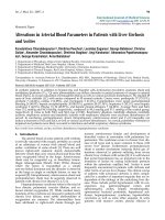
Structure, function and exon organization of CD44. (A) Structure and functions of CD44 glycoprotein [1–4]. CD44 structure. The heavy and
intermediate black tracks represent the conserved N-terminal region of the extracellular domain, the transmembrane region and the cytoplasmic tail.
The heavy black track at the N terminus represents the link module. The thin black track represents the nonconserved membrane-proximal region.
Filled circles, potential N-linked glycosylation sites; open circles, potential O-linked glycosylation sites; filled diamonds, sites for glycosaminoglycan
(chondroitin sulfate [CS] or heparan sulfate [HS]) attachments (the HS of exon v3 is involved in the binding of growth factors); P, potential sites for
phosphorylation. Insets: CD44 functions. Inset a, the basic cluster and the amino acids critical to HA binding. Inset b, binding sites for ezrin radixin
moesin (ERM) and for ankyrin as well as two phosphorylation sites; the location of the sugars and the functional sites is for illustrative purposes
only. (B) CD44 isoforms: exon map. Filled circles represent the constant-region exons; open circles represent exons that can be inserted by
alternative splicing, resulting in the generation of the variable region. Note that exon v1 is not expressed in human CD44. LP, leader peptide-
encoding exon; TM, transmembrane-encoding exon; CT, cytoplasmic tail-encoding exon. (C) Examples of alternatively spliced transcripts: 1,
standard CD44, which lacks the entire variable region; 2, pMeta-1 (CD44v4–v7; exons v4, v5, v6 and v7 are inserted in tandem between exons 5
and 17 [residues 204 and 205]); 3, pMeta-2 (CD44v6,v7) (pMeta-1 and pMeta-2 are known as metastatic CD44 because their cDNA confers,
upon transfection, metastatic potential on nonmetastatic rat tumor cells); 4, epithelial CD44 (CD44v8–v10); 5, keratinocyte CD44 (CD44v3–v10).
This figure is reproduced from Wiley Encyclopedia of Molecular Medicine, CD44 entry by D Naor and S Nedvetzki, vol 5, pp 619-624 (2002), by
permission of John Wiley and Sons, Inc.
108
types of hematopoietic cell [1–4]. Although alternative
splicing is a most efficient means of enriching the genetic
information stored in a single gene, post-translational mod-
ification by glycosylation and glycosaminoglycan (GAG)
attachments further modifies the CD44 protein, allowing
greater expansion of its variability and functions [1–4].
CD44 ligands
The multistructural nature of CD44 might also influence its
ligand repertoire. Indeed, CD44 has a wide range of
ligands, the principal one being hyaluronic acid (HA,
hyaluronate, hyaluronan), a linear polymer of repeating dis-
accharide units (
D-glucuronic acid-[1-β-3]-N-acetyl-D-glu-
cosamine-[1-β-4])
n
. The biological roles of hyaluronan
include the maintenance of water and protein homeosta-
sis, and the protection of cells from the potentially harmful
effects of other cells, microorganisms and macromole-
cules [15]. However, CD44 can interact with several addi-
tional molecules, such as collagen, fibronectin, fibrinogen,
laminin, chondroitin sulfate, mucosal vascular addressin,
serglycin/gp600, osteopontin and the MHC class II invari-
ant chain, as well as with L selectin and E selectin
(reviewed in [2] and [4]). In many cases CD44 does not
bind to its ligand unless activated by external stimuli. As
both CD44 and its ligand are ubiquitous, this mechanism
should avoid unnecessary engagement of the receptor. In
fact, three states of CD44 activation have been identified
in cell lines and normal cell populations [16]: active CD44,
which constitutively binds HA; inducible CD44, which
does not bind HA or binds it only weakly unless activated
by inducing mAbs, cytokines, growth factors or phorbol
ester [4]; and inactive CD44, which does not bind HA
even in the presence of inducing agents.
The involvement of CD44 in pathological activities might
be confined not only to certain CD44 isoforms but also to
their interaction with specific ligands. This interaction
might be dependent on the type of CD44 isoform or its
post-translational modification (glycosylation and GAG
attachments). Furthermore, the type of CD44 isoform
might dictate the pattern of the post-translational modifica-
tion. The rich ligand repertoire of CD44 is possibly related
to its multistructural nature. Discovery of novel CD44
ligands (as well as new isoforms) can be expected, as the
list of this receptor’s countermolecules is continuously
growing [2,4]. Nevertheless, the identification of existing
as well as novel CD44 ligands, especially those associ-
ated with pathological activities, might provide new targets
for therapy. If the CD44 countermolecule is preferentially
engaged with cell-surface CD44 involved in a pathological
activity, targeting of the ligand could also be a relatively
selective therapeutic modality.
CD44 involvement in cell extravasation
Circulating naïve lymphocytes entering the lymph node
through the high endothelial venule, and leukocytes enter-
ing inflamed tissues through venule capillaries, use a
similar mechanism of transendothelial migration, as shown
by the three-step Springer model [17]. In the first stage,
L selectins and sialomucin-like glycoproteins, expressed
on the cell surface of leukocytes flowing in the blood
vessels, form loose interactions (‘tethering’) with their
countermolecules (sialomucin-like molecules and E or
P selectins, respectively) expressed on the endothelial
cells, and then initiate rolling attachments mediated by an
adhesion–de-adhesion process. However, it has been
reported that in at least several cases, leukocytes entering
infected tissues through inflamed capillaries [5], and lym-
phoma cells infiltrating peripheral lymph nodes [8], exploit
cell-surface CD44 glycoprotein rather than selectin for
tethering and rolling on endothelial cells, using their cell-
surface HA as a countermolecule. In the second step,
chemoattractants produced by the endothelial cells or
cells located in the extravascular tissue interact with the
G-protein-like receptors of the rolling cells and deliver
intracellular signals through the G proteins. These signals
activate β2 (for example, lymphocyte function-associated
antigen-1; LFA-1) or α4 (very late antigen-4; VLA-4) cell-
surface integrins. In the third step, the activated integrins
of the rolling cells form strong attachments to the
immunoglobulin superfamily molecules (such as intercellu-
lar adhesion molecule-1 [ICAM-1] or vascular cell adhe-
sion molecule-1 [VCAM-1]) of endothelial cells, resulting
in cell arrest, transendothelial migration and localization in
extravascular tissue.
Cell migration in postcapillary venules can be simulated
in a parallel-plate flow chamber coated with the appropri-
ate ligand or an endothelial cell monolayer and mounted
on the stage of an inverted phase-contrast microscope.
The cells are perfused into the flow chamber under
physiological postcapillary venular wall shear stress
(1–4 dyn/cm
2
) and their rolling attachments are video-
taped and quantified directly from the monitor screen
[18]. Using the flow chamber technology, we showed [8]
that a cell-surface CD44 variant (CD44v4–v10), rather
than CD44s, mediates the rolling of mouse cells on HA
substrate. This suggests that the CD44 variant has an
intermediate affinity for the ligand-binding site required
for cell rolling. Too strong an affinity might avoid the
adhesion–de-adhesion-dependent cell rolling, a vital
step for transendothelial cell migration. Too weak an
affinity could reduce cell resistance to the shear stress
of the bloodstream, resulting in cell detachment from the
HA substrate. The inflammatory cascade might therefore
be dependent on the appropriate affinity of the circulat-
ing leukocyte CD44 variant. Furthermore, proinflamma-
tory cytokines (such as interleukin [IL]-1α) generated at
the inflammation site might induce alternative splicing
[19], forming inflammation-associated CD44 variants.
This implies that, once initiated, the inflammatory
cascade can be a self-sustained process.
Arthritis Research & Therapy Vol 5 No 3 Naor and Nedvetzki
109
Can CD44 serve as a potential therapeutic
target in rheumatoid arthritis?
As the ‘starter’ antigen of RA has not been identified, the
critical phase of disease initiation cannot be treated at
present by direct antigen-targeting therapy. However,
once induced, the disease must be fostered by multiple,
already-defined factors, at least part of which are essential
to but not sufficient for the development of the RA inflam-
matory cascade. The gradually increasing cell population
in RA joints includes neutrophils, macrophages and fibro-
blasts, as well as T (mostly CD4
+
and fewer CD8
+
) and
B lymphocytes. Some of these cells are derived locally,
whereas others are descendants of infiltrating leucocytes.
In principle, all these cells, and especially their proinflam-
matory cytokines and chemokines, can either be used or
are currently being used as therapeutic targets (for
example, anti-tumor necrosis factor [anti-TNF] antibody
and soluble TNF receptor [20,21]).
The list of potential or available targets is long. First,
TNF-α, a master cytokine, is secreted mostly by mono-
cytes and macrophages, which induces the synthesis of
other proinflammatory cytokines (IL-1, IL-6, IL-8 and granu-
locyte–monocyte colony stimulating factor [22–24]). In
addition, TNF-α stimulates the expression of ICAM-1
adhesion molecules on fibroblasts [25] and activates
chondrocytes to release tissue-destroying matrix metallo-
proteinase (collagenase) [26], leading to joint damage.
Second, interleukin-1, mainly produced by monocytes and
macrophages, stimulates the release of metalloproteinase
(collagenase) from chondrocytes [26]. Third, oncostatin
M, produced by macrophages, promotes, when syner-
gized with IL-1α, matrix metalloproteinase (collagenase)
synthesis by both chondrocytes and synovial fibroblasts
[27]. Fourth, IL-6, produced by T cells, macrophages and
fibroblasts, stimulates the proliferation of synovial fibro-
blasts [28], which, together with macrophage-like synovio-
cytes and B cells, generate the cartilage and
bone-invading pannus, enriched with metalloproteinases.
Fifth, fibroblast growth factor-2 (FGF-2) and vascular
endothelial growth factor (VEGF) induce the formation of a
new vascular network (angiogenesis) [29,30]. Notably,
FGF-2 generated by mast cells and endothelial cells of
arthritic joints [29] can induce VEGF expression [31] and
fibroblast proliferation. Eventually, cytokine and growth
factor receptors could also be therapeutically targeted.
However, in using this approach, careful measures should
be taken because anti-receptor agents can induce agonis-
tic rather than antagonistic effects.
Many of the joint pathological activities of patients with
RA, including the disease induction phase, are directly or
indirectly dependent on cell extravasation into the joint
tissue. If the extravasation is dependent on CD44, this
molecule should be ultimately considered a master target
in RA. Moreover, as CD44 is intensively alternatively
spliced, the CD44-mediating extravasation might in fact
be a CD44 variant that is not expressed, or expressed to a
smaller extent, on cells engaged in physiological activities,
leaving a handle for selective targeting. In this respect
CD44 has an advantage over other proinflammatory
factors, which are subjected, if at all, to much less alterna-
tive splicing.
The synovium of rheumatoid arthritis patients
contains both CD44 and its principal ligand, HA
The presence of CD44 and HA in the RA synovium is well
established although, quantitatively, considerable varia-
tions have been obtained in different studies. Western blot
analysis showed that the levels of CD44 in the synovial
tissue of patients with RA are 3.5-fold and 10.7-fold
higher than those of patients with osteoarthritis (OA) and
patients with joint trauma, respectively, and that the high
level of CD44 is related to the degree of inflammation
[32]. In contrast, other investigators [33] reported lower
levels of CD44 in RA synovial tissues or fibroblasts than in
the corresponding normal tissues or fibroblasts, using
immunostaining, Western blotting and enzyme-linked
immunosorbent assay.
In another study, histochemical immunostaining revealed
equal CD44 expression in both RA and OA synovial
tissues, including macrophages, fibroblasts and lining
cells, that was stronger than in normal synovial tissues
[34]. Enhanced expression of CD44 was also found on
synovial lymphocytes and macrophages of rats with adju-
vant arthritis [35]. In addition, CD44 expression was
markedly increased on lymphocytes from the synovial fluid
of patients with RA relative to that of lymphocytes from the
peripheral blood of the same subjects [36–38]. These dis-
parate findings can be attributed both to different
approaches in evaluating the data (for example, normal-
ized or non-normalized protein concentrations) and to vari-
ations in the sensitivity of the methodology used (Western
blot versus immunohistochemistry). Nevertheless, fibro-
blasts from RA synovia showed a high expression of
CD44 alternatively spliced variants, including long iso-
forms like CD44v3,v6–v10. This phenomenon was not
consistent in the synovial fibroblasts of patients with OA
and was not found in fibroblasts of non-inflamed synovia
[39,40]. These findings suggest either that joint inflamma-
tion activates the CD44 alternative splicing machinery or
that fibroblasts expressing CD44 variants are selected at
the inflammation site.
Hyaluronan, the principal countermolecule of CD44, is
present at lower concentrations in rheumatoid synovia
(0.71 ± 0.1 mg/cm
3
) than in the non-inflamed synovium
(1.07 ± 0.16 mg/cm
3
) [41,42], a tissue containing one of
the highest concentrations of HA in the entire human body
[15]. However, the ratio of extractable or ‘free’ HA to non-
extractable or ‘bound’ HA in rheumatoid synovium is
Available online />110
2.5-fold that in non-inflamed synovium [41], which explains
why the circulating HA is elevated in the serum of patients
with RA. The mean level of serum HA in patients with RA
was 3–7-fold [43–45], and in patients with OA 2-fold
[44], that in normal individuals (26–42 ng/ml). Serum HA
levels of patients with RA gradually increased during the
follow-up period [45], accounting for time-related varia-
tions in different reports. In addition, there is some dis-
agreement on the correlation between serum HA level and
the degree of inflammation [46].
Human fibroblasts enhance the synthesis of hyaluronan in
response to stimulation with IL-1β or TNF-α [47]. In its
native form, HA is present as a high-molecular-mass
polymer, but during inflammation smaller molecular frag-
ments accumulate. Fragmented HA (less than 500 kDa),
rather than the high-molecular-mass HA (more than
1 MDa) [48], stimulates the cell-surface CD44 receptor,
leading to intracellular signaling, gene activation and
expression of proinflammatory mediators such as NF-κB
[49], nitric oxide synthase [50] and chemokines [48]. Low-
molecular-mass fragments of HA also stimulate angiogen-
esis [51], an important factor in inflammation. Notably,
activated hyauronidase and reactive oxygen-derived free
radicals mediate the fragmentation of hyaluronan, as for
example in inflammatory joint disease, leading to the accu-
mulation of low-molecular-mass HA [52–55].
Evidence that CD44 and hyaluronate are
involved in the synovial inflammation of
patients with RA
The substantial presentation of CD44 and hyaluronate in
the inflamed synovium of patients with RA is a good
reason to explore the involvement of CD44 in this pathol-
ogy, but it cannot be considered conclusive evidence on
its own. Efforts should therefore be focused on targeting
in vitro and, more importantly, in vivo of CD44 or its
ligands and monitoring the influence of such targeting on
functions involved in the RA disease process. The arsenal
of targeting reagents could include anti-CD44 antibodies,
CD44 soluble peptides (such as CD44–immunoglobulin
conjugates), soluble ligands and ligand-cleaving enzymes.
The consequences of targeting cell-surface molecules
could be signal transmission and promoting disease
development (for example the release of proinflammatory
cytokines), or, in contrast, blockade of the proinflammatory
molecules.
Studies
in vitro
When CD44 molecules expressed on fibroblast-like syn-
ovial cells from patients with RA were cross-linked with
anti-CD44 mAb, VCAM-1 was autocrinically upregulated
by the activation of activator protein-1 transcription factor,
which controls the VCAM-1 gene promoter. HA, espe-
cially when fragmented, also upregulated VCAM-1.
Fibroblast-like synovial cells, expressing VCAM-1 after
being cross-linked with anti-CD44 mAb, displayed
enhanced adhesion to activated T cells, mediated by
VCAM-1–VLA-4 and LFA-1–ICAM-1 interactions [56].
Hence, the cross-talk between cell-surface CD44 and
VCAM-1, and the consequent interaction between fibrob-
lasts and T cells, might lead to the release of proinflamma-
tory factors from both partners (for example, enzymes from
fibroblasts and cytokines from T cells).
Activated T cells (for example those stimulated by phorbol
12-myristate 13-acetate plus ionomycin, anti-CD3 plus
anti-CD28 mAbs or simply by tetanus or staphylococcal
enterotoxin) acquired the ability to bind soluble fluores-
cein-labeled HA and to roll on immobilized HA under phys-
iological shear stress (2.0 dyn/cm
2
), as shown by
videotaping from a flow chamber. Rolling was blocked
with anti-CD44 mAb and soluble HA, suggesting a depen-
dence on CD44–HA interaction [5,57–59]. CD44-depen-
dent rolling on HA under physiological shear stress was
also detected in T cells from inflamed human tonsils and
from the blood of patients with pediatric rheumatology,
systemic lupus erythematosus and chronic arthropathies.
Cells with rolling capability were found mainly in the blood
of patients with active diseases, but not in the blood of
those with inactive diseases [59]. These results suggest
that the extravasation of T cells into arthritic tissue, which
is dependent on rolling, is mediated by the interaction of
cell-surface CD44 with endothelial cell hyaluronan.
To simulate cartilage generation in the joint, a three-dimen-
sional culture system has been constructed by using
human chondrocytes cultivated in collagen sponges pre-
treated with bovine embryonic extracellular matrix (ECM).
The production of a cartilaginous matrix by the chondro-
cytes was monitored by the incorporation of
35
S into the
proteoglycan [60]. The addition of RA synovial fibroblasts
caused destruction (presumably enzyme-mediated) of the
cartilage, as indicated by release of
35
S. When the fibrob-
lasts were co-cultured with a monocyte cell line or mono-
cyte-derived cytokines (TNF-α, IL-1β), cartilage damage
was enhanced, whereas the addition of IL-1 receptor
antagonist or anti-IL-1β mAb decreased the destruction of
the cartilaginous matrix. If the fibroblasts were pretreated
with anti-CD44 mAb and then added to the three-dimen-
sional culture, cartilaginous matrix destruction was also
markedly inhibited [60]. This suggests that the deleterious
interaction between RA fibroblasts and cartilage is medi-
ated by CD44 on the fibroblast cell surface.
It would be of interest to know which CD44 isoform is
involved, and which CD44 countermolecules are present
in the cartilaginous matrix. In this context, it should be
mentioned that RA-like synovial fibroblasts, which invade
Matrigel, as monitored by transwell assay in vitro, are
enriched in v3- and v6-containing CD44 isoforms. This
invasion was significantly inhibited by anti-CD44v3 and
Arthritis Research & Therapy Vol 5 No 3 Naor and Nedvetzki
111
anti-CD44v6 mAbs, rather than by mAbs directed against
constant (pan) CD44 epitopes or against epitopes
included in the v7/v8 exon products [40]. Hence, v3 and
v6 encoded epitopes confer an invasive advantage on RA
fibroblast-like fibroblasts. In contrast, the anti-CD44v7/v8
mAb impeded the proliferation of RA fibroblast-like syn-
oviocytes [61], indicating that epitopes in the v7/v8 region
provide a proliferative advantage, possibly after interaction
with unknown matrix components.
Studies
in vivo
Studies in vitro suggest that CD44 is associated with
various RA manifestations. If this is true, injection of anti-
CD44 mAbs into animals with experimental human-like
arthritis should abolish the disease or markedly hinder its
development. Several research groups [9–11,62,63] have
taken up this experimental challenge. The administration of
anti-CD44 mAbs to DBA/1 or BALB/c mice at the onset
of CIA or proteoglycan (cartilage-derived)-induced arthritis
decreased the arthritic activity as evaluated by joint
swelling [9,11,62], incidence of arthritis [63], clinical
score [11], histopathology [9,11,62] and the degree of
ankle joint extension [9]. A substantial decrease in the
accumulation of arthritic fluorochrome-labeled leukocytes
in inflamed synovial tissues was observed after their intra-
venous injection into arthritic mice administered with anti-
CD44 mAb [9]. This implies that the antibody interferes
with CD44-dependent cell migration into the inflammatory
site. We confirmed this conclusion by transferring spleno-
cytes from arthritic mice into naïve SCID (severe com-
bined immunodeficiency) mice that had been administered
with IRAWB14 anti-CD44 mAb. Injection of the antibody
completely abolished the generation of arthritis in the
recipient mice [11].
The anti-arthritic mechanism of anti-CD44 mAbs in these
animal models can be interpreted in several ways. Mikecz
and colleagues [9,38,62] maintain that the interaction of
IM7.8.1 anti-CD44 mAb with CD44 induces shedding of
this cell-surface glycoprotein, which is subsequently
detected in the circulation. However, they themselves
show that although KM201 anti-CD44 mAb did not
induce loss of CD44 from the cell surface, it inhibited
further development of the experimental arthritis. In con-
trast, IRAWB14 anti-CD44 mAb, which induced the loss
of CD44 from the cell surface, enhanced the arthritic
activity [62]. These inconsistent findings, detected by the
use of different mAbs, imply that additional mechanisms
must exist. We ([11] and Nedvetzki and Naor, unpublished
data) suggest that IM7.8.1 anti-CD44 interferes with the
interaction between cell-surface CD44 and HA, which is
essential for leukocyte accumulation in the inflamed site.
This conclusion is based on the fact that Fab′ fragments of
anti-CD44 mAb induced partial resistance to CIA [11].
This finding supports the notion that anti-CD44 mAb
blocks CD44 function rather than modulates CD44
expression, because modulation requires intact antibody,
or at least F(ab′)
2
fragments. Furthermore, we found that
hyaluronidase also markedly reduced the arthritic activity
in DBA/1 mice (Nedvetzki and Naor, unpublished data)
and diabetogenic activity in NOD mice [6].
Serum interferon-γ (IFN-γ) was markedly elevated after an
injection of type II collagen, together with IM7.8.1 anti-
CD44 mAb, which was used to decrease CIA severity in
DBA/1 mice [10]. Amelioration of the disease after this
treatment was attributed to the antiproliferative action of
IFN-γ and to the ability of this cytokine to downregulate
IL-1, which is involved in bone resorption [10]. Finally,
IM7.8.1 anti-CD44 mAb inhibited the formation of a
hyaluronan-rich pericellular matrix around synovial cells
in vitro and reduced joint oedema in vivo. This suggests
that IM7.8.1 can inhibit the accumulation of pericellular
HA-bound water in the ECM [9] (the ability of hyaluronan
to retain water is a well-established feature of this GAG).
Several anti-mouse CD44 mAbs can induce partial or
even almost complete resistance to experimental arthritis.
Some of these antibodies, including KM201 [62], KM81
[11] and IRAWB14 [11], recognize CD44 constant epi-
topes within or near the HA-binding domain [64]. Another
anti-mouse CD44 mAb, IM7.8.1 [9,11,62], which inter-
acts with a constant epitope outside the HA-binding
domain [64], can, at least in some cells, decrease HA
binding to CD44 [64,65], perhaps by affecting the con-
figuration of the cell surface. Interestingly, IM7.8.1 (which
also cross-reacts with human CD44), rather than mAbs
recognizing the HA-binding site, is the most efficient
inducer of resistance to arthritis in experimental animal
models [11, 62]. This suggests that the IM7.8.1-binding
site is the most potent proinflammatory epitope or, alter-
natively, that the ligand affinity of IM7.8.1 mAb is stronger
than that of the other mAbs. We showed [11] that KM81
Fab′ fragments induced partial resistance to CIA in
DBA/1 mice. Although this response was weaker than
that induced by the intact mAb, the interpretation of the
effect is of both academic and practical importance. The
finding suggests that at least part of the KM81 anti-arthri-
togenic effect is related neither to Fc-dependent activities
(complement-dependent cytotoxicity or antibody-depen-
dent cellular clearance) nor to shedding of the cell-
surface receptor [11], as these activities are attributable
to intact antibodies or, as far as receptor modulation is
concerned, at least to F(ab′)
2
fragments.
Our IRAWB14 data [11] are incompatible with the find-
ings of Mikecz et al. [62]. We did not detect CD44 loss in
mouse leukocytes 24 h after injection of IRAWB14 anti-
CD44 mAb [11], indicating that even if they lost their cell-
surface CD44 they could recycle it within this period.
Furthermore, although our group and Mikecz et al. used a
similar treatment strategy, we found [11] that IRAWB14
Available online />112
mAb inhibited experimental arthritis, whereas the latter
claimed [62] that the same anti-CD44 mAb aggravated the
arthritic activity. Differences in mouse strains (DBA/1
versus BALB/c) and type of disease (CIA versus proteogly-
can-induced arthritis), as well as small variations in experi-
mental techniques, might account for this discrepancy.
Injection of anti-CD44 mAb into mice with CIA or proteo-
glycan-induced arthritis did not influence their humoral and
cellular responses to collagen or proteoglycan [10,62]. It
was further reported [10] that the delayed type hypersen-
sitivity (DTH) to oxazolone (T cell-dependent response),
but not olive oil-induced inflammation (T cell-independent
response), was reduced in mice treated with IM7.8.1 anti-
CD44 mAb. Using the NOD transfer model and a treat-
ment protocol almost identical to the one reported for CIA,
we succeeded in inducing resistance against diabetes by
injecting IM7.8.1 anti-CD44 mAb into male recipient mice
infused with diabetogenic female splenocytes [6].
However, in contrast to the above-mentioned report [10],
the DTH to oxazolone was not influenced by this treatment
in our experiments. Despite this discrepancy, which can
be related to the different experimental models, it seems
that at least some arms of the immune response are not
substantially affected by injection with anti-CD44 mAb,
whereas the antibody has a significant effect on autoim-
mune inflammation. It is conceivable that some conven-
tional immune responses require higher concentrations of
anti-CD44 mAb than do autoimmune inflammatory
responses in order to reach the threshold of sensitivity to
the treatment.
The reduced destructive inflammatory activities in mice
treated with anti-CD44 mAb [6,9–13] suggest that the
related diseases, including CIA, are dependent on CD44.
If this is true, CD44 knockout mice should resist the devel-
opment of arthritis after being injected with type II colla-
gen. Although anti-CD44 mAb interferes with embryonic
development in mice [66], CD44 knockout mice display
an almost normal phenotype. No gross developmental or
neurological abnormalities were evident; neither were
there deficits in hematopoiesis, leukocyte count, cellular
composition or CD4
+
/CD8
+
distribution in these animals.
The levels of total serum Ig and isotype subclasses, as
well as immune responses to mitogens and foreign anti-
gens, including type II collagen, are normal in CD44-defi-
cient mice [67–70]. The expression of adhesion
molecules, except for a decrease in L selectin level, is also
normal in such mice [69]. However, augmented levels of
granulocyte–macrophage colony-forming units in bone
marrow and lower numbers of these progenitors in the
spleen and peripheral blood were noted [67]. In addition,
the CD44-deficient mice showed delayed lymphocyte
homing to the lymph nodes and inefficient homing to the
thymus [68], although these anomalies do not cause any
major visible defect.
To determine the influence of CD44 deficiency on CIA,
Mikecz and colleagues [69] used a targeting vector
designed to delete, by homologous recombination, most
of exons 4 and 5 in the murine CD44 gene, including a
substantial part of the HA-binding domain and the coding
site of the IM7.8.1 epitope. The linearized targeting vector
was introduced into DBA/1 embryonic stem cells, which
were then microinjected into C57BL/6 blastocytes to gen-
erate chimeric offspring. Wild-type and homozygous
CD44-deficient mice, backcrossed to DBA/1 mice, were
identified by polymerase chain reaction, with the use of
primers specific for CD44 and/or neomycin genes.
Although the investigators emphasized the reduced
arthritic activity (both incidence and severity) in CD44
knockout mice treated with type II collagen and the
delayed infiltration of CD44-deficient arthritic lymphocytes
into the joint tissue of wild-type arthritic recipients [69,70],
it is very clear from their own data that the bulk of the joint
inflammatory reaction was persistent in these animals. This
finding suggests that CD44 is not an essential factor in
CIA. However, if this is so, why does anti-CD44 mAb sub-
stantially impede CIA even when administered after
disease onset [9,11]? We must therefore conclude that a
lack of CD44 activity during embryogenesis, rather than its
targeting in adulthood, exerts a survival pressure leading
to a compensatory process that later supports CIA.
A similar situation might exist in the experimental lung
inflammation induced by bleomycin administration.
Whereas in wild-type mice the inflammatory response is
resolved, in CD44 knockout mice the inflammation is ele-
vated in an uncontrolled manner, leading to an impaired
clearance of apoptotic neutrophils, a persistent accumula-
tion of fragmented HA, an impaired activation of transform-
ing growth factor-β
1
, an increase in total cell count, and
death [71]. The redundancy in CD44-deficient mice might
be associated with a decline in cell-surface L selectin that
decreases cell homing to the lymph nodes, allowing more
intensive cell infiltration into the joints [69]. Alternatively, it
is possible that a different molecule, possessing at least
some of the CD44 functions, is upregulated during the
development of the CD44-deficient embryo and is later
used to support the generation of CIA. We are now focus-
ing our efforts on identifying the replacement molecule in
CD44-deficient mice with CIA.
Conclusions
CD44, which is expressed on both local and infiltrating
cells from joints of patients with RA, has a substantial role
in the development of CIA, the animal model that mimics
several aspects of human rheumatoid arthritis. Consider-
able levels of hyaluronic acid, the principal ligand of
CD44, are also found in RA joints and it is functionally
associated with CIA. It was suggested that cell-surface
CD44 is involved in HA-mediated cell rolling on the
endothelium of blood vessels, an essential step preceding
Arthritis Research & Therapy Vol 5 No 3 Naor and Nedvetzki
113
the integrin-dependent transendothelial migration, leading
to an accumulation of destructive inflammatory cells in the
synovial tissues [59]. In addition, a human culture model
suggests that fibroblast cell-surface CD44 mediates the
interaction between fibroblasts and cartilage, possibly by
recognizing collagen and/or one or more other ECM com-
ponents [60]. This interaction might allow the focal release
of proteolytic enzymes from the former, causing damage
to the collagenous tissue. It was further proposed that the
CD44 of macrophages from RA synovium presents fibrob-
last growth factor to the cognate receptor [72], leading to
a proliferation of endothelial cells and fibroblasts. This list
might include additional CD44-dependent biological activ-
ities (see the section on CD44 structure and function) that
could potentially support the RA inflammatory cascade;
however, this awaits formal evidence.
The disruption of CD44–ligand interaction by targeting
one of these partners should therefore interfere with the
development of arthritic inflammation, even if this process
has already been initiated. Indeed, we and others [9,11]
have shown that injection of anti-CD44 mAb after the
onset of CIA markedly reduced the inflammatory activity.
However, in all experiments, the antibody was directed
against constant epitopes shared by all CD44 isoforms,
including those expressed on cells engaged in physiologi-
cal functions, rendering the use of this kind of antibody
less attractive for clinical therapeutic trials. The ultimate
alternative would be to focus efforts on the identification
of CD44 splicing variants that are exclusively or preferen-
tially expressed on the CD44 of synovial fluid cells from
patients with RA and to produce mAbs recognizing the
RA-associated epitopes. Alternative splicing might gener-
ate, in addition to multiple CD44 variants, sequence alter-
nations at the splicing junctions of the pre-mRNA, a
process that could be enhanced by the inflammatory envi-
ronment. If the translated protein is also modified and a
configurational change results, an RA-specific CD44
epitope should be generated that could then be targeted
by specific antibodies. This approach would be substan-
tially bolstered if it were found that the RA-associated
CD44 variant, or the RA-associated CD44 modified
epitope, has a biological function essential to the inflam-
matory cascade. Validation of all these predictions should
be a major goal for future studies.
Competing interests
None declared.
Acknowledgements
We thank Dr Alexandra Mahler for editorial assistance and Sharon
Saunders for typing the manuscript. The research of our group was
supported by the associates of The Lautenberg Center, New York, NY.
References
1 Lesley J, Hyman R, Kincade PW: CD44 and its interaction with
extracellular matrix. Adv Immunol 1993, 54:271-335.
2 Naor D, Vogt Sionov R, Ish-Shalom D: CD44: structure, function
and association with malignant process. Adv Cancer Res
1997, 71:241-319.
3 Lesley J, Hyman R: CD44 structure and function. Frontiers
Biosci 1998, 3:d616-d630.
4 Naor D, Nedvetzki S, Golan I, Melnik L, Faitelson Y: CD44 in
cancer. Crit Rev Clin Lab Sci 2002, 39:527-579.
5 DeGrendele HC, Estess P, Siegelman MH: Requirement for
CD44 in activated T cell extravasation into an inflammatory
site. Science 1997, 278:672-675.
6 Weiss L, Slavin S, Reich S, Cohen P, Shuster S, Stern R,
Kaganovsky E, Okon E, Rubinstein AM, Naor D: Induction of
resistance to diabetes in non-obese diabetic mice by target-
ing CD44 with a specific monoclonal antibody. Proc Natl Acad
Sci USA 2000, 97:285-290.
7 Zahalka MA, Okon E, Gosslar U, Holzmann B, Naor D: Lymph
node (but not spleen) invasion by murine lymphoma is both
CD44- and hyaluronate-dependent. J Immunol 1995, 154:
5345-5355.
8 Wallach-Dayan SB, Grabovsky V, Moll J, Sleeman J, Herrlich P,
Alon R, Naor D: CD44-dependent lymphoma cell dissemina-
tion: a cell surface CD44 variant, rather than standard CD44,
supports in vitro lymphoma cell rolling on hyaluronic acid
substrate and its in vivo accumulation in the peripheral lymph
nodes. J Cell Sci 2001, 114:3463-3477.
9 Mikecz K, Brennan FR, Kim JH, Glant TT: Anti-CD44 treatment
abrogates tissue oedema and leukocyte infiltration in murine
arthritis. Nat Med 1995, 1:558-563.
10 Verdrengh M, Holmdahl R, Tarkowski A: Administration of anti-
bodies to hyaluronan receptor (CD44) delays the start and
ameliorates the severity of collagen II arthritis. Scand J
Immunol 1995, 42:353-358.
11 Nedvetzki S, Walmsley M, Alpert E, Williams RO, Feldmann M,
Naor D: CD44 involvement in experimental collagen-induced
arthritis (CIA). J Autoimmun 1999, 13:39-47.
12 Wittig B, Schwärzler C, Föhr N, Günthert U, Zöller M: Cutting
edge: curative treatment of an experimentally induced colitis
by a CD44 variant V7-specific antibody. J Immunol 1998, 161:
1069-1073.
13 Brocke S, Piercy C, Steinman L, Weissman IL, Veromaa T: Anti-
bodies to CD44 and integrin
αα
4
, but not L-selectin, prevent
central nervous system inflammation and experimental
encephalomyelitis by blocking secondary leukocyte recruit-
ment. Proc Natl Acad Sci USA 1999, 96:5896-6901.
14 van Weering DHJ, Baas PD, Bos JL: A PCR-based method for
the analysis of human CD44 splice products. PCR Methods
Appl 1993, 3:100-106.
15 Laurent TC, Fraser JR: Hyaluronan. FASEB J 1992, 6:2397-
2404.
16 Lesley J, English N, Perschl A, Gregoroff J, Hyman R: Variant cell
lines selected for alterations in the function of the hyaluronan
receptor CD44 show differences in glycosylation. J Exp Med
1995, 182:431-437.
17 Springer TA: Traffic signals for lymphocyte recirculation and
leukocyte emigration: the multistep paradigm. Cell 1994, 76:
301-314.
18 Alon R, Kassner PD, Carr MW, Finger EB, Hemler ME, Springer
TA: The integrin VLA-4 supports tethering and rolling in flow
on VCAM-1. J Cell Biol 1995, 128:1243-1253.
19 Fitzgerald KA, O’Neill LAJ: Characterization of CD44 induction
by IL-1: a critical role for Egr-1. J Immunol 1999, 162:4920-
4927.
20 Elliot MJ, Maini RN, Feldmann M, Kalden JR, Antoni C, Smolen JS,
Buckhard L, Breedveld FC, Macfarlane JD, Bijl H, Woody JN:
Randomised double-blind comparison of chimeric mono-
clonal antibody to tumour necrosis factor
αα
(cA2) versus
placebo in rheumatoid arthritis. Lancet 1994, 344:1105-1110.
21 Moreland LW, Baumgartner SW, Schiff MH, Tindall EA, Fleis-
chmann RM, Weaver AL, Ettlinger RE, Cohen S, Koopman WJ,
Mohler K, Widmer MB, Blosch CM: Treatment of rheumatoid
arthritis with a recombinant human tumor necrosis factor
receptor (p75)-Fc fusion protein. N Engl J Med 1997, 337:141-
147.
22 Nawroth PP, Bank I, Handley D, Cassimeris J, Chess L, Stern D:
Tumor necrosis factor/cachectin interacts with endothelial
cell receptors to induce release of interleukin 1. J Exp Med
1986, 163:1363-1375.
Available online />114
23 Butler DM, Maini RN, Feldmann M, Brennan FM: Modulation of
proinflammatory cytokine release in rheumatoid synovial
membrane cell cultures. Comparison of monoclonal anti TNF-
alpha antibody with the interleukin 1 receptor antagonist. Eur
Cytokine Netw 1995, 6:225-230.
24 Haworth C, Brennan FM, Chantry D, Turner M, Maini RN, Feld-
mann M: Expression of granulocyte-macrophage colony-stim-
ulating factor in rheumatoid arthritis: regulation by tumor
necrosis factor-
αα
. Eur J Immunol 1991, 21:2575-2579.
25 Chin JE, Winterrowd GE, Krzesicki RF, Sanders ME: Role of
cytokines in inflammatory synovitis. The coordinate regulation
of intercellular adhesion molecule 1 and HLA class I and class
II antigens in rheumatoid synovial fibroblasts. Arthritis Rheum
1990, 33:1776-1786.
26 Shingu M, Nagai Y, Isayama T, Naono T, Nobunaga M, Nagai Y:
The effects of cytokines on metalloproteinase inhibitors
(TIMP) and collagenase production by human chondrocytes
and TIMP production by synovial cells and endothelial cells.
Clin Exp Immunol 1993, 94:145-149.
27 Cawston TE, Curry VA, Summers CA, Clark IM, Riley GP, Life PF,
Spaull JR, Goldring MB, Koshy PJT, Rowan AD, Shingleton WD:
The role of oncostatin M in animal and human connective
tissue collagen turnover and its localization within the
rheumatoid joint. Arthritis Rheum 1998, 41:1760-1771.
28 Van Snick J: Interleukin-6: an overview. Annu Rev Immunol
1990, 8:253-278.
29 Qu Z, Huang X-N, Ahmadi P, Andresevic J, Planck SR, Hart CE,
Rosenbaum JT: Expression of basic fibroblast growth factor in
synovial tissue from patients with rheumatoid arthritis and
degenerative joint disease. Lab Invest 1995, 73:339-346.
30 Yamashita A, Yonemitsu Y, Okano S, Nakagawa K, Nakashima Y,
Irisa T, Iwamoto Y, Nagai Y, Hasegawa M, Sueishi K: Fibroblast
growth factor-2 determines severity of joint disease in adju-
vant-induced arthritis in rats. J Immunol 2002, 168:450-457.
31 Seghezzi G, Patel S, Ren CJ, Gualandris A, Pintucci G, Robbins
ES, Shapiro RL, Galloway AC, Rifkin DB, Mignatti P: Fibroblast
growth factor-2 (FGF-2) induces vascular endothelial growth
factor (VEGF) expression in the endothelial cells of forming
capillaries: an autocrine mechanism contributing to angiogen-
esis. J Cell Biol 1998, 141:1659-1673.
32 Haynes BF, Hale LP, Patton KL, Martin ME, McCallum RM: Mea-
surement of an adhesion molecule as an indicator of inflam-
matory disease activity. Up-regulation of the receptor for
hyaluronate (CD44) in rheumatoid arthritis. Arthritis Rheum
1991, 34:1434-1443.
33 Henderson KJ, Edwards JCW, Worrall JG: Expression of CD44
in normal and rheumatoid synovium and cultured synovial
fibroblasts. Ann Rheum Dis 1994, 53:729-734.
34 Johnson BA, Haines GK, Harlow LA, Koch AE: Adhesion mole-
cule expression in human synovial tissue. Arthritis Rheum
1993, 36:137-146.
35 Halloran MM, Szekanecz Z, Barquin N, Haines GK, Koch AE: Cel-
lular adhesion molecules in rat adjuvant arthritis. Arthritis
Rheum 1996, 39:810-819.
36 Takahashi H, Söderström K, Nilsson E, Kiessling R, Patarroyo M:
Integrins and other adhesion molecules on lymphocytes from
synovial fluid and peripheral blood of rheumatoid arthritis
patients. Eur J Immunol 1992, 22:2879-2885.
37 Kelleher D, Murphy A, Hall N, Omary MB, Kearns G, Long A,
Casey EB: Expression of CD44 on rheumatoid synovial fluid
lymphocytes. Ann Rheum Dis 1995, 54:566-570.
38 Brennan FR, Mikecz K, Glant TT, Jobanputra P, Pinder S, Baving-
ton C, Morrison P, Nuki G: CD44 expression by leucocytes in
rheumatoid arthritis and modulation by specific antibody:
implications for lymphocyte adhesion to endothelial cells and
synoviocytes in vitro. Scand J Immunol 1997, 45:213-220.
39 Croft DR, Dall P, Davies D, Jackson DG, McIntyre P, Kramer IM:
Complex CD44 splicing combinations in synovial fibroblasts
from arthritic joints. Eur J Immunol 1997, 27:1680-1684.
40 Wibulswas A, Croft D, Pitsillides AA, Bacarese-Hamilton I, McIn-
tyre P, Genot E, Kramer IM: Influence of epitopes CD44v3 and
CD44v6 in the invasive behavior of fibroblast-like synovio-
cytes derived from rheumatoid arthritic joints. Arthritis Rheum
2002, 46:2059-2064.
41 Pitsillides AA, Worrall JG, Wilkinson LS, Bayliss MT, Edwards
JCW: Hyaluronan concentrations in non-inflamed and
rheumatoid synovium. Br J Rheum 1994, 33:5-10.
42 Dahl LB, Dahl IM, Engström-Laurent A, Granath K: Concentration
and molecular weight of sodium hyaluronate in synovial fluid
from patients with rheumatoid arthritis and other
arthropathies. Ann Rheum Dis 1985, 44:817-822.
43 Engström-Laurent A, Hällgren R: Circulating hyaluronate in
rheumatoid arthritis: relationship to inflammatory activity and
the effect of corticosteroid therapy. Ann Rheum Dis 1985, 44:
83-88.
44 Goldberg RL, Huff JP, Lenz ME, Glickman P, Katz R, Thonar EJ-
MA: Elevated plasma levels of hyaluronate in patients with
osteoarthritis and rheumatoid arthritis. Arthritis Rheum 1991,
34:799-807.
45 Paimela L, Heiskanen A, Kurki P, Helve T, Leirisalo-Repo M:
Serum hyaluronate level as a predictor of radiologic progres-
sions in early rheumatoid arthritis. Arthritis Rheum 1991, 34:
815-821.
46 Woessner JF Jr: Serum hyaluronan: a status report from the
joint. Arthritis Rheum 1991, 34:927-930.
47 Wells AF, Klareskog L, Lindblad S, Laurent TC: Correlation
between increased hyaluronan localized in arthritic synovium
and the presence of proliferating cells: a role for macrophage-
derived factors. Arthritis Rheum 1992, 35:391-396.
48 McKee CM, Penno MB, Cowman M, Burdick MD, Strieter RM,
Bao C, Noble PW: Hyaluronan (HA) fragments induce
chemokine gene expression in alveolar macrophages. J Clin
Invest 1996, 98:2403-2413.
49 Noble PW, McKee CM, Cowman M, Shin HS: Hyaluronan frag-
ments activate an NF-
κκ
B/I-
κκ
B
αα
autoregulatory loop in murine
macrophages. J Exp Med 1996, 183:2373-2378.
50 McKee CM, Lowenstein CJ, Horton MR, Wu J, Bao C, Chin BY,
Choi AMK, Noble PW: Hyaluronan fragments induce nitric-
oxide synthase in murine macrophages through a nuclear
factor
κκ
B-dependent mechanism. J Biol Chem 1997, 272:
8013-8018.
51 West DC, Hampson IN, Arnold F, Kumar S: Angiogenesis
induced by degradation products of hyaluronic acid. Science
1985, 228:1324-1326.
52 Saari H: Oxygen derived free radicals and synovial fluid
hyaluronate. Ann Rheum Dis 1991, 50:389-392.
53 Prehm P: Release of hyaluronate from eukaryotic cells.
Biochem J 1990, 267:185-189.
54 Greenwald RA, Moy WW: Effect of oxygen-derived free radi-
cals on hyaluronic acid. Arthritis Rheum 1980, 23:455-463.
55 McNeil JD, Wiebkin OW, Betts WH, Cleland LG: Depolymerisa-
tion products of hyaluronic acid after exposure to oxygen-
derived free radicals. Ann Rheum Dis 1985, 44:780-789.
56 Fujii K, Tanaka Y, Hubscher S, Saito K, Ota T, Eto S: Cross-
linking of CD44 on rheumatoid synovial cells up-regulates
VCAM-1. J Immunol 1999, 162:2391-2398.
57 DeGrendele HC, Estess P, Picker LJ, Siegelman MH: CD44 and
its ligand hyaluronate mediate rolling under physiologic flow:
a novel lymphocyte-endothelial cell primary adhesion
pathway. J Exp Med 1996, 183:1119-1130.
58 DeGrendele HC, Kosfiszer M, Estess P, Siegelman MH: CD44
activation and associated primary adhesion is inducible via T
cell receptor stimulation. J Immunol 1997, 159:2549-2553.
59 Estess P, DeGrendele HC, Pascual V, Siegelman MH: Functional
activation of lymphocyte CD44 in peripheral blood is a marker
of autoimmune disease activity. J Clin Invest 1998, 102:1173-
1182.
60 Neidhart M, Gay RE, Gay S: Anti-interleukin-1 and anti-CD44
interventions producing significant inhibition of cartilage
destruction in an in vitro model of cartilage invasion by
rheumatoid arthritis synovial fibroblasts. Arthritis Rheum 2000,
43:1719-1728.
61 Wibulswas A, Croft D, Bacarese-Hamilton I, McIntyre P, Genot E,
Kramer IM: The CD44v7/8 epitope as a target to restrain prolif-
eration of fibroblast-like synoviocytes in rheumatoid arthritis.
Am J Pathol 2000, 157:2037-2044.
62 Mikecz K, Dennis K, Shi M, Kim JH: Modulation of hyaluronan
receptor (CD44) function in vivo in a murine model of
rheumatoid arthritis. Arthritis Rheum 1999, 42:659-668.
63 Zeidler A, Bräuer R, Thoss K, Bahnsen J, Heinrichs V, Jablonski-
Westrich D, Wroblewski M, Rebstock S, Hamann A: Therapeutic
effects of antibodies against adhesion molecules in murine
collagen type II-induced arthritis. Autoimmunity 1995, 21:245-
252.
Arthritis Research & Therapy Vol 5 No 3 Naor and Nedvetzki
115
64 Zheng Z, Katoh S, He Q, Oritani K, Miyake K, Lesley J, Hyman R,
Hamik A, Parkhouse RME, Farr AG, Kincade PW: Monoclonal
antibodies to CD44 and their influence on hyaluronan recog-
nition. J Cell Biol 1995, 130:485-495.
65 Vogt Sionov R, Naor D: Hyaluronan-independent lodgment of
CD44
+
lymphoma cells in lymphoid organs. Int J Cancer 1997,
71:462-469.
66 Sherman L, Wainwright D, Ponta H, Herrlich P: A splice variant
of CD44 expressed in the apical ectodermal ridge presents
fibroblast growth factors to limb mesenchyme and is required
for limb outgrowth. Genes Dev 1998, 12:1058-1071.
67 Schmits R, Filmus J, Gerwin N, Senaldi G, Kiefer F, Kundig T,
Wakeham A, Shahinian A, Catzavelos C, Rak J, Furlonger C,
Zakarian A, Simard JJL, Ohashi PS, Paige CJ, Gutierrez-Ramos
JC, Mak TW: CD44 regulates hematopoietic progenitor distrib-
ution, granuloma formation, and tumorigenicity. Blood 1997,
90:2217-2233.
68 Protin U, Schweighoffer T, Jochum W, Hilberg F: CD44-deficient
mice develop normally with changes in subpopulations and
recirculation of lymphocyte subsets. J Immunol 1999, 163:
4917-4923.
69 Stoop R, Kotani H, McNeish JD, Otterness IG, Mikecz K:
Increased resistance to collagen-induced arthritis in CD44-
deficient DBA/1 mice. Arthritis Rheum 2001, 44:2922-2931.
70 Stoop R, Gál I, Glant TT, McNeish JD, Mikecz K: Trafficking of
CD44-deficient murine lymphocytes under normal and inflam-
matory conditions. Eur J Immunol 2002, 32:2532-2542.
71 Teder P, Vandivier RW, Jiang D, Liang J, Cohn L, Puré E, Henson
PM, Noble PW: Resolution of lung inflammation by CD44.
Science 2002, 296:155-158.
72 Jones M, Tussey L, Athanasou N, Jackson DG: Heparan sulfate
proteoglycan isoforms of the CD44 hyaluronan receptor
induced in human inflammatory macrophages can function as
paracrine regulators of fibroblast growth factor action. J Biol
Chem 2000, 275:7964-7974.
Correspondence
David Naor PhD, The Lautenberg Center for General and Tumor
Immunology, The Hebrew University-Hadassah Medical School,
Jerusalem 91120, Israel. Tel: +972 2 675 8722; fax: +972 2 642
4653; e-mail:
Available online />