Báo cáo y học: "α α Upregulated hypoxia inducible factor-1α and -2α pathway in rheumatoid arthritis and osteoarthritis" pps
Bạn đang xem bản rút gọn của tài liệu. Xem và tải ngay bản đầy đủ của tài liệu tại đây (1.5 MB, 9 trang )
Introduction
Rheumatoid arthritis (RA), a polyarticular disease of
autoimmune nature [1], and osteoarthritis (OA) a nonin-
flammatory degenerative disease of the articular cartilage
[2], have in common an increased tendency for new blood
vessel formation [3–5]. This phenomenon, however, may
not necessarily proceed in a similar manner in the two
conditions. Neoangiogenesis is important in the develop-
ment of new cartilage and mineralization in OA [6],
whereas the same process contributes to synovitis,
pannus formation and articular cartilage destruction in RA
[7]. In a previous study [8], we showed increased levels of
expression of the angiogenic factors vascular endothelial
growth factor (VEGF) and platelet-derived endothelial cell
growth factor (PD-ECGF; also known as thymidine phos-
phorylase) and, as a consequence, an increased
APAAP = alkaline phosphatase/antialkaline phosphatase; CI = confidence interval; HIF = hypoxia inducible factor; KDR = kinase insert domain
protein receptor; mAb = monoclonal antibody; MVD = microvessel density; OA = osteoarthritis; PD-ECGF = platelet-derived endothelial cell
growth factor; RA = rheumatoid arthritis; TBS = tris-buffered saline; VEGF = vascular endothelial growth factor.
Available online />Research article
Upregulated hypoxia inducible factor-1
αα
and -2
αα
pathway in
rheumatoid arthritis and osteoarthritis
Alexandra Giatromanolaki
1
, Efthimios Sivridis
1
, Efstratios Maltezos
2
, Nick Athanassou
3
,
Dimitrios Papazoglou
2
, Kevin C Gatter
3
, Adrian L Harris
4
and Michael I Koukourakis
5
1
Department of Pathology, Democritus University of Thrace, Alexandroupolis, Greece
2
Department of Internal Medicine, Democritus University of Thrace, Alexandroupolis, Greece
3
Department of Pathology, Nuffield Department of Clinical Laboratory Sciences, John Radcliffe Hospital, Oxford, UK
4
Cancer Research UK, Molecular Oncology Laboratories, Institute of Molecular Medicine, John Radcliffe Hospital, Oxford, UK
5
Department of Radiotherapy/Oncology Democritus University of Thrace, Alexandroupolis, Greece
Corresponding author: Michael I Koukourakis (e-mail: )
Received: 20 Dec 2002 Revisions requested: 17 Feb 2003 Revisions received: 26 Feb 2003 Accepted: 10 Mar 2003 Published: 29 Apr 2003
Arthritis Res Ther 2003, 5:R193-R201 (DOI 10.1186/ar756)
© 2003 Giatromanolaki et al., licensee BioMed Central Ltd (Print ISSN 1478-6354; Online ISSN 1478-6362). This is an Open Access article:
verbatim copying and redistribution of this article are permitted in all media for any purpose, provided this notice is preserved along with the
article's original URL.
Abstract
The pathogenesis of rheumatoid arthritis (RA) and
osteoarthritis (OA) remains obscure, although angiogenesis
appears to play an important role. We recently confirmed an
overexpression of two angiogenic factors, namely vascular
endothelial growth factor (VEGF) and platelet-derived
endothelial cell growth factor (PD-ECGF), by the lining and
stromal cells of the synovium in both conditions. Because
hypoxia inducible factor (HIF)-1α and HIF-2α are essential in
regulating transcription of the VEGF gene, active participation
of HIF-α molecules in the pathogenesis of these arthritides is
anticipated. We investigated the immunohistochemical
expression of HIF-1α and HIF-2α in the synovium of
22 patients with RA, 34 patients with OA and 22 ‘normal’
nonarthritic individuals, in relation to VEGF, VEGF/KDR (kinase
insert domain protein receptor) vascular activation, PD-ECGF
and bcl-2. A significant cytoplasmic and nuclear
overexpression of HIF-1α and HIF-2α was noted in the synovial
lining and stromal cells of both diseases relative to normal.
Overexpression of HIF-αs was related to high microvessel
density, high PD-ECGF expression and high VEGF/KDR
receptor activation, suggesting HIF-α-dependent synovial
angiogenesis in OA. By contrast, the activation of the
angiogenic VEGF/KDR pathway was persistently increased in
RA, as indeed was microvessel density and the expression of
PD-ECGF, irrespective of the extent of HIF-α expression,
indicating a cytokine-dependent angiogenesis. In all cases, the
VEGF/KDR vascular activation was significantly lower in OA
than in RA, suggesting a relative failure of the HIF-α pathway to
effectively produce a viable vasculature for OA, which is
consistent with the degenerative nature of the disease. The
activation of the HIF-α pathway occurs in both RA and OA,
although for unrelated reasons.
Keywords: hypoxia inducible factors, osteoarthritis, rheumatoid arthritis, thymidine phosphorylase, VEGF
Open Access
R193
R194
Arthritis Research & Therapy Vol 5 No 4 Giatromanolaki et al.
microvessel density (MVD) of the entire synovial vascula-
ture in both RA and OA, relative to normal. However, the
presence of an activated synovial vasculature
(‘VEGF/kinase insert domain protein receptor [KDR]
complex’ expression) was high only in the case of RA. This
failure of OA to activate the VEGF/KDR pathway, in the
presence of increased VEGF expression, is consistent
with the degenerative nature of the disease, whereas the
profoundly upregulated VEGF/KDR pathway in RA
pursues a destructive angiogenesis-related course.
Hypoxia inducible factor (HIF)-1α and HIF-2α are impor-
tant transcription factors, regulating VEGF gene
responses to hypoxic stimuli. Reduction in the degradation
rate of HIF-αs, as occurs under hypoxic stress, results in
accumulation of HIF-1α and HIF-2α proteins and upregu-
lation of the angiogenic process [9,10]. The direct link
between accumulation of HIF-αs and overexpression of
VEGF [11,12], and the important role of the VEGF angio-
genic pathway in arthritides suggest a central role for
HIF-αs in the pathogenesis of RA and OA.
In the present study, we investigated the immunohisto-
chemical expression of HIF-1α and HIF-2α in synovial
tissues in RA and OA. The results were related to the
angiogenic process in the synovial membrane and to the
anti-apoptotic protein bcl-2. More specifically, the results
were analyzed with reference to MVD, VEGF, and the acti-
vation of the angiogenic pathways VEGF/KDR and
PD-ECGF.
Material and methods
Formalin-fixed paraffin-embedded synovial tissues were
retrieved from the files of the Departments of Pathology,
Democritus University of Thrace, Alexandroupolis, Greece,
and Nuffield Orthopaedic Centre, Oxford, UK. The material
was from 22 cases of active RA, 34 cases of OA and
22 nonarthritic cases derived from hip joint replacement
following fracture. Table 1 shows the patient characteris-
tics. Histological confirmation of the arthritic pathology was
performed on haematoxylin and eosin stained sections.
Immunohistochemistry for HIF-1
αα
and HIF-2
αα
expression
The HIF-1α and HIF-2α proteins were detected using
ESEE 122 (IgG
1
monoclonal antibody [mAb]; dilution
1:20) and the EP190b (IgG
1
mAb; neat), as previously
described [13,14]. Sections were deparaffinized and per-
oxidase was quenched with methanol and 3% H
2
O
2
for
15 min. Microwaving for antigen retrieval was used
(3 × 5 min). The primary antibodies were applied for 90
min. Following washing with tris-buffered saline (TBS),
sections were incubated with a secondary antirabbit anti-
mouse antibody (Kwik Biotinylated Secondary, 0.69A
Shandon-Upshaw, Pittsburgh, PA, USA) for 15 min and
washed in tris-buffered saline. Kwik Streptavidin peroxi-
dase reagent (039A Shandon-Upshaw) was applied for
15 min and sections were again washed in TBS. The
colour was developed by 15 min of incubation with
diamone benzidine solution, and sections were weakly
counterstained with haematoxylin. Breast cancer tissues
with strong nuclear HIF-1α and HIF-2α expression were
used as positive controls. Normal mouse IgG was substi-
tuted for primary antibody as negative control at the same
concentration as the test antibody.
Immunohistochemistry for VEGF, VEGF/KDR, PD-ECGF,
and endothelial cell expression
VEGF expression and that of the VEGF/KDR complex was
assessed using the 11B5 mAb, an IgM isotype produced
using the VEGF amino-terminus as an immunogen [15].
VEGF expression was also assessed using the VG1 mAb
(VEGF blocking mAb, IgG isotype), which recognizes the
121, 165, and 189 isoforms of VEGF [16]. Assessment of
PD-ECGF expression was performed using the P-GF.44C
mAb [17]. Paraffin embedded sections, 3 µm thick, were
stained using the alkaline phosphatase/antialkaline phos-
phatase (APAAP) procedure, following microwaving for
antigen retrieval. The primary antibodies were applied at
room temperature as follows: 11B5 at dilution 1:3 for
120 min; VG1 at dilution 1:3 for 90 min; and P-GF.44C at
dilution 1:3 for 30 min. They were subsequently washed in
TBS. Rabbit antimouse antibody 1:50 (vol:vol) was
applied for 30 min, followed by application of APAAP
complex 1:1 (vol:vol) for 30 min. After washing in TBS, the
last two steps were repeated for 10 min each. This step
was not required for the PGF-44c staining. The colour
was developed by 15 min of incubation with new fuchsin
solution and sections were weakly counterstained with
haematoxylin. Non-specific immunoglobulins were substi-
tuted for primary antibody as negative controls at the same
concentration as the test antibody.
Table 1
Patient characteristics
Rheumatoid
Fracture arthritis Osteoarthritis
Number of patients 22 22 34
Age range (median; years) 55–64 (58) 28–72 (62) 69–78 (72)
Sex
Male 8 12 14
Female 14 10 20
Location
Hip 22 8 25
Knee 0 10 9
Wrist 0 4 0
R195
The JC70 monoclonal antibody (DAKO, Glostrup,
Denmark), which recognizes the CD31 pan-endothelial
antigen (platelet/endothelial cell adhesion molecule-1) [18],
was used for microvessel staining (dilution 1:50 for 30 min)
on 3 µm thick paraffin embedded sections. The above-men-
tioned APAAP technique was applied using protease diges-
tion, rather than microwaving, for antigen retrieval.
Immunohistochemistry for bcl-2 expression
The clone 124 (dilution 1:20; DAKO) was used for bcl-2
assessment. Sections were dewaxed and endogenous
peroxidase was quenched by 30 min of incubation in
0.6% H
2
O
2
in methanol. Sections were rehydrated and
heated in citrate buffer (pH 6.0) in a microwave oven for
10 min. The primary antibody was applied at a dilution
1:80 and the sections were incubated overnight at 4°C.
Thereafter, tissues were treated with the ABC technique
using the ABC kit (DAKO). The peroxidase reaction was
developed using diaminobenzidine as chromogen and
sections were counterstained with haematoxylin. For nega-
tive controls we used a nonspecific IgG (normal rabbit
IgG) instead of the primary antibody.
Assessment of synovial lining and stromal cell reactivity
The expression levels of HIF-1α, HIF-2α, VEGF, VEGF/KDR,
PD-ECGF, and bcl-2 were assessed at the synovial lining
and the underlying stromal cells. Morphologic criteria were
used to distinguish lymphocytes and macrophages on a
background of stromal fibroblasts. The staining was
assessed by two independent observers (AG and ES) using
a ×200 magnification. The percentage of immunoreactive
synovial membrane cells was recorded in all optical fields.
Assessment of standard and activated microvessel
densities
The standard MVD, which corresponds to the entire tissue
vascular network of the tissue, was detected using mAb
CD31. The ‘activated MVD’ (i.e. the VEGF bound to its
receptor KDR [VEGF/KDR complex]) was identified using
mAb 11B5.
The method used for microvessel counting was the same
for both standard MVD and activated MVD. Sections were
scanned at low power (×40 and ×100). The MVD was
assessed in all ×200 optical fields by counting vascular
structures with clearly defined lumens or a linear shape.
The final MVD was the mean score obtained from three
fields with the highest individual scores, in order to assess
the maximum angiogenic activity in each case.
Statistical analysis
Statistical analysis and graphical presentation were per-
formed using the GraphPad Prism 2.01 package (Graph-
Pad, San Diego, CA, USA; www.graphpad.com). The
Fisher’s exact test of the unpaired two-tailed t-test was
used for testing relationships between categoric variables
as appropriate. Linear regression analysis was used to
assess correlation between continuous variables. P < 0.05
was considered statistically significant.
Results
HIF-
αα
expression in synovial lining cells
HIF-1α and HIF-2α expression was both cytoplasmic and
nuclear in arthritic synovial tissues (Fig. 1a,b). The median
percentage of synovial lining cells exhibiting cytoplasmic
and/or nuclear HIF-1α expression was 50% (range
10–80%; 95% confidence interval [CI] 40–61%) in RA
and 60% (range 20–100%; 95% CI 53–73%) in OA. For
HIF-2α, the median percentage of positive synovial cells
was 60% (range 10–80%; 95% CI 40–63%) in RA and
50% (range 0–100%; 95% CI 43–66%) in OA (Table 2).
Normal synovia were, in most cases, unreactive, and only
in a small percentage of cases (6/22 and 4/22 for HIF-1α
and HIF-2α, respectively) exhibited a weak and focal cyto-
plasmic reactivity that in no case exceeded 20% of the
cells present. This difference in the frequency of HIF-1α
and HIF-2α between the arthritides and the normal tissues
was statistically significant (Fig. 2; P < 0.0001).
The pathologic synovial tissues were grouped into cate-
gories of high and low HIF-α reactivity (Table 3), using a
40% cytoplasmic/nuclear reactivity as a cut-off point. This
represents the percentage of HIF positive synovial cells
corresponding to the lowest 95% CI value of HIF reactivity
noted in the arthritic material.
HIF-
αα
expression in synovial stromal cells
Normal synovial stromal cells were, by and large, negative
to HIF-1α and HIF-2α proteins, although focal cytoplasmic
reactivity was noted in some cases. In contrast, fibroblastic
HIF-α positivity was noted in both RA and OA, which, in
most cases, was strong and diffuse. Macrophages and
blood vessels (Fig. 1c), together with the lymphoid and
plasma cell component of RA, were also reactive to HIF-αs.
VEGF and PD-ECGF reactivity
VEGF expression was invariably cytoplasmic, and usually
strong and diffuse in the synovial lining cells of RA and
OA. Normal synovium reacted only weakly with VEGF anti-
bodies. PD-ECGF expression was mixed cytoplasmic and
nuclear, and was noted in varying percentages of synovial
lining cells in both RA and OA. PD-ECGF expression was
absent or focally weak in the normal synovium.
VEGF expression was diffuse in the fibroblasts of both RA
and OA. Normal material was either negative or focally
weak. PD-ECGF expression was diffuse in all cases of RA,
but such reactivity was either focal or diffuse in OA mater-
ial. Fibroblasts did not express PD-ECGF in normal
tissues. The lymphocytic component of RA showed
PD-ECGF and VEGF reactivity. Foamy macrophages
exhibited strong VEGF expression.
Available online />Analysis of the extent of HIF-
αα
expression
Table 4 shows the association between the extent of
HIF-αs expression in RA and OA and the various parame-
ters investigated. The most important findings are as
follows. First, In OA a high HIF-1α, but not HIF-2α, syn-
ovial lining/stromal cell expression was significantly asso-
ciated with increased standard MVD, VEGF/KDR
activated MVD, and PD-ECGF expression. Second, the
activated MVD and synovial stromal cell PD-ECGF reactiv-
ity were significantly higher in RA than in OA (P < 0.001),
and this was independent of HIF-α expression (Fig. 3a,b).
Finally, extensive bcl-2 expression by synovial membrane
cells was noted only in OA, and this was directly associ-
ated with HIF-1α expression (Fig. 4).
Discussion
RA and OA, two common conditions with different clinical
features [1,2], have in common an increased tendency for
new blood vessel formation [3–5]. The importance of VEGF
in the pathogenesis of RA and OA has been emphasized in
several recent reports [7,19]. Following chronic inflamma-
tion, an upregulation of VEGF increases vascular permeabil-
ity [20], resulting in edema, protein leakage, and probably
granulation tissue formation (pannus) with erosion of the
articular cartilage and progressive destruction of the joint. In
a previous study [8], we showed that VEGF is overex-
pressed in RA and OA, and such pathologic synovia are
highly vascularized as compared with normal controls. Simi-
larly, the expression of PD-ECGF, another potent factor for
Arthritis Research & Therapy Vol 5 No 4 Giatromanolaki et al.
R196
Table 2
Percentage of synovial cells expressing HIF-1
αα
and HIF-2
αα
proteins in normal, rheumatoid, and osteoarthritic synovium
Normal (n = 22) OA (n = 34) RA (n = 22)
HIF-1α HIF-2α HIF-1α HIF-2α HIF-1α HIF-2α
Minimum 0.00 0.00 20.00 0.00 10.00 10.00
25th percentile 0.00 0.00 40.00 30.00 35.00 25.00
Median 0.00 0.00 60.00 50.00 50.00 60.00
75th percentile 10.00 0.00 90.00 90.00 75.00 80.00
Maximum 20.00 10.00 100.0 100.0 80.00 80.00
Mean 5.45 1.81 63.53 55.29 50.91 50.91
Standard deviation 9.11 3.94 29.12 32.50 24.08 28.10
Standard error 1.94 0.84 4.99 5.57 5.13 5.99
Lower 95% CI 1.41 0.06 53.37 43.96 40.23 39.55
Upper 95% CI 9.49 3.56 73.69 66.63 61.59 63.37
CI, confidence interval; HIF, hypoxia inducible factor; OA, osteoarthritis; RA, rheumatoid arthritis.
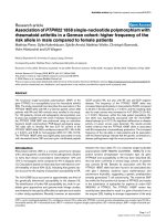
Figure 1
(a) Mixed nuclear and cytoplasmic expression of synovial lining and stromal cells in a case of rheumatoid arthritis (RA). Note a similar reactivity in the
lymphoid and plasma cell component. (b) Mixed nuclear and cytoplasmic expression of synovial lining cells, stromal cells, and endothelial cells in a
case of osteoarthritis (OA). (c) Mixed nuclear and cytoplasmic expression of endothelial cells in a case of RA.
(a) (b) (c)
angiogenesis, was considerably enhanced in the arthritic
synovial membranes. In the present study, we found a
varying degree of expression of HIF-αs in the synovial lining
and stromal cells of RA and OA, whereas normal synovium
was persistently negative. The lack of HIF-1α expression by
the normal synovium was also reported by Hollander and
coworkers [21]. In the latter study, however, HIF-1α was
more prominent in RA than in OA, which was not confirmed
in our study, which included a larger number of specimens.
As HIF-αs are directly involved in the upregulation of VEGF,
it might be suggested that VEGF overexpression in arthri-
tides is probably a result of HIF pathway activation.
There were differences in the expression of HIF-αs
between RA and OA. Thus, although the extent of detec-
tion of HIF-αs was more or less similar in both conditions,
HIF-1α expression in OA only was significantly associated
with standard MVD, VEGF/KDR activated MVD,
Available online />R197
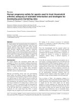
Figure 2
Overexpression of hypoxia inducible factor (HIF)-1α and HIF-2α in
osteoarthritic (OA) and rheumatoid arthritic (RA) synovium, relative to
normal (N).
NOARA NOARA
0
10
20
30
40
50
60
70
80
90
100
P <0.0001 P <0.0001
HIF-1α HIF-2α
% cells with strong HIF+
cytoplasmic expression
Table 3
Expression of HIF-1
αα
and HIF-2
αα
in normal, rheumatoid, and
osteoarthritic synovium
Fractures RA OA P
HIF-1α
Negative 16 0 0
Low 6 10 13 <0.0001
High 0 12 21
HIF-2α
Negative 18 0 0
Low 4 8 14 <0.0001
High 0 14 20
HIF, hypoxia inducible factor; OA, osteoarthritis; RA, rheumatoid
arthritis.
Table 4
Correlation of HIF-1α and HIF-2α expression with MVD, VEGF/KDR activated MVD, and PD-ECGF expression by synovial lining and
stromal cells, and with bcl-2 expression, in the rheumatoid and osteoarthritic synovium
HIF-1α HIF-2α
Low High P Low High P
Rheumatoid arthritis
Standard MVD 57 ±10 63 ± 7 0.16 61 ±6 59 ±10 0.63
Activated MVD 27±12 34 ±10 0.14 31 ±10 31 ± 12 0.97
% PD-ECGF lin. cells 50 ±13 55 ±24 0.57 50 ±30 54 ±12 0.65
% PD-ECGF str. cells 82 ±10 81 ± 9 0.95 80 ±10 82 ± 9 0.51
% bcl-2 4 ±8 2 ±4 0.54 0 ±0 5 ±7 –
Osteoarthritis
Standard MVD 58 ±10 73 ± 18 0.01 68 ± 23 66 ± 11 0.71
Activated MVD 15±3 18 ±4 0.01 16 ±3 17 ±4 0.46
% PD-ECGF lin. cells 51 ±14 68 ±21 0.01 55 ±18 66 ±21 0.14
% PD-ECGF str. cells 30 ±17 49 ± 26 0.01 34 ± 18 47 ± 21 0.13
% bcl-2 5 ±8 32 ±25 0.0007 18 ± 25 25 ± 23 0.46
HIF, hypoxia inducible factor; lin., synovial lining; MVD, microvessel density; OA, osteoarthritis; PD-ECGF, platelet-derived endothelial cell growth
factor; RA, rheumatoid arthritis; str., stromal.
PD-ECGF expression and with the antiapoptotic protein
bcl-2. In the case of RA, the high standard MVD was inde-
pendent of the extent of HIF-1α expression, whereas the
VEGF/KDR activated MVD was persistently higher than
that in OA, irrespective of the extent of HIF-1α staining.
Similarly, the extent of PD-ECGF expression in the syn-
ovial rheumatoid stroma was significantly higher than that
in the osteoarthritic, regardless of the magnitude of
HIF-1α reactivity.
Hypoxic stimulation is the primary cause for intracellular
HIF-1α accumulation, not because of increased mRNA
transcription or translation but rather as a result of a redox-
sensitive stabilization [22]. Following HIF-α heterodimer-
ization with the HIF-1β unit, the complex enters into the
nucleus, binds to DNA at the hypoxia response elements
of target genes (i.e. VEGF), and induces transcription.
Although upregulation of the HIF pathway may also occur
as a result of a genetic alteration [23,24], the nonmalig-
nant nature of RA and OA suggests that hypoxic signaling
may be implicated in the pathogenesis of these diseases.
However, a range of growth factor signalling pathways
and cytokines can also upregulate HIF-αs (e.g. tumor
necrosis factor-α, epidermal growth factor, insulin growth
factor-II and thrombin) [25–28].
The direct association of the extent of HIF-1α with MVD and
the VEGF/KDR activated MVD in OA is consistent with the
notion that VEGF is induced by hypoxia after activation of the
HIF-α pathway. This is further reinforced by the direct associ-
ation between HIF-1α expression and the expression of pro-
teins PD-ECGF and bcl-2. Oxidative stress is probably a
major stimulus for the expression of PD-ECGF [29], whereas
hypoxic induction of bcl-2 was shown to prevent apoptotic
cell death induced by hypoxia [30–33]. It is possible then
that within the degenerative context of the osteoarthritic
disease, impaired vascular homeostasis results in focal, still
progressively expanding, hypoxic regions in the synovium. In
these areas, upregulation of HIF-αs leads to overexpression
of VEGF and PD-ECGF by the synovial lining and stromal
cells, and to the genesis of a defective vascular network with
poor survival ability. As previously shown, the activation of
the OA vasculature is low, despite the over-production of
VEGF [8]. Although a relationship between HIF-1α and
VEGF/KDR activated MVD was observed in the present
study, the magnitude of the increase was limited. Given that
the importance of the VEGF/KDR pathway in mediating
Arthritis Research & Therapy Vol 5 No 4 Giatromanolaki et al.
R198
Figure 3
(a) Relationship of hypoxia inducible factor (HIF)-1α expression in
osteoarthritis (OA) and rheumatoid arthritis (RA) with vascular
endothelial growth factor (VEGF)/KDR vascular activation pathway.
Note that the degree of VEGF/KDR microvessel density is directly
correlated with the degree of HIF-1α expression only in the case of
OA; VEGF/KDR is consistently high in RA, and higher than in OA.
(b) Relationship of HIF-1α expression in osteoarthritis (OA) and
rheumatoid arthritis (RA) with stromal cell thymidine phosphorylase
(TP; referred to in the text as platelet-derived endothelial cell growth
factor [PD-ECGF]) reactivity. Note that the degree of TP expression is
directly correlated with the degree of HIF-1α expression only in the
case of OA; TP expression is consistently high in RA, and higher than
in OA.
0
10
20
30
40
P =0.01
P =0.14
OA
low
OA
high
A
low
RA
high
P < 0.001
HIF-1α expression
VEGF/KDR activated
microvessel density
0
25
50
75
100
0.01
P =0.
95
OA
low
OA
high
RA
low
RA
high
% of stromal cells
with TP reactivity
0. 001
HIF-1α expression
(a)
(b)
P <
P =
Figure 4
Relationship of HIF-1α expression in osteoarthritis (OA) and
rheumatoid arthritis (RA) with bcl-2 reactivity. Bcl-2 protein is almost
exclusively expressed in OA and is significantly related to the extent of
HIF-1α expression.
0
10
20
30
40
P = 0.0007
OA
low
OA
high
R
A
low
RA
high
P =0.54
HIF-1α expr
ession
% of lining cells with
bcl-2 reactivity
endothelial cell survival has repeatedly been confirmed
[34–36], the survival ability of the OA vasculature may be
hindered despite an upregulated HIF/VEGF system.
In RA, high MVD, high activated VEGF/KDR pathway, and
upregulated PD-ECGF expression were constant features,
independent of the extent of the activated HIF-α pathway.
This may mean that angiogenesis is not exclusively depen-
dent on the extent of HIF reactivity and that hypoxia is not
the only factor that upregulates the HIF/VEGF pathway. It
is well known that a variety of cytokines are produced by
lymphocytes in the context of the rheumatoid pathology
(i.e. interleukin-1 and tumor necrosis factor [37,38]) and
that blocking such cytokines induces clinical remission of
the disease [39]. Cytokines released by the immune
response system may directly stimulate both HIF-α depen-
dent mRNA transcription [40] and HIF-independent VEGF
or PD-ECGF overexpression [41–44]. The latter mecha-
nism is less likely to be engaged in OA, where the synovial
tissues bear reduced cytokine expression as compared
with RA [45]. By contrast, a dense vasculature, character-
ized by a VEGF/KDR-activated status [8] and pannus for-
mation, is probably a primary event in RA.
Our finding that bcl-2 is predominantly expressed in OA
whereas rheumatoid synovium lacks expression of this
anti-apoptotic protein is in direct contrast to a previously
reported study by Perlman and coworkers [46]. Although
this discrepancy is difficult to explain, forced bcl-2 down-
regulation failed to induce cell death in rheumatoid fibrob-
lasts, suggesting that bcl-2 is probably of minor
importance in the pathology of the synovium [46]. An
experimental study from the same group concluded that
the expression of bcl-2 is temporally expressed, so that its
role in RA may be confined to just a step in the develop-
ment of rheumatic pathology [47]. In accordance with the
diminished role of bcl-2 in RA is a study conducted by
Chu and coworkers [48], which showed lack of bcl-2
involvement in apoptosis in RA.
It is concluded that activation of the HIF-α pathway occurs
in both RA and OA, although for unrelated reasons.
Hypoxia, consistent with an impaired vascular homeosta-
sis, may hinder the angiogenic effect of the upregulated
HIF/VEGF pathway in OA. Deranged vascular homeosta-
sis should not be attributed to a defective HIF pathway,
but rather to a defective communication between VEGF
Available online />R199
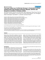
Figure 5
Schematic representation of the suggested pathogenetic model in osteoarthritis (OA) and rheumatoid arthritis (RA). HIF, hypoxia inducible factor;
TP, thymidine phosphorylase (referred to in the text as platelet-derived endothelial cell growth factor [PD-ECGF]); VEGF, vascular endothelial
growth factor.
Aging – Degeneration Immunogen ?
Reduced vascular survival ability Lymphocyte and Macrophage aggregation
Random areas of vascular deprivation Fibroblast activation
Focal Hypoxia (synovium and stroma) Cytokine release
(protein stabilization) HIFα (mRNA transcription)
(expanding with disease progression)
Synovium/Fibroblasts/Reactive cells:
VEGF and TP production
Angiogenic attempt on the background Angiogenic attempt on the background
of a reduced responsiveness of vessels to VEGF of an intact VEGF/KDR pathway
Chaotic genesis of a dense, still with Chaotic genesis of a dense, viable
poor viability, vascular system and hyperpermeable vascular system
OSTEOARTHRITIC PATHOLOGY RHEUMATOID PATHOLOGY
and vascular receptors. Furthermore, the intact VEGF-
dependent angiogenic and vascular survival pathway of
RA appears to be cytokine, rather than hypoxia, stimu-
lated. This premise is schematically represented in Fig. 5.
Competing interests
None declared.
References
1. Maini RN: Autoimmunity in rheumatoid arthritis. An approach
via a study of B lymphocytes. Rheum Dis Clin North Am 1987,
13:319.
2. Hough AJ: Pathology of osteoarthritis. In Arthritis and Allied
Conditions. Edited by McCarty DJ, Koopman WJ. Philadelphia:
Lea and Febiger; 1993:1135-1153.
3. Brown RA, Weiss JB: Neovascularisation and its role in the
osteoarthritic process. Ann Rheum Dis 1998, 47:881-885.
4. Sattar A, Kumar P, Kumar S: Rheumatoid- and osteoarthritis:
quantitation of ultrastructural features of capillary endothelial
cells. J Pathol 1986, 148:45-53.
5. Semble EL, Turner RA, McCrickard EL: Rheumatoid arthritis and
osteoarthritis synovial fluid effects on primary human
endothelial cell cultures. J Rheumatol 1985, 12: 237-241.
6. Brown RA, Weiss JB, Tomlinson IW, Philip P, Kumar S: Angio-
genic factor from synovial fluid resembling that from tumours.
Lancet 1980, 1:682-685.
7. Walsh DA: Angiogenesis and arthritis. Rheumatology 1999, 38:
103-112.
8. Giatromanolaki A, Sivridis E, Athanassou N, Zois E, Thorpe PE,
Brekken RA, Gatter KC, Harris AL, Koukourakis MI: The angio-
genic pathway ‘vascular endothelial growth factor/flk-1(KDR)-
receptor’ in rheumatoid arthritis and osteoarthritis. J Pathol
2001, 194:101-108.
9. Blancher C, Harris AL: The molecular basis of the hypoxia
response pathway: tumour hypoxia as a therapy target.
Cancer Metast Rev 1998, 17:187-194.
10. Semenza GL: Hypoxia-inducible factor 1: master regulator of
O
2
homeostasis. Curr Opin Genet Dev 1998, 8:588-594.
11. Forsythe JA, Jiang BH, Iyer NV, Agani F, Leung SW, Koos RD,
Semenza GL: Activation of vascular endothelial growth factor
gene transcription by hypoxia-inducible factor 1. Mol Cell Biol
1996, 16:4604-4613.
12. Ema M, Taya S, Yokotani N, Sogawa K, Matsuda Y, Fujii-Kuriyama
Y: A novel bHLH-PAS factor with close sequence similarity to
hypoxia-inducible factor 1
αα
regulates the VEGF expression
and is potentially involved in lung and vascular development.
Proc Natl Acad Sci USA 1997, 94:4273-4278.
13. Wiesener MS, Turley H, Allen WE, Willam C, Eckardt KU, Talks
KL, Wood SM, Gatter KC, Harris AL, Pugh CW, Ratcliffe PJ,
Maxwell PH: Induction of endothelial PAS domain protein-1 by
hypoxia: characterization and comparison with hypoxia-
inducible factor-1
αα
. Blood 1998, 92:2260-2268.
14. Talks KL, Turley H, Gatter KC, Maxwell PH, Pugh CW, Ratcliffe
PJ, Harris AL: The expression and distribution of the hypoxia
inducible factors HIF-1
αα
and HIF-2
αα
in normal human tissues,
cancers and tumor associated macrophages. Am J Pathol
2000, 157:411-421.
15. Brekken RA, Huang X, King SW, Thorpe PE: Vascular endothe-
lial growth factor as a marker of tumor endothelium. Cancer
Res 1998, 58:1952-1959.
16. Zhang L, Scott P, Turley H, Leek R, Lewis CE, Gatter KC, Harris
AL, Mackenzie IZ, Rees MP, Bicknell R: Validation of anti-vascu-
lar endothelial growth factor (anti-VEGF) antibodies for
immunohistochemical localization of VEGF in tissue sections:
expression of VEGF in the human endometrium. J Pathol
1998, 185:402-408.
17. Fox SB, Moghaddam A, Westwood M, Turley H, Bicknell R,
Gatter KC, Harris AL: Platelet derived endothelial cell growth
factor/thymidine phosphorylase expression in normal tissues
an immunohistochemical study. J Pathol 1995, 176:183-190.
18. Parums DV, Cordell JL, Micklem K, Heryet AR, Gatter KC, Mason
DY: JC70: a new monoclonal antibody that detects vascular
endothelium associated antigen on routinely processed
tissue sections. J Clin Pathol 1990, 43:752-757.
19. Koch AE, Harlow LA, Haines GK, Amento EP, Unemori EN, Wong
PL, Pope RM, Ferrara N: Vascular endothelial growth factor. A
cytokine modulating endothelial function in rheumatoid arthri-
tis. J Immunol 1994, 152:4149-4156.
20. Ferrara N, Houck K, Jakeman L, Leung DW: Molecular and bio-
logical properties of the vascular endothelial growth factor
family of proteins. Endocr Rev 1992, 13:18-32.
21. Hollander AP, Corke KP, Freemont AJ, Lewis CE: Expression of
hypoxia-inducible factor 1alpha by macrophages in the
rheumatoid synovium: implications for targeting of therapeu-
tic genes to the inflamed joint. Arthritis Rheum 2001, 44:1540-
1544.
22. Huang LE, Gu J, Schau M, Bunn F: Regulation of hypoxia-
inducible factor 1a is mediated by an O2-dependent degrada-
tion domain via the ubiquitine-proteasome pathway. Proc Natl
Acad Sci USA 1998, 95:7987-7992.
23. Mazure NM, Chen EY, Laderoute KR, Giaccia AJ: Induction of
vascular endothelial growth factor by hypoxia is modulated by
a phosphatidylinositol 3-kinase/Akt signaling pathway in Ha-
ras-transformed cells through a hypoxia inducible factor-1
transcriptional element. Blood 1997, 90:3322-3331.
24. Zundel W, Schindler C, Haas-Kogan D, Koong A, Kaper F, Chen
E, Gottschalk AR, Ryan HE, Johnson RS, Jefferson AB, Stokoe D,
Giaccia AJ: Loss of PTEN facilitates HIF-1-mediated gene
expression. Genes Dev 2000, 14:391-396.
25. Albina JE, Mastrofrancesco B, Vessella JA, Louis CA, Henry WL
Jr, Reichner JS: HIF-1 expression in healing wounds: HIF-
1alpha induction in primary inflammatory cells by TNF-alpha.
Am J Physiol Cell Physiol 2000, 281:1971-1977.
26. Feldser D, Agani F, Iyer NV, Pak B, Ferreira G, Semenza GL: Rec-
iprocal positive regulation of hypoxia-inducible factor 1alpha
and insulin-like growth factor 2. Cancer Res 1999, 59:3915-
3918.
27. Gorlach A, Diebold I, Schini-Kerth VB, Berchner-Pfannschmidt U,
Roth U, Brandes RP, Kietzmann T, Busse R: Thrombin activates
the hypoxia-inducible factor-1 signaling pathway in vascular
smooth muscle cells: Role of the p22(phox)-containing
NADPH oxidase. Circ Res 2001, 89:47-54.
28. Zhong H, Chiles K, Feldser D, Laughner E, Hanrahan C,
Georgescu MM, Simons JW, Semenza GL: Modulation of
hypoxia-inducible factor 1alpha expression by the epidermal
growthfactor/phosphatidylinositol 3-kinase/PTEN/AKT/FRAP
pathway in human prostate cancer cells: implications for
tumor angiogenesis and therapeutics. Cancer Res 2000, 60:
1541-1545.
29. Griffiths L, Dachs GU, Bicknell R, Harris AL, Stratford IJ: The
influence of oxygen-tension and pH on the expression of
platelet-derived endothelial cell growth factor thymidine
phosphorylase in human breast-tumor cells grown in vitro
and in vivo. Cancer Res 1997, 57:570-572.
30. Freeland K, Boxer LM, Latchman DS: The cyclic AMP response
element in the Bcl-2 promoter confers inducibility by hypoxia
in neuronal cells. Brain Res Mol Brain Res 2001, 92:98-106.
31. Oehler MK, Norbury C, Hague S, Rees MC, Bicknell R:
Adrenomedullin inhibits hypoxic cell death by upregulation of
bcl-2 in endometrial cancer cells: a possible promotion mech-
anism for tumour growth. Oncogene 2001, 20:2937-2945.
32. Shimizu S, Eguchi Y, Kosaka H, Kamiike W, Matsuda H, Tsujimoto
Y: Prevention of hypoxia-induced cell death by Bcl-2 and Bcl-
xL. Nature 1995, 374:811-813.
33. Yamabe K, Shimizu S, Kamiike W, Waguri S, Eguchi Y, Hasegawa
J, Okuno S, Yoshioka Y, Ito T, Sawa Y, Uchiyama Y, Tsujimoto Y,
Matsuda H: Prevention of hypoxic liver cell necrosis by in vivo
human bcl-2 gene transfection. Biochem Biophys Res
Commun 1998, 243:217-223.
34. Alon T, Hemo I, Itin A, Peer J, Stone J, Keshet E: Vascular
endothelial growth factor acts as a survival factor for newly
formed retinal vessels and has implications for retinopathy of
prematurity. Nat Med 1995, 1:1024-1018.
35. Gerber HP, Dixit V, Ferrara N: Vascular endothelial growth
factor induces expression of the antiapoptotic proteins Bcl-2
and A1 in vascular endothelial cells. J Biol Chem 1998, 273:
13313-13316.
36. Watanabe Y, Dvorak HF: Vascular permeability factor/vascular
endothelial growth factor inhibits anchorage-disruption-
induced apoptosis in microvessel endothelial cells by induc-
ing scaffold formation. Exp Cell Res 1997, 233:340-349.
Arthritis Research & Therapy Vol 5 No 4 Giatromanolaki et al.
R200
37. Paleolog EM, Young S, Stark AC, McCloskey RV, Feldmann M,
Maini RN: Modulation of angiogenic vascular endothelial
growth factor by tumor necrosis factor alpha and interleukin-1
in rheumatoid arthritis. Arthritis Rheum 1998, 41:1258-1265.
38. Szekanecz Z, Kim J, Koch AE: Chemokines and chemokine
receptors in rheumatoid arthritis. Semin Immunol 2003, 15:15-
21.
39. Gorman JD, Sack KE, Davis JC Jr: Treatment of ankylosing
spondylitis by inhibition of tumor necrosis factor alpha. N Engl
J Med 2002, 346:1349-1356.
40. Thornton RD, Lane P, Borghaei RC, Pease EA, Caro J, Mochan E:
Interleukin 1 induces hypoxia-inducible factor 1 in human gin-
gival and synovial fibroblasts. Biochem J 2000, 15:307-312.
41. Bucht A, Larsson P, Weisbrot L, Thorne C, Pisa P, Smedegard G,
Keystone EC, Gronberg-A: Expression of interferon-gamma
(IFN-gamma), IL-10, IL-12 and transforming growth factor-
beta (TGF-beta) mRNA in synovial fluid cells from patients in
the early and late phases of rheumatoid arthritis (RA). Clin
Exp Immunol 1996, 103:357-367.
42. Hossain MA, Bouton CM, Pevsner J, Laterra J: Induction of vas-
cular endothelial growth factor in human astrocytes by lead.
Involvement of a protein kinase C/activator protein-1
complex-dependent and hypoxia-inducible factor 1-indepen-
dent signaling pathway. J Biol Chem 2000, 275:27874-2782.
43. Matsumoto K, Kanmatsuse K: Interleukin-18 and interleukin-12
synergize to stimulate the production of vascular permeability
factor by T lymphocytes in normal subjects and in patients
with minimal-change nephrotic syndrome. Nephron 2000, 85:
127-133.
44. Schwartz EL, Hoffman M, O’Connor CJ, Wadler S: Stimulation
of 5-fluorouracil metabolic activation by interferon-
αα
in human
colon carcinoma cells. Biochem Biophys Res Commun 1992,
182:1232-1239.
45. Dolhain RJ, ter Haar NT, Hoefakker S, Tak PP, de Ley M, Claassen
E, Breedveld FC, Miltenburg AM: Increased expression of inter-
feron (IFN) gamma together with IFN gamma receptor in the
rheumatoid synovial membrane compared with synovium of
patients with osteoarthritis. Br J Rheumatol 1996, 35:24-32.
46. Perlman H, Liu H, Georganas C, Koch AE, Shamiyeh E, Haines
GK III, Pope RM: Differential expression pattern of the anti-
apoptotic proteins, Bcl-2 and FLIP, in experimental arthritis.
Arthritis Rheum 2001, 44:2899-2908.
47. Perlman H, Georganas C, Pagliari LJ, Koch AE, Haines K III, Pope
RM: Bcl-2 expression in synovial fibroblasts is essential for
maintaining mitochondrial homeostasis and cell viability. J
Immunol 2000, 164:5227-5235.
48. Chou CT, Yang JS, Lee MR: Apoptosis in rheumatoid arthritis:
expression of Fas, Fas-L, p53, and Bcl-2 in rheumatoid syn-
ovial tissues. J Pathol 2001, 193:110-116.
Correspondence
Michael I Koukourakis, MD, Tumour and Angiogenesis Research
Group, P.O. Box 12, Alexandroupolis 68100, Greece. Tel: +30 6932
480808; fax: +30 25510 74623; e-mail:
Available online />R201