Báo cáo y học: "Transcription profiling of rheumatic diseases" doc
Bạn đang xem bản rút gọn của tài liệu. Xem và tải ngay bản đầy đủ của tài liệu tại đây (865.6 KB, 13 trang )
Page 1 of 13
(page number not for citation purposes)
Available online />Abstract
Rheumatic diseases are a diverse group of disorders. Most of
these diseases are heterogeneous in nature and show varying
responsiveness to treatment. Because our understanding of the
molecular complexity of rheumatic diseases is incomplete and
criteria for categorization are limited, we mainly refer to them in
terms of group averages. The advent of DNA microarray
technology has provided a powerful tool to gain insight into the
molecular complexity of these diseases; this technology facilitates
open-ended survey to identify comprehensively the genes and
biological pathways that are associated with clinically defined
conditions. During the past decade, encouraging results have been
generated in the molecular description of complex rheumatic
diseases, such as rheumatoid arthritis, systemic lupus erythe-
matosus, Sjögren syndrome and systemic sclerosis. Here, we
describe developments in genomics research during the past
decade that have contributed to our knowledge of pathogenesis,
and to the identification of biomarkers for diagnosis, patient
stratification and prognostication.
Introduction
Rheumatic diseases are a diverse group of disorders that
involve the musculoskeletal system. Generally, the cause of
these disorders is unknown and their pathogenesis poorly
understood. Although these diseases involve the synovial
joints, they also have many systemic features. For example,
rheumatoid arthritis (RA) is a chronic inflammatory disease
that - in addition to its systemic manifestations - primarily
affects the joints. On the other hand, systemic lupus
erythematosus (SLE) is a typical systemic disease with
secondary involvement of multiple organs.
The aetiology of the rheumatic diseases is largely unknown.
Clinical and laboratory observations suggest an immune-
mediated attack directed against self-antigens in a number of
these diseases. This is highlighted by the association
between many of these diseases and human leucocyte
antigen (HLA) loci, and by the expression of autoantibodies
such as antibodies against nuclear components in SLE,
Sjögren’s syndrome (SS) and systemic sclerosis (SSc), and
rheumatoid factor (RF) and anti-citrullinated protein anti-
bodies (ACPAs) in RA. That these diseases have an immune-
mediated background is corroborated by the ameliorative
effect of immunosuppressive therapies.
Most of the rheumatic disorders are heterogeneous diseases
with a clinical spectrum that ranges from mild to severe, and
variability in secondary organ system involvement (for example,
heart failure). The heterogeneous nature is reflected by
variation in responsiveness to virtually all treatment modalities.
The heterogeneity probably has its origin in the mutifactorial
nature of the diseases, in which it is likely that specific
combinations of environmental factor(s) and varying poly-
genic background influence not only susceptibility but also
severity and disease outcome. The fact that we generally
refer to these diseases in terms of group averages may
hamper progress in our understanding of pathogenic mecha-
nisms, genetic background and the efficacy of treatment in
subsets of patients. Unfortunately, our understanding of the
molecular complexity of these disorders is incomplete, and
criteria for subtyping patients (for example, in order to select
those patients who will benefit from a specific treatment) are
currently lacking.
By definition, nearly every aspect of a disease phenotype
should be represented in the pattern of genes and proteins
that are expressed in the patient. This molecular signature
typically represents the contributions made by and
interactions between specific factors and distinct cells that
are associated with disease characteristics and subtypes,
Review
Transcription profiling of rheumatic diseases
Lisa GM van Baarsen
1
, Carina L Bos
1
, Tineke CTM van der Pouw Kraan
2
and Cornelis L Verweij
1,3
1
Department of Pathology, VU University Medical Center, 1007 MB Amsterdam, The Netherlands
2
Department of Molecular Cell Biology and Immunology, VU University Medical Center, 1007 MB Amsterdam, The Netherlands
3
Department of Rheumatology, VU University Medical Center, 1007 MB Amsterdam, The Netherlands
Corresponding author: Cornelis L Verweij,
Published: 30 January 2009 Arthritis Research & Therapy 2009, 11:207 (doi:10.1186/ar2557)
This article is online at />© 2009 BioMed Central Ltd
ACPA = anti-citrullinated protein antibody; DAS28 = Disease Activity Score using 28 joint counts; DC = dendritic cell; FLS = fibroblast-like
synoviocyte; HLA = human leucocyte antigen; IFN = interferon; IL = interleukin; MMP = matrix metalloproteinase; OA = osteoarthritis; PBMC =
peripheral blood mononuclear cell; RA = rheumatoid arthritis; RF = rheumatoid factor; SLE = systemic lupus erythematosus; SoJIA = systemic
onset juvenile idiopathic arthritis; SS = Sjögren’s syndrome; SSc = systemic sclerosis; STAT = signal transducer and activator of transcription; TNF =
tumour necrosis factor.
Page 2 of 13
(page number not for citation purposes)
Arthritis Research & Therapy Vol 11 No 1 van Baarsen et al.
and thus it defines the samples’ unique biology. A very
powerful way to gain insight into the molecular complexity of
cells and tissues has arisen with the advent of DNA
microarray technology, which facilitates open-ended survey to
identify comprehensively the fraction of genes that are
differentially expressed among patients with clinically defined
disease. The differentially expressed gene sets may then be
used to determine the involvement of a particular biological
pathway in disease, and may serve to identify disease
classifiers for diagnosis, prognosis, prediction analysis and
patient stratification (Figure 1). Hence, the identification of
differentially expressed genes and proteins may provide a
comprehensive molecular description of disease hetero-
geneity that can reveal clinically relevant biomarkers.
Initially, several pitfalls were experienced in the use this
multistage and relatively expensive technology, which
depends critically on perfectly standardized conditions. First
of all, handling of blood and tissue samples may differ
considerably between laboratories. Usage of different
platforms and the lack of standardized procedures limit
consistency of study results. For example, variability in the
amount and quality of starting RNA; amplification and
labelling strategies employed; and dyes, probe sequences
and hybridization conditions may all influence the sensitivity,
reproducibility and compatibility of datasets. In addition, lack
of standardized approaches to normalization and data
analysis can influence the outcome of research. Moreover,
the high costs associated with use of this technology can
impede ability to conduct well powered studies. Therefore,
verification of results became an essential step in microarray
studies. In order to establish quality criteria for performing
and publishing microarray studies, standards for microarray
experiments and data analysis were created [1].
Now, after a decade of technical and analytical improvement,
the technology and algorithms for data analysis have been
shown to be robust and reproducible across properly
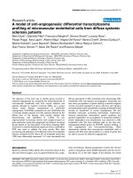
Figure 1
Schematic outline for genomics in rheumatic diseases. Patients with rheumatic diseases exhibited striking heterogeneity, based on clinical,
biological and molecular criteria. Categorization of patients is expected to be of the utmost importance for decision making in clinical practice.
Application of high-throughput screening technologies such as genomics allows us to characterize patients based on their molecular profile. The
procedure starts with collecting different types of material such as serum, peripheral blood (PB) cells, RNA from blood (using, for example,
Paxgene tubes), tissue biopsies and isolated mesenchymal cells from the same patients. Gene expression profiles of this material can be
determined using genomics technology. When associated with clinical readouts, we could select the clinically useful molecular markers and apply
these in routine clinical practice. In addition, these data may help to elucidate the distinct pathological mechanisms that are at play, potentially
explaining the inter-patient variation in clinical presentation, disease progression and treatment response. Ultimately, knowledge of the different
pathogenic mechanisms may help us to identify new drug targets for selected patient subgroups.
Page 3 of 13
(page number not for citation purposes)
designed and controlled experiments, and different research
groups. The Paxgene (PreAnalytix, GmbH, Germany) whole
blood isolation system, which directly lyses cells and stabilizes
the RNA in the aspiration tube, excludes ex vivo processing
artifacts and forms a crucial step in the standardization of
procedures. Although this approach does not a priori account
for cell subset differences, the gene expression data generated
may provide important information from which extrapolations
regarding relative distributions and phenotypic differences can
be made. Careful standardization is still required for cell
subsets and tissues that are obtained via ex vivo manipulation.
Encouraging results have been generated with the use of
microarray technology in the identification of predictors for
disease outcome and metastasis, and underlying pathways in
breast cancer and lymphoma [2,3]. The perceived importance
and support for large-scale and well powered gene expression
profiling studies in oncology have been considerable, and this
may account for the success in this area. However,
transcriptomics approaches have lagged behind in the field of
rheumatology. We believe that collaborative efforts between
groups to increase samples size in order to create high-power
studies are of critical importance to move the field forward.
Equally important is implementation of standardized sample
processing procedures and use of the technology, and data
analysis and algorithms between different sites. Moreover, to
maximize the usage of information from different laboratories,
full and open access to genomics data is essential.
Here, we describe novel developments in genomics research
conducted to identify biological pathways that contribute to
disease and biomarkers for diagnosis, prognosis and patient
stratification in rheumatic diseases. An overview of the
genomics studies in rheumatic diseases discussed in this
review is provided in Table 1. The findings of these studies will
also improve our understanding of the underlying biology of the
diseases and refine their clinical management. Ultimately, this
information may help clinicians to optimise treatment by
identifying subgroups of patients who are most likely to respond.
Gene expression profiling in affected target
tissues
One of the first studies of gene expression profiles in
rheumatic diseases was conducted in RA biopsy tissues, and
used a combination of subtractive hybridization and high-
density cDNA arrays [4]. This study identified increased
expression of genes involved in chronic inflammation, such as
immunoglobulins and HLA-DR, in RA synovium as compared
with normal synovium. However, because the investigators
used pooled tissues from three patients with RA and three
healthy control individuals, it was not possible to consider
heterogeneity in RA.
Devauchelle and coworkers [5] studied differences in gene
expression profiles between the synovial tissue of patients with
RA (n = 5) and those with osteoarthritis (OA; n = 10). A total of
63 (48 known genes and 15 expressed sequence tags) were
differentially regulated between RA and OA samples.
Comparative analysis of synovial biopsy tissue from RA, OA
and SLE patients with active disease partly confirmed and
extended previous observations that distinct diseases were
characterized by distinct molecular signatures [6]. Whereas
genes involved in T-cell and B-cell regulation were
upregulated in RA tissues, in SLE tissues IFN-induced genes
were more highly expressed and genes involved in homeo-
stasis of the extracellular matrix were downregulated.
Histological analysis confirmed that in RA the synovium was
characterized by greater numbers of infiltrating T cells and B
cells as compared with SLE and OA synovium.
Molecular tissue markers for heterogeneity
within rheumatic diseases
Recently, Lindberg and coworkers [7] studied variability in
gene expression levels in synovial tissues within and between
RA patients. This study demonstrated that different arthro-
scopic biopsies taken from one joint yield gene expression
signatures that are more similar within the joint of one patient
than between patients.
A large-scale gene expression profiling study of synovial
tissues from patients with erosive RA revealed considerable
heterogeneity between different patients [8,9]. A systematic
characterization of the differentially expressed genes
highlighted the existence of at least two molecularly distinct
forms of RA tissues. One group exhibited abundant expres-
sion of clusters of genes indicative of ongoing inflammation
and involvement of the adaptive immune response. This
subgroup was referred to as the RA high inflammation group.
The increased expression of immunoglobulin genes was
shown to be one of the main discriminators between high and
low inflammatory tissues. Further analyses of the genes in-
volved in the high inflammation tissues provided evidence for
a prominent role for genes indicative of an activated IFN/
signal transducer and activator of transcription (STAT)-1
pathway. These findings were confirmed at the protein level
[10,11]. From the 16 genes that overlapped between the
microarray used in this study and the one used by
Devauchelle and colleagues [5], seven had comparable gene
expression profiles (TIMP2, PDGFRA, GBP1, Fos, CTSL,
TUBB and BHLHB2). Two of these (GBP1 and CTSL) are
known to be regulated by type I IFN.
The expression profiles of the second group of RA tissues
were reminiscent of those of tissues from patients with OA.
These profiles exhibited a low inflammatory gene expression
signature and increased expression of genes involved in
tissue remodelling activity, which is associated with fibroblast
dedifferentiation. In contrast to the high inflammation tissues,
these tissues had increased levels of matrix metalloproteinase
(MMP)11 and MMP13 expression, and low expression levels
of MMP1 and MMP3 [9].
Available online />Arthritis Research & Therapy Vol 11 No 1 van Baarsen et al.
Page 4 of 13
(page number not for citation purposes)
Table 1
Genomics studies in rheumatic diseases
Number of Approximate number
Disease Tissue samples of genes on array Comparison Results Reference
RA Synovium 13 RA 16,164 Intra- and interindividual Gene expression differences between patients are [7]
patients greater than between biopsies obtained from the same joint
RA Synovium 5 RA and 10 OA 5,760 RA versus OA Genes differentially expressed between RA and OA [5]
RA Synovium 21 RA and 9 OA 11,500 and 18,000 Within RA and versus OA Evidence for the existence of multiple pathways of tissue [8,9]
destruction and repair
RA Synovium 12 early and 4 late 23,040 Early versus longstanding Early RA fell into two groups based on differences in genes [16]
RA critical for proliferative inflammation
RA Synovium 10 RA 30,000 cDNA spots Before versus after about Genes specifically changed in patients who have a good [53]
9 weeks of infliximab response to infliximab treatment.
RA Synovium 18 RA 18,000 Responders versus Patients with high expression levels of genes involved in [54]
nonresponders to infliximab tissue inflammation before treatment are more likely to
treatment benefit from Infliximab therapy.
RA Synovium 12 RA 11,500 and 18,000 Within RA Identification of IL-7 signalling pathway in tissues [15]
characterized by lymphoid neogenesis
RA FLS 19 RA 18,000 Within RA Heterogeneity between synovial tissues is reflected in FLSs [27]
RA FLS 2 RA 12,600 Resting versus TNF-α or Identification of TNF-α and IL-1β regulated genes in RA FLSs [26]
IL-1β 4-hour stimulated cells
RA FLS 5 RA and 5 HC 588 RA versus HC Over-expression of genes responsible for tumor-like [24]
growth in RA FLSs
RA Whole blood 35 RA and 15 HC 18,000 Within RA and versus HC Assignment of a type I IFN signature in a subpopulation of [39,40]
patients
RA PBMC 19 4,300 Early versus longstanding Gene signature in early disease overlaps with normal [38]
RA response to virus
RA PBMC 29 RA and 21 HC 12,626 RA versus HC Monocyte associated gene signature increased in RA [36]
RA PBMC 33 10,000 Before versus 3 months Gene expression profile correlating with treatment response [51]
after infliximab
RA PBMC 8 RF
+
, 6 RF
-
10,000 RF
+
versus RF
-
and No genes differentially expressed between RF
+
and RF
-
[35]
and 7 HC versus HC RA patients. Increased expression of immunoinflammatory
response genes, especially those related to phagocytic
functions, in RA
RA PBMC 19 18,500 Before versus 72 hours Gene pairs and triplets predictive for response to treatment [52]
after etanercept at an early stage of treatment
RA B-cells 8 RA versus 8 HC 21,329 RA versus HC Dysregulated B-cell biology in RA is multifaceted [37]
SSc Skin biopsies 24 SSc and 6 HC 33,000 Within SSc and versus HC A 177-gene signature associated with severity of skin [14]
disease in diffuse SSc
Continued overleaf
Available online />Page 5 of 13
(page number not for citation purposes)
Table 1 (continued)
Number of Approximate number
Disease Tissue samples of genes on array Comparison Results Reference
SSc Dermal fibroblasts 15 SSc twins 16,659 Lesional versus nonlesional At the molecular level, concordance for the SSc fibroblast [31]
and 5 HC and versus twin pair and phenotype is high in MZ twins and greatly exceeds that
versus HC in DZ twins
SSc Dermal non-lesional 21 SSc and 16,659 Lesional versus nonlesional Fibroblasts from nonlesional sites in SSc have detectable [30]
fibroblasts 18 HC and versus HC abnormalities in a variety of cellular processes, including
ECM formation, fibrillogenesis, angiogenesis and
complement activation
SSc PBMC 18 SSc and 18 HC 16,659 SSc versus HC Differentially regulated expression of genes involved in [42]
IFN and vasculopathy
SSc PBMC 9 early diffuse SSc 38,500 SSc versus HC Type I IFN induced Siglec-1 is increased on circulating [43]
and 4 HC SSc CD14
+
monocytes
SS Minor salivary glands 10 SS and 10 HC 6,803 SS versus HC Increased expression of genes involved in chronic [13]
inflammation and type I IFN
SS Minor salivary glands 7 SS and 7 HC 7,261 SS versus HC Activation of IFN pathways in SS [17]
SS Whole saliva 10 SS and 8 HC 38,500 SS versus HC Activation of IFN pathway in SS [18]
SLE Synovium 6 SLE, 7 RA and 38,500 SLE versus RA versus OA The different diseases were characterized by distinct [6]
6 OA molecular signatures. Upregulation of IFN-induced genes and
downregulation of genes involved in ECM homeostasis in SLE
SLE Glomeruli 12 SLE and 4 HC 3,602 and 4,030 SLE versus HC and Characterization of heterogeneity in the molecular [12]
within SLE pathogenesis of lupus nephritis
Paediatric PBMC 30 SLE, 12 JCA 12,626 SLE versus JCA versus IFN signature in the majority of SLE patients and [32]
SLE and 9 controls controls upregulation of granulocyte specific transcripts
SLE PBMC 48 SLE and 42 HC 10,260 Within SLE and versus HC About half of the patients studied exhibited dysregulated [33]
expression of genes in the IFN pathway associated with
more severe disease
SLE Whole blood 269 patients 256 Within SLE Categorization of SLE patients into two groups based on [34]
a high or low IFN signature. Disease activity correlates with
the high IFN signature
Paediatric PBMC 44 SoJIA, 94 17,454 SoJIA versus controls A SoJIA-specific gene signature containing 88 genes. [45]
SoJIA infected patients, Blood transcriptional patterns in the systemic phase of SoJIA
38 SLE, 6 PAPA are more similar to those of patients with infections than to
and 39 healthy controls those of SoJIA patients in a later arthritic stage of disease
Paediatric PBMC 8 untreated and 17,454 Treated versus untreated Increased expression of type I IFN regulated genes in the [55]
SoJIA 5 infliximab treated patients anti-TNF treated SoJIA patients, suggesting cross-regulation
SoJIA between TNF and type I IFN
Autoimmune PBMC 20 RA, 24 SLE, 4,329 Between autoimmune Overlapping gene expression profiles in RA, SLE, type I [48,58,59]
diseases 5 type I diabetes, disease diabetes and MS, which is distinct from a normal immune
4 MS and 9 HC response profile
RA, SLE Whole blood 6 HC, 4 RA, 4 SLE 4,000 RA versus SLE versus Shared autoimmune gene expression signature in patients [47]
and 5 family members HC versus family and unaffected first-degree relatives
DZ, dizygotic twin; ECM, extracellular matrix; FLS, fibroblast-like synoviocyte; HC, healthy control individuals; OA, osteoarthritis; IFN, interferon; IL, interleukin; JCA, juvenile chronic arthritis; MS,
multiple sclerosis; MZ, monozygotic; PAPA syndrome, a familial autoinflammatory disease that causes pyogenic sterile arthritis, pyoderma gangrenosum and acne; PBMC, peripheral blood
mononuclear cell; RA, rheumatoid arthritis; RF, rheumatoid factor; SLE, systemic lupus erythematosus; SoJIA, systemic onset juvenile idiopathic arthritis; SS, Sjögren’s syndrome; SSc,
scleroderma; TNF, tumour necrosis factor.
Histological analyses revealed that the differences observed
in global gene expression between the different groups of
patients are related to differences in cell distribution. Tissues
that contain germinal centre-like structures were selectively
found among the high inflammation tissues. The increased
immunoglobulin transcript expression is in accordance with
the presence of B cells and/or plasma cells, and may reflect
local production of antibodies. Increased immunoglobulin
transcripts were also found in target tissues of other
rheumatic diseases such as SLE [12], SS [13] and SSc [14].
Germinal centre-containing tissues in RA also exhibited
enhanced expression of the chemokines C-X-C chemokine
ligand-12 and C-C chemokine ligand-19 and the associated
receptors C-X-C chemokine receptor-4 and C-X-C chemo-
kine receptor-5, which are important for the attraction of
T cells, B cells and dendritic cells. Pathway analysis revealed
increased expression of genes involved in Janus kinase/STAT
signalling, T-cell and B-cell specific pathways, Fc receptor
type I signalling in mast cells, and IL-7 signal transduction in
the tissues with ectopic lymphoid follicles, accompanied by
increased expression of IL-7 receptor α, IL-2 receptor γ
chains and IL-7. Protein expression of IL-7 in RA tissues was
localized within fibroblast-like synoviocytes, macrophages
and blood vessels, and was co-localized with extracellular
matrix structures around the B-cell follicles. These findings
indicate that activation of the IL-7 pathway may play an
important role in lymphoid neogenesis, analogous to its role in
the development of normal lymphoid tissue [15]. Tissues with
a diffuse type of infiltrate exhibited a profile that indicated
repression of angiogenesis and increased extracellular matrix
remodelling.
Tsubaki and colleagues [16] demonstrated that tissue hetero-
geneity within RA can already be observed in the early phase
of RA. In this study, gene expression profiles were analyzed
from synovial lining tissues from 12 patients with early RA
(duration <1 year after diagnosis) and four with longstanding
RA (duration >3 years after diagnosis). As seen in the
previous study using biopsies from longstanding RA patients,
the early RA patients could be divided into at least two
different groups based on their gene expression profiles.
A study conducted in minor salivary gland tissue from 10
patients with primary SS and 10 healthy control individuals
identified 200 genes that were differentially expressed [13].
Clear upregulation of IFN-inducible genes (ISGF3G, IFIT3,
G1P2 and IRF1) was identified, besides increased expres-
sion of genes related to lymphocyte development and
activation, and antigen processing and signal transduction.
Other studies confirmed that genes in the IFN pathway were
upregulated in salivary glands of SS patients [17,18].
Upregulated IFN-induced gene expression has also been
reported in affected skin of SSc patients [19]. In addition,
Milano and coworkers [14] described distinct patterns of
gene expression profiles in skin tissues when patients were
grouped into those with diffuse SSc and those with limited
SSc. Moreover, these data provided evidence for the
existence of three different subgroups of patients with SSc:
one in those with diffuse SSc and two among those with
limited SSc.
Two main subgroups of lupus nephritis biopsies were
identified based on clustering of genes with the highest
interbiopsy variance [12]. One patient subgroup was
characterized by high expression of fibrosis-related genes in
the absence of an IFN signature. The other subgroup had
high expression of IFN signature genes but low expression of
the fibrosis cluster. The clinical features of the patients were
not significantly different, although the fibrosis subgroup
tended to have higher indices of activity (acute, reversible
damage) and chronicity (irreversible damage), whereas the
IFN subgroup generally had lower activity/chronicity indices.
These results hint at a molecular and biological explanation
for severity of renal injury.
Overall, tissue profiling in rheumatic diseases has led to an
increase in our understanding of disease pathogenesis. In
particular, an IFN signature was observed in target tissues of
subsets of patients with RA, SLE, SS and SSc. This provides
insights that will facilitate assessment of disease activity and
identification of therapeutic targets. Moreover, this informa-
tion will provide a basis for categorization of patients with
rheumatic diseases.
Gene expression in mesenchymal cells
derived from affected target tissues
Fibroblasts are ubiquitous mesenchymal cells that play
important roles in organ development, inflammation, wound
healing, fibrosis and pathology [20]. In chronic inflammation,
fibroblasts are considered sentinel cells that contribute to
leucocyte migration and local immune response through the
production of various immune modulators [21]. These
observations suggest that these fibroblasts may acquire the
capacity to modulate the immune response [22,23].
Fibroblast-like synoviocytes (FLSs) are major players in joint
destruction in RA. One of the first gene expression profile
analyses of FLSs revealed over-expression of genes
responsible for tumour-like growth of rheumatoid synovium
[24]. In this study a cDNA array membrane containing 588
cDNA fragments of known cancer-related genes was used to
compare the gene expression profiles of FLSs from five
patients with RA with those of five traumatic control patients.
Increased expression levels were found for PDGFR
α
, PAI-1
and SDF1A in FLSs derived from rheumatoid synovium when
compared with normal FLSs. Because the sample size was
very small in this study, heterogeneity between FLSs derived
from different RA patients was not considered. Other
investigators studied the influence of tumour necrosis factor
(TNF) on FLSs [25,26]. TNF has been shown to be of primary
importance in the pathogenesis of chronic inflammatory
Arthritis Research & Therapy Vol 11 No 1 van Baarsen et al.
Page 6 of 13
(page number not for citation purposes)
diseases. These studies are instrumental in defining TNF-α
response signatures for application in pharmacology studies
to monitor the effects of TNF blockade.
We recently profiled FLSs derived from 19 RA patients using
microarrays with a complexity of 24,000 cDNA elements.
Correlation studies of paired synovial tissue and FLS
clustering revealed that heterogeneity at the synovial tissue
level is associated with a specific phenotypic characteristic of
the cultured resident FLSs [27]. The high inflammation
tissues were associated with an FLS subtype that exhibits
similarity with so-called myofibroblasts. The myofibroblast is a
specialized fibroblast that has acquired the capacity to
express α-smooth muscle actin, an actin isoform that is
typical of vascular smooth muscle cells. It is now well
accepted that the myofibroblast is a key cell for connective
tissue remodelling and contributes to cell infiltration. These
cells are characterized by a markedly increased expression of
genes that represent the transforming growth factor (TGF)-β
response programme. Among these response genes were
SMA, SERPINE1, COL4A1 (type IV collagen-α chain), IER3
(immediate early response 3), TAGLN (transgelin) and the
gene encoding activin A, which is a potential agonist for the
induction of the TGF-β response programme. Similar cells
were recently identified in the human TNF
+/-
transgenic
mouse model of arthritis [28]. Studies in the field of oncology
indicate that myofibroblasts present in tumours play a crucial
role in angiogenesis through the production of extracellular
matrix proteins, chemokines and growth factors. Hence, it is
hypothesized that myofibroblast-like synoviocytes in RA
synovial tissue contribute to angiogenesis.
These data support the notion that cellular variation between
target tissues is reflected in the stromal cells, and provide
evidence for a link between an increased myofibroblast-like
phenotype and high inflammation in the target tissue.
Genes characteristically expressed in fibroblasts are differen-
tially expressed between SSc and normal tissue biopsies [29].
Detectable abnormalities in the expression of genes involved
extracellular matrix formation, fibrillogenesis, complement
activation and angiogenesis are also present in dermal
fibroblasts cultured from nonlesional skin of SSc patients [30].
No significant differences in gene expression levels were
observed between lesional and nonlesional fibroblasts [31].
The finding that fibroblasts from discordant monozygotic SSc
twin pairs were not significantly different indicates that there is
a strong genetic predisposition to the SSc phenotype [31].
Gene expression in peripheral blood cells
Although the gene expression analysis of tissue samples of
affected organs offers insights into the genes that are
instrumental in patient stratification and primarily involved in
disease activity and pathogenesis, it is not feasible to use this
approach to study large cohorts of patients. Because of the
systemic nature of a number of rheumatic diseases and the
communication between the systemic and organ-specific
compartments, we and others also have studied whole blood
and/or peripheral blood mononuclear cells (PBMCs) to
obtain disease-related gene expression profiles. The
peripheral blood may not have direct implications for our
understanding of disease pathogenesis, but it is especially
suitable for analyzing gene expression profiles that can be
used as biomarkers to permit improved diagnosis and
individualized therapy.
Gene expression profiling in the peripheral blood of patients
with SLE revealed the presence of an IFN signature in
approximately half of the patients studied [32-34]. This
signature included well known IFN-regulated genes (for
example, the anti-viral MX1 [myxovirus {influenza virus}
resistance 1, interferon-inducible protein p78 {mouse}]) as
well as additional IFN response genes. The group of patients
carrying the IFN signature had a significant higher frequency
of certain severe manifestations of disease (renal, central
nervous system and haematological involvement) as
compared with those who did not. Furthermore, the
expression of these genes was significantly correlated with
the number of American College of Rheumatology criteria for
SLE. Pascual and colleagues [32] also noted that IFN genes
were among those most highly correlated with the Systemic
Lupus Erythematosus Disease Activity Index. The same
molecular signature is found in SLE synovial tissue [6]. The
imbalance between IFN molecules and other molecules in
SLE synovial tissue might be of interest pathophysiologically
during the course of SLE arthritis.
RA has systemic manifestations, and a number of
investigators have studied gene expression levels in
peripheral blood cells to address the issue of whether
disease characteristics correlate with gene expression levels
in peripheral blood cells. Bovin and colleagues [35] studied
the gene expression profiles of PBMCs in RA patients
(n = 14; seven RF positive and seven RF negative) and
healthy control individuals (n = 7) using DNA microarrays.
Using two independent mathematical methods, 25 genes
were selected that discriminated between RA patients and
healthy control individuals. These genes reflected changes in
the immune/inflammatory responses in RA patients, and
among these were the genes encoding the calcium-binding
proteins S100A8 and S100A12. No significant differences
between RF-positive and RF-negative RA were observed.
Batliwalla and colleagues [36] studied gene expression
differences between PBMCs from RA patients (n = 29) and
those from healthy control individuals (n = 21). They identified
81 differentially expressed genes, including those encoding
glutaminyl cyclase, IL-1 receptor antagonist, S100A12 and
Grb2-associated binding protein, as the main discriminators.
This profile was associated with increased monocyte count in
RA. Szodoray and colleagues [37] studied gene expression
differences in peripheral blood B cells from eight RA patients
Available online />Page 7 of 13
(page number not for citation purposes)
and eight healthy control individuals. A total of 305 genes
were upregulated, whereas 231 genes were downregulated
in RA B cells. However, the investigators did not address
heterogeneity in peripheral blood gene expression profiles
among patients with RA.
Olsen and colleagues [38] studied gene expression levels in
PBMCs in order to identify differentially expressed genes
between early (disease duration <2 years) and established
RA (with an average disease duration of 10 years). Out of
4,300 genes analyzed, nine were expressed at threefold
higher levels in the early RA group, including the genes
encoding colony stimulating factor 3 receptor, cleavage
stimulation factor, and TGF-β receptor II, which affect B-cell
function. A total of 44 genes were expressed at threefold
lower levels. These genes were involved in immunity and cell
cycle regulation. The observation that a quarter of the early
arthritis genes overlapped with an influenza-induced gene set
led the authors to suggest that the early arthritis signature may
partly reflect the response to an unknown infectious agent.
We examined the gene expression profiles of whole blood
cells and also identified clear and significant differences
between RA patients (n = 35) and healthy individuals (n = 15)
[39]. The microarray data confirmed previous observations of
increased expression of, for instance, the calcium-binding
proteins S100A8 and S100A12. Application of pathway
analysis algorithms revealed increased expression of immune
defence genes, including type I IFN response genes, which
indicates that this pathway is also activated systemically in
RA. This type I IFN signature may be a direct reflection of
increased activity of type I IFN. However, it cannot be
excluded that another ligand known to activate the IFN/STAT-1
pathway is involved. The increased expression of the type I
IFN response genes was characteristic of not all but
approximately half of the patients. Moreover, the immune
defence gene programme that was activated in a subgroup of
RA patients was reminiscent of that of poxvirus-infected
macaques [40]. This subgroup of RA patients expressed
significantly increased titres of anti-cyclic citrullinated peptide
antibodies (anti-CCP/ACPA). Based on these findings, we
conclude that activation of an immune response, with a type I
IFN signature among the gene sets, defines a subgroup of
RA patients characterized by increased autoreactivity against
citrullinated proteins.
The gene expression analyses in peripheral blood of
individuals at high risk for developing RA (RF and/or ACPA
positive arthralgia patients) that we performed provide a
framework for the identification of predictive biomarkers that
may permit identification of individuals who will develop
arthritis within 2 years [41].
Tan and coworkers reported increased IFN-response gene
expression in SSc [42]. Similar observations were made by
York and coworkers [43], who described increased expres-
sion of Siglec-1, an IFN-response gene, in both the diffuse
and the limited cutaneous type of disease as compared with
healthy individuals. Recent findings from our group indicate
an association between the IFN response signature and anti-
centromer autoantibodies and digital ulcers in SSc [44].
An analysis of significance across several febrile inflammatory
disease (44 paediatric systemic onset juvenile idiopathic
arthritis [SoJIA], 94 paediatric infections, 38 paediatric SLE,
six PAPA [a familial autoinflammatory disease that causes
pyogenic sterile arthritis, pyoderma gangrenosum and acne]
and 39 healthy children) revealed a SoJIA-specific signature
composed of 88 genes in peripheral blood [45].
Common denominators
Upregulation of IFN-response genes has now been observed
in peripheral blood cells and/or target tissues of (a subset of)
patients with autoimmune diseases such as RA, SLE, SSc,
SS, multiple sclerosis and type 1 diabetes. These findings
suggest that an activated IFN response gene expression
programme is a common denominator in rheumatic diseases,
and autoimmune diseases in general.
Type I IFNs, which are the early mediators of the innate immune
response that influences the adaptive immune response
through direct and indirect actions on dendritic cells (DCs), T
and B cells, and natural killer cells, could affect the initiation
or amplification of autoimmunity and tissue damage through
their diverse and broad actions on almost every cell type and
promotion of T-helper-1 responses. It is speculated that the
IFN response programme could be associated with activation
of immature monocyte-derived DCs, which regulate deletion
of autoreactive lymphocytes. Subsequently, IFN-matured DCs
may activate autoreactive T cells, leading to autoreactive B-
cell development, representing the first level of autoimmunity
[46]. Loss of tolerance may lead to autoantibody production.
In the case of SLE, autoantigen/autoantibody complexes may
trigger pathogen recognition receptors (such as Toll-like
receptors) that induce IFN-α production and thereby per-
petuate the IFN response programme.
Apart from a role for the IFN response programme as a
common denominator in autoimmune diseases, other gene
profiles have been identified that are shared by autoimmune
diseases. In particular, Maas and colleagues [47] studied the
overlap of gene expression profiles between different
diseases. They identified 95 genes that were increased and
117 genes that were decreased in the PBMCs of all patients
with RA, SLE, type 1 diabetes and multiple sclerosis. These
genes were involved in, for example, inflammation, signalling,
apoptosis, ubiquitin/proteasome function and cell cycle. Hier-
archical cluster analysis on the basis of gene signatures in
PBMCs revealed that RA and SLE patients were intermixed
with one another. Moreover, they reported that from the
genes that were differentially expressed between PBMCs
from patients and those from unrelated unaffected individuals,
Arthritis Research & Therapy Vol 11 No 1 van Baarsen et al.
Page 8 of 13
(page number not for citation purposes)
the gene expression profile of 127 genes was shared
between patients with autoimmune diseases and unaffected
first-degree relatives. This commonality between affected and
unaffected first-degree relatives suggests a genetic basis for
these shared gene expression profiles. Accordingly, the
investigators showed that these genes are clustered in
chromosomal domains, supporting the hypothesis that there
is some genetic logic to this commonality [48].
Pharmacogenomics in rheumatic diseases
Given the destructive nature of most rheumatic diseases, it
would be highly desirable to predict at an early stage the
most beneficial treatment for those patients at risk. If we rely
solely on clinical or radiographic manifestations, we will
probably be responding too late and failing to maximize
protection. Ideally, it would be desirable to make predictions
on success before the start of therapy. Ultimately, this may
lead to a personalized form of medicine, whereby a specific
therapy will be applied that is best suited to an individual
patient.
TNF antagonists are approved worldwide for the treatment of
various rheumatic diseases. Clinical experience indicates that
there are ‘responders’ as well as ‘nonresponders’, but clear
criteria for such classification are still lacking. For RA,
treatment is only effective for approximately two-thirds of
patients [49], which has attracted interest in the pharma-
cology and mechanisms of action of the available therapies.
We present the results of studies assessing progress in
exploiting pharmacogenomics (in particular transcriptomics
for disease profiling) and pharmacodynamics to predict
response to therapy. The term ‘pharmacogenomics’ emerged
in the late 1990s and pertains to the application of genomics
in drug development. ‘Pharmacogenomics’ is defined as, ‘The
investigation of variations of DNA and RNA characteristics as
related to drug response’. Here, we focus on transcriptomics
studies.
Until now a few pharmacogenomics studies have been
conducted to gain insight into pharmacodynamics and to
identify genes predictive of responsiveness to TNF blockers.
The pharmacogenomics of RA patients (n = 15) before and
1 month after the start of infliximab treatment revealed a
similar change in the expression of a pharmacogenomic
response gene set in the peripheral blood compartment of all
patients treated, irrespective of clinical response. This result
indicates that all RA patients exhibit an active TNF response
programme that contributes to disease pathogenesis [50].
Lequerre and colleagues [51] studied 13 patients (six res-
ponders and seven nonresponders) who began treatment
with an infliximab/methotrexate combination. Treatment res-
ponse, determined after 3 months, was based on a difference
in Disease Activity Score using 28 joint counts (DAS28) of
1.2 or more. Gene expression analysis of the PBMCs identi-
fied a preselected set of 2,239 transcripts out of 10,000
transcripts screened, which exhibited abnormal expression in
at least one out of the 13 patients. Subsequent statistical (t-
test and serial analysis of microarrays) analysis identified a
total of 41 transcripts, covering a diverse set of proteins and
functions, which discriminated between responders and
nonresponders. In a validation study conducted in 20 patients
(10 responders and 10 nonresponders) and with a set of 20
transcripts, correct classification of 16 out of the 20 patients
was found (90% sensitivity and 70% specificity). Koczan and
colleagues [52] determined pharmacogenomic differences
after 72 hours in 19 RA patients (12 responders and seven
nonresponders) using a microarray with a complexity of about
18,400 genuine transcripts after administration of etanercept.
They identified an informative set of genes, including
NFKBIA, CCLA4, IL8, IL1B, TNFAIP3, PDE4B, PP1R15
and ADM, which are involved in nuclear factor-κB and cAMP
signalling, whose expression changes after 72 hours was
associated with good clinical responses (DAS28 >1.2).
Comparative analysis did not reveal an overlap between the
two gene sets.
Lindberg and colleagues [53] studied synovial tissue gene
expression profiles in 10 infliximab-treated patients (three
responders, five with moderate response and two non-
responders). The data revealed 279 genes that were signifi-
cantly differentially expressed between the good responding
and nonresponding patients (false discovery rate <0.025).
Among the identified genes was that encoding MMP3.
Moreover, their data revealed that TNF-α could be an
important biomarker for successful infliximab treatment.
We conducted a gene expression profiling study in synovial
biopsies from 18 patients (12 responders and six non-
responders, based on DAS28 ≥ 1.2 after 16 weeks). Several
biological processes related to inflammation that were
upregulated in patients who responded to therapy, as
compared with those who did not show clinical improvement,
were identified. These findings indicate that patients with a
high level of tissue inflammation are more likely to benefit
from anti-TNF-α treatment [54].
Overall, identification of biomarkers before treatment to
predict response to anti-TNF treatment in RA has not yet
yielded consistent results. Therefore, additional studies using
large cohorts of patients and more stringent response criteria
are necessary.
A comparative microarray analysis of PBMCs from eight
SoJIA patients without anti-TNF therapy and five SoJIA
patients undergoing therapy with infliximab [55] revealed
over-expression of IFN-α-regulated genes after TNF block-
ade. Conversely, the addition of IFN to stimulated human
PBMCs inhibits the production of both IL-1 and TNF, and
induces the production of IL-1 receptor antagonist [56].
These findings indicate that cross-regulation of type I IFNs
Available online />Page 9 of 13
(page number not for citation purposes)
and TNF plays an important role in the regulation of
pathological inflammatory responses. Because TNF plays a
critical role in the pathogenesis of certain rheumatic diseases
(such as RA) and because IFN-α plays a pivotal role in
another set of diseases (including SLE), the cross-regulation
of TNF and IFN might have clinical relevance for the blockade
of TNF in, for instance, patients with RA. It is speculated that
these results provide a mechanistic explanation for the
development of anti-double-stranded DNA antibodies and
lupus-like syndrome in patients undergoing anti-TNF therapy.
However, recent gene expression studies in whole blood of
RA patients before and 1, 2 and 3 months after the start of
TNF blockade (infliximab) revealed a variable effect on the
expression of IFN response genes upon treatment. Therefore,
the positive effect of TNF blockade on IFN is not consistently
observed in RA [57].
Conclusion
Genomic profiling approaches have fuelled insight to the
possibility of finding expression patterns that correlate with
disease characteristics and therefore provide a promising
tool for future clinical applications. Molecular profiling of
blood cells and affected target tissues has already revealed
important pathways that contribute to the spectrum of
rheumatic diseases (Figure 2). Both disease-specific and
subgroup-specific signatures and common signatures are
emerging. The latter is reflected by the observation that
clinically distinct rheumatic diseases, and even autoimmune
diseases in general, all show evidence of a dysregulation of
the type I IFN response pathway. Together, the developments
support the notion that there is a basis for a molecular
subcategorization of clinically defined rheumatic diseases.
Moreover, the results indicate that innate immune pathways
remain of critical importance throughout the course of
rheumatic diseases. The clinical implications of these
observations require further definition and independent
validation.
Pharmacogenomics studies are just emerging, and the
results obtained thus far indicate promise for the future. The
finding of biomarkers and gene signatures before the start of
targeted therapies paves the way to more individualized
treatment strategies. However, caution must be exercised in
the interpretation of these results because of small sample
sizes and differences in measures of treatment response. To
increase the sample sizes, collaborative efforts from different
groups are essential. Moreover, agreement on usage of
standardized objective measures of treatment responses is of
Arthritis Research & Therapy Vol 11 No 1 van Baarsen et al.
Page 10 of 13
(page number not for citation purposes)
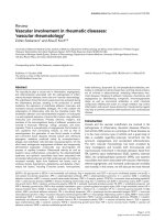
Figure 2
Discovery of molecular rheumatic disease subtypes. Schematic overview of the discovery of rheumatic disease subtypes in peripheral blood cells
and affected target tissues. Heterogeneity in rheumatic diseases have been demonstrated at peripheral blood as well as tissue level using high-
throughput genomics technology. Several studies have described the presence of at least two subgroups of patients based on the presence or
absence of an activated type I interferon (IFN) induced gene expression profile in peripheral blood as well as in affected tissues. In addition,
peripheral blood cells of rheumatic patients exhibit heterogeneous expression levels for genes involved in granulopoiesis and monocyte activation,
as well as for genes encoding the inflammatory S100 proteins. Moreover, subsets of patients exhibit gene expression profiles similar to pathogen-
induced profiles. Apart from type I IFN, tissue heterogeneity is reflected at the level of lymphoid neogenesis, fibrosis, myofibroblasts, tissue
remodelling and transforming growth factor (TGF)-β signalling. The exact relationship between the peripheral blood profile and tissue profile needs
to be further investigated.
critical importance because this will make data from different
studies comparable.
To maximize the usage of information from different
laboratories, full and open access to genomics data is
important. Moreover, standardization of sample processing
procedures and use of the technology, and data analysis and
algorithms used are of critical importance. This will ultimately
allow a systems biology approach, whereby genomics,
proteomics and clinical datasets from different sources are
integrated to assign and validate clinically relevant markers
that reflect disease pathogenesis (diagnosis), prognosis and
heterogeneity, and will facilitate selection of patients with a
high likelihood of responding to therapy.
Competing interests
The VU University Medical Center has filed a patent
applications that is based on the present work (Patent file no.
P086657EP00, ‘Predicting clinical response to treatment
with a soluble TNF-antagonist or TNF, or a TNF receptor
agonist’; patent file no. EP08167570.4, ‘Preclinical bio-
markers for predicting the development of chronic auto-
immune diseases’. CL Verweij, W Bos, LGM van Baarsen, D
van Schaardenburg). CV and LvB are listed as inventors on
the patent applications, and are stakeholders in Preselect
Diagnostics BV.
Acknowledgements
We are grateful to Drs Pat Brown and David Botstein, in whose labora-
tories part of the work described in this report was performed.
Supported in part by the Howard Hughes Medical Institute, EU Marie
Curie trainings network EURO-RA, EU-integrated programme AUTO-
CURE, and the Centre for Medical Systems Biology (a centre of excel-
lence approved by the Netherlands Genomics Initiative/Netherlands
Organization for Scientific Research), and grants from the National
Cancer Institute, the Netherlands Organization for Scientific Research
(NWO) and the Dutch Arthritis Foundation.
References
1. Brazma A, Hingamp P, Quackenbush J, Sherlock G, Spellman P,
Stoeckert C, Aach J, Ansorge W, Ball CA, Causton HC, Gaaster-
land T, Glenisson P, Holstege FC, Kim IF, Markowitz V, Matese
JC, Parkinson H, Robinson A, Sarkans U, Schulze-Kremer S,
Stewart J, Taylor R, Vilo J, Vingron M: Minimum information
about a microarray experiment (MIAME)-toward standards for
microarray data. Nat Genet 2001, 29:365-371.
2. Alizadeh AA, Eisen MB, Davis RE, Ma C, Lossos IS, Rosenwald A,
Boldrick JC, Sabet H, Tran T, Yu X, Powell JI, Yang L, Marti GE,
Moore T, Hudson J Jr, Lu L, Lewis DB, Tibshirani R, Sherlock G,
Chan WC, Greiner TC, Weisenburger DD, Armitage JO, Warnke
R, Levy R, Wilson W, Grever MR, Byrd JC, Botstein D, Brown PO,
Staudt LM: Distinct types of diffuse large B-cell lymphoma
identified by gene expression profiling. Nature 2000, 403:503-
511.
3. van de Vijver MJ, He YD, van’t Veer LJ, Dai H, Hart AA, Voskuil
DW, Schreiber GJ, Peterse JL, Roberts C, Marton MJ, Parrish M,
Atsma D, Witteveen A, Glas A, Delahaye L, van der Velde T,
Bartelink H, Rodenhuis S, Rutgers ET, Friend SH, Bernards R: A
gene-expression signature as a predictor of survival in breast
cancer. N Engl J Med 2002, 347:1999-2009.
4. Zanders ED, Goulden MG, Kennedy TC, Kempsell KE: Analysis
of immune system gene expression in small rheumatoid
arthritis biopsies using a combination of subtractive
hybridization and high-density cDNA arrays. J Immunol
Methods 2000, 233:131-140.
5. Devauchelle V, Marion S, Cagnard N, Mistou S, Falgarone G,
Breban M, Letourneur F, Pitaval A, Alibert O, Lucchesi C, Anract
P, Hamadouche M, Ayral X, Dougados M, Gidrol X, Fournier C,
Chiocchia G: DNA microarray allows molecular profiling of
rheumatoid arthritis and identification of pathophysiological
targets. Genes Immun 2004, 5:597-608.
6. Nzeusseu TA, Galant C, Theate I, Maudoux AL, Lories RJ, Hous-
siau FA, Lauwerys BR: Identification of distinct gene expres-
sion profiles in the synovium of patients with systemic lupus
erythematosus. Arthritis Rheum 2007, 56:1579-1588.
7. Lindberg J, af KE, Ulfgren AK, Stark A, Andersson T, Nilsson P,
Klareskog L, Lundeberg J: Variability in synovial inflammation in
rheumatoid arthritis investigated by microarray technology.
Arthritis Res Ther 2006, 8:R47.
8. van der Pouw Kraan TC, van Gaalen FA, Huizinga TW, Pieterman
E, Breedveld FC, Verweij CL: Discovery of distinctive gene
expression profiles in rheumatoid synovium using cDNA
microarray technology: evidence for the existence of multiple
pathways of tissue destruction and repair. Genes Immun
2003, 4:187-196.
9. van der Pouw Kraan TC, van Gaalen FA, Kasperkovitz PV, Verbeet
NL, Smeets TJ, Kraan MC, Fero M, Tak PP, Huizinga TW, Pieter-
man E, Breedveld FC, Alizadeh AA, Verweij CL: Rheumatoid
arthritis is a heterogeneous disease: evidence for differences
in the activation of the STAT-1 pathway between rheumatoid
tissues. Arthritis Rheum 2003, 48:2132-2145.
10. Kasperkovitz PV, Verbeet NL, Smeets TJ, van Rietschoten JG,
Kraan MC, van der Pouw Kraan TC, Tak PP, Verweij CL: Activa-
tion of the STAT1 pathway in rheumatoid arthritis. Ann Rheum
Dis 2004, 63:233-239.
11. Lorenz P, Ruschpler P, Koczan D, Stiehl P, Thiesen HJ: From
transcriptome to proteome: differentially expressed proteins
identified in synovial tissue of patients suffering from
rheumatoid arthritis and osteoarthritis by an initial screen
with a panel of 791 antibodies. Proteomics 2003, 3:991-1002.
12. Peterson KS, Huang JF, Zhu J, D’Agati V, Liu X, Miller N, Erlander
MG, Jackson MR, Winchester RJ: Characterization of hetero-
geneity in the molecular pathogenesis of lupus nephritis from
transcriptional profiles of laser-captured glomeruli. J Clin
Invest 2004, 113:1722-1733.
13. Hjelmervik TO, Petersen K, Jonassen I, Jonsson R, Bolstad AI:
Gene expression profiling of minor salivary glands clearly dis-
tinguishes primary Sjogren’s syndrome patients from healthy
control subjects. Arthritis Rheum
2005, 52:1534-1544.
14. Milano A, Pendergrass SA, Sargent JL, George LK, McCalmont
TH, Connolly MK, Whitfield ML: Molecular subsets in the gene
expression signatures of scleroderma skin. PLoS ONE 2008,
3:e2696.
15. Timmer TC, Baltus B, Vondenhoff M, Huizinga TW, Tak PP,
Verweij CL, Mebius RE, van der Pouw Kraan TC: Inflammation
and ectopic lymphoid structures in rheumatoid arthritis syn-
ovial tissues dissected by genomics technology: identification
of the interleukin-7 signaling pathway in tissues with lym-
phoid neogenesis. Arthritis Rheum 2007, 56:2492-2502.
16. Tsubaki T, Arita N, Kawakami T, Shiratsuchi T, Yamamoto H,
Available online />Page 11 of 13
(page number not for citation purposes)
This article is part of a special collection of reviews, The
Scientific Basis of Rheumatology: A Decade of
Progress, published to mark Arthritis Research &
Therapy’s 10th anniversary.
Other articles in this series can be found at:
/>The Scientific Basis
of Rheumatology:
A Decade of Progress
Takubo N, Yamada K, Nakata S, Yamamoto S, Nose M: Charac-
terization of histopathology and gene-expression profiles of
synovitis in early rheumatoid arthritis using targeted biopsy
specimens. Arthritis Res Ther 2005, 7:R825-R836.
17. Gottenberg JE, Cagnard N, Lucchesi C, Letourneur F, Mistou S,
Lazure T, Jacques S, Ba N, Ittah M, Lepajolec C, Labetoulle M,
Ardizzone M, Sibilia J, Fournier C, Chiocchia G, Mariette X: Acti-
vation of IFN pathways and plasmacytoid dendritic cell
recruitment in target organs of primary Sjogren’s syndrome.
Proc Natl Acad Sci USA 2006, 103:2770-2775.
18. Hu S, Wang J, Meijer J, Ieong S, Xie Y, Yu T, Zhou H, Henry S,
Vissink A, Pijpe J, Kallenberg C, Elashoff D, Loo JA, Wong DT:
Salivary proteomic and genomic biomarkers for primary Sjo-
gren’s syndrome. Arthritis Rheum 2007, 56:3588-3600.
19. Mondini M, Vidali M, De Andrea M, Azzimonti B, Airò P, D’Ambro-
sio R, Riboldi P, Meroni PL, Albano E, Shoenfeld Y, Gariglio M,
Landolfo S: A novel autoantigen to differentiate limited cuta-
neous systemic sclerosis from diffuse cutaneous systemic
sclerosis: the interferon-inducible gene IFI16. Arthritis Rheum
2006, 54:3939-3944.
20. Chang HY, Chi JT, Dudoit S, Bondre C, van de RM, Botstein D,
Brown PO: Diversity, topographic differentiation, and posi-
tional memory in human fibroblasts. Proc Natl Acad Sci USA
2002, 99:12877-12882.
21. Smith RS, Smith TJ, Blieden TM, Phipps RP: Fibroblasts as sen-
tinel cells. Synthesis of chemokines and regulation of inflam-
mation. Am J Pathol 1997, 151:317-322.
22. Brouty-Boye D, Pottin-Clemenceau C, Doucet C, Jasmin C,
Azzarone B: Chemokines and CD40 expression in human
fibroblasts. Eur J Immunol 2000, 30:914-919.
23. Hogaboam CM, Steinhauser ML, Chensue SW, Kunkel SL: Novel
roles for chemokines and fibroblasts in interstitial fibrosis.
Kidney Int 1998, 54:2152-2159.
24. Watanabe N, Ando K, Yoshida S, Inuzuka S, Kobayashi M, Matsui
N, Okamoto T: Gene expression profile analysis of rheumatoid
synovial fibroblast cultures revealing the overexpression of
genes responsible for tumor-like growth of rheumatoid syn-
ovium. Biochem Biophys Res Commun 2002, 294:1121-1129.
25. Gallagher J, Howlin J, McCarthy C, Murphy EP, Bresnihan B,
FitzGerald O, Godson C, Brady HR, Martin F: Identification of
Naf1/ABIN-1 among TNF-alpha-induced expressed genes in
human synoviocytes using oligonucleotide microarrays. FEBS
Lett 2003, 551:8-12.
26. Taberner M, Scott KF, Weininger L, Mackay CR, Rolph MS: Over-
lapping gene expression profiles in rheumatoid fibroblast-like
synoviocytes induced by the proinflammatory cytokines inter-
leukin-1 beta and tumor necrosis factor. Inflamm Res 2005,
54:10-16.
27. Kasperkovitz PV, Timmer TC, Smeets TJ, Verbeet NL, Tak PP, van
Baarsen LG, Baltus B, Huizinga TW, Pieterman E, Fero M,
Firestein GS, van der Pouw Kraan TC, Verweij CL: Fibroblast-
like synoviocytes derived from patients with rheumatoid
arthritis show the imprint of synovial tissue heterogeneity:
evidence of a link between an increased myofibroblast-like
phenotype and high-inflammation synovitis. Arthritis Rheum
2005, 52:430-441.
28. Aidinis V, Carninci P, Armaka M, Witke W, Harokopos V, Pavelka
N, Koczan D, Argyropoulos C, Thwin MM, Möller S, Waki K,
Gopalakrishnakone P, Ricciardi-Castagnoli P, Thiesen HJ,
Hayashizaki Y, Kollias G: Cytoskeletal rearrangements in syn-
ovial fibroblasts as a novel pathophysiological determinant of
modeled rheumatoid arthritis. PLoS Genet 2005, 1:e48.
29. Whitfield ML, Finlay DR, Murray JI, Troyanskaya OG, Chi JT,
Pergamenschikov A, McCalmont TH, Brown PO, Botstein D, Con-
nolly MK: Systemic and cell type-specific gene expression pat-
terns in scleroderma skin. Proc Natl Acad Sci USA 2003, 100:
12319-12324.
30. Tan FK, Hildebrand BA, Lester MS, Stivers DN, Pounds S, Zhou
X, Wallis DD, Milewicz DM, Reveille JD, Mayes MD, Jin L, Arnett
FC Jr: Classification analysis of the transcriptosome of nonle-
sional cultured dermal fibroblasts from systemic sclerosis
patients with early disease. Arthritis Rheum 2005, 52:865-876.
31. Zhou X, Tan FK, Xiong M, Arnett FC, Feghali-Bostwick CA:
Monozygotic twins clinically discordant for scleroderma show
concordance for fibroblast gene expression profiles. Arthritis
Rheum 2005, 52:3305-3314.
32. Bennett L, Palucka AK, Arce E, Cantrell V, Borvak J, Banchereau
J, Pascual V: Interferon and granulopoiesis signatures in sys-
temic lupus erythematosus blood. J Exp Med 2003, 197:711-
723.
33. Baechler EC, Batliwalla FM, Karypis G, Gaffney PM, Ortmann
WA, Espe KJ, Shark KB, Grande WJ, Hughes KM, Kapur V,
Gregersen PK, Behrens TW: Interferon-inducible gene expres-
sion signature in peripheral blood cells of patients with
severe lupus. Proc Natl Acad Sci USA 2003, 100:2610-2615.
34. Nikpour M, Dempsey AA, Urowitz MB, Gladman DD, Barnes DA:
Association of a gene expression profile from whole blood
with disease activity in systemic lupus erythaematosus. Ann
Rheum Dis 2008, 67:1069-1075.
35. Bovin LF, Rieneck K, Workman C, Nielsen H, Sorensen SF,
Skjodt H, Florescu A, Brunak S, Bendtzen K: Blood cell gene
expression profiling in rheumatoid arthritis. Discriminative
genes and effect of rheumatoid factor. Immunol Lett 2004, 93:
217-226.
36. Batliwalla FM, Baechler EC, Xiao X, Li W, Balasubramanian S,
Khalili H, Damle A, Ortmann WA, Perrone A, Kantor AB, Gulko
PS, Kern M, Furie R, Behrens TW, Gregersen PK: Peripheral
blood gene expression profiling in rheumatoid arthritis. Genes
Immun 2005, 6:388-397.
37. Szodoray P, Alex P, Frank MB, Turner M, Turner S, Knowlton N,
Cadwell C, Dozmorov I, Tang Y, Wilson PC, Jonsson R, Centola
M: A genome-scale assessment of peripheral blood B-cell
molecular homeostasis in patients with rheumatoid arthritis.
Rheumatology (Oxford) 2006, 45:1466-1476.
38. Olsen N, Sokka T, Seehorn CL, Kraft B, Maas K, Moore J, Aune
TM: A gene expression signature for recent onset rheumatoid
arthritis in peripheral blood mononuclear cells. Ann Rheum
Dis 2004, 63:1387-1392.
39. van der Pouw Kraan TC, Wijbrandts CA, van Baarsen LG, Voskuyl
AE, Rustenburg F, Baggen JM, Ibrahim SM, Fero M, Dijkmans BA,
Tak PP, Verweij CL: Rheumatoid arthritis subtypes identified
by genomic profiling of peripheral blood cells: assignment of
a type I interferon signature in a subpopulation of patients.
Ann Rheum Dis 2007, 66:1008-1014.
40. van der Pouw Kraan TC, van Baarsen LG, Wijbrandts CA, Voskuyl
AE, Rustenburg F, Baggen JM, Dijkmans BA, Tak PP, Verweij CL:
Expression of a pathogen-response program in peripheral
blood cells defines a subgroup of rheumatoid arthritis
patients. Genes Immun 2008, 9:16-22.
41. van Baarsen EGM, Bos WH, Rustenburg F, van der Pouw Kraan
TCTM, Wolbink GJ, van der Horst-Bruinsma IE, Dijkmans BAC,
van Schaardenburg D, Verweij CL: Altered innate immune
response in a subgroup of individuals at risk for rheumatoid
arthritis [abstract 1206]. Arthritis Rheum 2008, 58:S616.
42. Tan FK, Zhou X, Mayes MD, Gourh P, Guo X, Marcum C, Jin L,
Arnett FC Jr: Signatures of differentially regulated interferon
gene expression and vasculotrophism in the peripheral blood
cells of systemic sclerosis patients. Rheumatology (Oxford)
2006, 45:694-702.
43. York MR, Nagai T, Mangini AJ, Lemaire R, van Seventer JM, Lafy-
atis R: A macrophage marker, Siglec-1, is increased on circu-
lating monocytes in patients with systemic sclerosis and
induced by type I interferons and toll-like receptor agonists.
Arthritis Rheum 2007, 56:1010-1020.
44. Bos CL, van Baarsen LGM, Timmer TCG, Basoski NM, Rusten-
burg F, Baggen JMC, Thiesen HJ, Dijkmans BAC, van der Pouw
Kraan TCTM, Voskuyl AE, Verweij CL: Molecular subtypes of
systemic sclerosis in association with anti-centromer antibod-
ies and digital ulcers. Genes Immun 2009, in press.
45. Allantaz F, Chaussabel D, Stichweh D, Bennett L, Allman W,
Mejias A, Ardura M, Chung W, Wise C, Palucka K, Ramilo O,
Punaro M, Banchereau J, Pascual V: Blood leukocyte microar-
rays to diagnose systemic onset juvenile idiopathic arthritis
and follow the response to IL-1 blockade. J Exp Med 2007,
204:2131-2144.
46. Banchereau J, Pascual V: Type I interferon in systemic lupus
erythematosus and other autoimmune diseases. Immunity
2006, 25:383-392.
47. Maas K, Chen H, Shyr Y, Olsen NJ, Aune T: Shared gene
expression profiles in individuals with autoimmune disease
and unaffected first-degree relatives of individuals with
autoimmune disease. Hum Mol Genet 2005, 14:1305-1314.
48. Aune TM, Maas K, Parker J, Moore JH, Olsen NJ: Profiles of gene
expression in human autoimmune disease. Cell Biochem
Arthritis Research & Therapy Vol 11 No 1 van Baarsen et al.
Page 12 of 13
(page number not for citation purposes)
Biophys 2004, 40:81-96.
49. Maini R, St Clair EW, Breedveld F, Furst D, Kalden J, Weisman M,
Smolen J, Emery P, Harriman G, Feldmann M, Lipsky P: Infliximab
(chimeric anti-tumour necrosis factor alpha monoclonal anti-
body) versus placebo in rheumatoid arthritis patients receiv-
ing concomitant methotrexate: a randomised phase III trial.
Lancet 1999, 354:1932-1939.
50. van Baarsen EGM, Wijbrandts CA, Rustenburg F, van der Pouw
Kraan TCTM, Dijkmans BAC, Tak PP, Verweij CL: Pharmacoge-
nomics of anti-TNF treatment in rheumatoid arthritis reveals
an active baseline TNF response profile in all patients
[abstract 1650]. Arthritis Rheum 2008, 58:S776.
51. Lequerré T, Gauthier-Jauneau AC, Bansard C, Derambure C,
Hiron M, Vittecoq O, Daveau M, Mejjad O, Daragon A, Tron F, Le
Loët X, Salier JP: Gene profiling in white blood cells predicts
infliximab responsiveness in rheumatoid arthritis. Arthritis Res
Ther 2006, 8:R105.
52. Koczan D, Drynda S, Hecker M, Drynda A, Guthke R, Kekow J,
Thiesen HJ: Molecular discrimination of responders and non-
responders to anti-TNFalpha therapy in rheumatoid arthritis
by etanercept. Arthritis Res Ther 2008, 10:R50.
53. Lindberg J, af KE, Catrina AI, Nilsson P, Klareskog L, Ulfgren AK,
Lundeberg J: Effect of infliximab on mRNA expression profiles
in synovial tissue of rheumatoid arthritis patients. Arthritis Res
Ther 2006, 8:R179.
54. van der Pouw Kraan TC, Wijbrandts CA, van Baarsen LG, Rusten-
burg F, Baggen JM, Verweij CL, Tak P: Responsiveness to anti-
tumour necrosis factor alpha therapy is related to
pre-treatment tissue inflammation levels in rheumatoid arthri-
tis patients. Ann Rheum Dis 2008, 67:563-566.
55. Palucka AK, Blanck JP, Bennett L, Pascual V, Banchereau J:
Cross-regulation of TNF and IFN-alpha in autoimmune dis-
eases. Proc Natl Acad Sci USA 2005, 102:3372-3377.
56. Rothuizen LE, Buclin T, Spertini F, Trinchard I, Munafo A, Buch-
walder PA, Ythier A, Biollaz J: Influence of interferon beta-1a
dose frequency on PBMC cytokine secretion and biological
effect markers. J Neuroimmunol 1999, 99:131-141.
57. van Baarsen EGM, Wijbrandts CA, Rustenburg F, Cantaert T, van
der Pouw Kraan TC, Baeten D, Dijkmans B, Tak PP, Verweij CL:
IFN/TNF cross-regulation in vivo during infliximab treatment
in rheumatoid arthritis [abstract 1356]. Arthritis Rheum 2008,
58:S670.
58. Aune TM, Maas K, Moore JH, Olsen NJ: Gene expression pro-
files in human autoimmune disease. Curr Pharm Des 2003, 9:
1905-1917.
59. Maas K, Chan S, Parker J, Slater A, Moore J, Olsen N, Aune TM:
Cutting edge: molecular portrait of human autoimmune
disease. J Immunol 2002, 169:5-9.
Available online />Page 13 of 13
(page number not for citation purposes)