Báo cáo y học: " Monocytes are essential for inhibition of synovial T-cell glucocorticoid-mediated apoptosis in rheumatoid arthritis" docx
Bạn đang xem bản rút gọn của tài liệu. Xem và tải ngay bản đầy đủ của tài liệu tại đây (3.46 MB, 9 trang )
Open Access
Available online />Page 1 of 9
(page number not for citation purposes)
Vol 10 No 6
Research article
Monocytes are essential for inhibition of synovial T-cell
glucocorticoid-mediated apoptosis in rheumatoid arthritis
Dimitrios Makrygiannakis, Shankar Revu, Petra Neregård, Erik af Klint, Omri Snir,
Cecilia Grundtman and Anca Irinel Catrina
Department of Rheumatology, Karolinska University Hospital and Karolinska Institutet, Stockholm, S-17176, Sweden
Corresponding author: Anca Irinel Catrina,
Received: 14 Jul 2008 Revisions requested: 21 Aug 2008 Revisions received: 22 Nov 2008 Accepted: 19 Dec 2008 Published: 19 Dec 2008
Arthritis Research & Therapy 2008, 10:R147 (doi:10.1186/ar2582)
This article is online at: />© 2008 Makrygiannakis et al.; licensee BioMed Central Ltd.
This is an open access article distributed under the terms of the Creative Commons Attribution License ( />),
which permits unrestricted use, distribution, and reproduction in any medium, provided the original work is properly cited.
Abstract
Introduction Rheumatoid arthritis (RA) is characterized by
synovial inflammation with local accumulation of mononuclear
cells such as macrophages and lymphocytes. We previously
demonstrated that intra-articular glucocorticoids decrease the
synovial tissue (ST) T-cell population and therefore aimed to
investigate whether this is mediated through modulation of
apoptosis.
Methods Apoptosis and cell phenotype were evaluated by
immunohistochemistry and dual-immunofluorescence in
synovial biopsy sections from 12 RA patients before and after a
mean of 11 days of an intra-articular triamcinolone knee
injection. In vitro, RA synovial fluid (SF)-derived T cells were
evaluated for Annexin V expression by multicolor flow cytometry
after 24-hour exposure to dexamethasone, methylprednisolone,
or triamcinolone. We also tested induction of apoptosis by
dexamethasone on psoriatic arthritis SF-derived T cells using
the same method.
Results Intra-articular glucocorticoids reduced ST T cells but
not macrophage number. ST apoptosis levels were unchanged
following treatment, virtually absent from lymphoid aggregates,
and minimal in CD3
+
cells both before and after treatment. RA
SF T cells were resistant to glucocorticoid-induced apoptosis
when cultured in the presence of monocytes but were rendered
sensitive to all three tested compounds upon SF isolation.
Furthermore, transwell coculture of monocytes and T cells
demonstrated that soluble factor(s) and not cellular contact are
essential for T-cell resistance to glucocorticoid-mediated
apoptosis. This feature is RA-specific as far as dexamethasone-
induced apoptosis in nonisolated SF T cells obtained from
psoriatic arthritis patients is concerned.
Conclusions We demonstrate that monocytes rescue synovial
T cells from glucocorticoid-induced apoptosis, a feature that is
specific for RA. To overcome this, we propose the use of
monocyte-targeted therapies rather than T-cell apoptosis-
inducing therapies.
Introduction
Rheumatoid arthritis (RA) is a chronic inflammatory disease
that is characterized by excessive synovial infiltration and pro-
liferation of mononuclear cells (MCs) partly due to a defective
apoptotic process [1]. RA synovial T cells express a pheno-
type suggesting chronic immune activation but have been
found to be anergic [2] and resistant to apoptosis [3,4]. It has
been suggested that factors such as chronic exposure to
tumor necrosis factor (TNF) [5], exposure to interleukin-2
receptor (IL-2R) chain cytokines, and inhibitory signals
received through interaction with stromal cells [3] might con-
tribute to the T cell-specific phenotype of the rheumatoid syn-
ovium. This phenotype has been associated with the
overexpression of two intracellular molecules, Bcl-2 and Bcl-xl
[3,6,7], capable of blocking mitochondria-induced apoptosis.
Glucocorticoids are potent anti-inflammatory agents that mod-
ulate apoptosis of immune cells. Glucocorticoid activities can
be divided in (a) genomic effects mediated through cytosolic
glucocorticoid receptors (GRs) that need hours to become
evident at the cellular and tissue levels and (b) nongenomic
effects mediated through membrane-bound GR or nonspecific
physicochemical interaction with the cell membrane which
might explain some of the immediate effects observed with
7-AAD: 7-amino-actinomycin D; FITC: fluorescein isothiocyanate; GR: glucocorticoid receptor; hGR: human glucocorticoid receptor; IL: interleukin;
IL-2R: interleukin-2 receptor; MC: mononuclear cell; RA: rheumatoid arthritis; SEM: standard error of the mean; SF: synovial fluid; ST: synovial tissue;
TNF: tumor necrosis factor; TUNEL: terminal deoxynucleotidyl transferase-mediated dUTP-biotin nick end-labeling.
Arthritis Research & Therapy Vol 10 No 6 Makrygiannakis et al.
Page 2 of 9
(page number not for citation purposes)
glucocorticoid administration in vivo [8]. One of the classic
effects of glucocorticoids is induction of apoptosis. In vitro,
synthetic glucocorticoids induce apoptosis of human thymo-
cytes and activated T cells of human peripheral blood [9,10].
The mechanism of T-cell glucocorticoid-induced apoptosis is
primarily mediated through the mitochondrial cell death path-
way [11] and is thought to be essentially dependent on
genomic effects [12]. Two of the main mechanisms for resist-
ance to glucocorticoid apoptosis are defects in the GR sign-
aling and/or defects of the cell apoptotic machinery, such as
disregulation of the Bcl-2 rheostat [13]. To date, several syn-
thetic glucocorticoids such as triamcinolone (for local intra-
articular administration) and methylprednisolone (for both local
and systemic administration) are currently used in clinical
practice. Differences in the mechanisms of action of these two
compounds have been previously reported [14].
We have previously demonstrated that treatment with intra-
articular glucocorticoids reduces the number of synovial tissue
(ST) T cells in a wide range of arthritis types and suggested
that this finding might be the consequence of reduced inflam-
matory cell trafficking to the joints [15]. However, apoptosis
induction by glucocorticoids might be an additional mecha-
nism. In this study, we used sequential arthroscopic biopsies
to characterize the effect of glucocorticoids on synovial cellu-
larity and apoptosis levels in patients with RA. We further
investigated ex vivo the link between synovial-derived immune
cell interactions and sensitivity to glucocorticoid-induced
apoptosis. We demonstrate that monocytes rescue synovial T
cells from glucocorticoid-induced apoptosis through a soluble
factor(s)-mediated mechanism, a feature that is specific for
RA.
Materials and methods
Patients
Twelve patients (10 women and 2 men with a median age of
57 years and range of 34 to 83 years) with active knee arthritis
(mean duration of current knee arthritis episode of 2 months
and mean disease duration of 84 months) who fulfilled the
1987 American College of Rheumatology criteria for RA [16]
were recruited for this study. All patients received an intra-
articular injection of 40 mg of triamcinolone hexacetonide.
Synovial biopsy samples from areas close to cartilage were
obtained prior to and a median of 11 days (range of 8 to 14
days) after injection. All other associated treatments (including
disease-modifying drugs, biologic agents, nonsteroidal anti-
inflammatory drugs, and oral glucocorticoids) were maintained
at constant levels for at least 2 weeks before and throughout
the whole study period. The ethics committee at the Karolinska
University Hospital (Stockholm, Sweden) approved all experi-
ments on human cells and tissues. Informed consent was
obtained from all study subjects.
Tissue preparation and immunohistochemical analysis
Serial cryostat sections (7 m) were fixed for 20 minutes with
2% (vol/vol) formaldehyde or for 10 minutes with 100% ace-
tone and stored at -70°C. We evaluated synovial apoptosis
using the TUNEL (terminal deoxynucleotidyl transferase-medi-
ated dUTP-biotin nick end-labeling) technique and staining for
the active form of caspase-3 in 2% formaldehyde-fixed sec-
tions as previously described [17]. We characterized the ST-
cell phenotype in acetone-fixed sections using the following
primary antibodies: mouse IgG1 anti-human CD3 (SK7; BD
Bioscences, San Jose, CA, USA), mouse IgG1 anti-human
CD68 (KP1; DakoCytomation, Glostrup, Denmark), and
mouse IgG1 anti-human CD163 (Ber-MAC3; DakoCytoma-
tion) as previously described [17]. Matched controls were
included for all markers.
Immunofluorescence staining
Two percent formaldehyde-fixed sections were first developed
with a fluorescein-labeled TUNEL kit (11684817910; Roche,
Basel, Switzerland) for 1 hour at 37°C. Sections were further
incubated with the polyclonal rabbit anti-human CD3 antibody
(A0452; DakoCytomation) for 3 hours followed by the addition
of secondary biotinylated goat anti-rabbit antibody (S0123;
Vector Laboratories, Burlingame, CA, USA), which was fol-
lowed by the addition of streptavidin-conjugated rhodamin red
(61751; Jackson ImmunoResearch Laboratories, Inc., West
Grove, PA, USA). Sections were mounted with Mowiol 4–88
mounting medium (475904; Calbiochem, now part of EMD
Biosciences, Inc., San Diego, CA, USA).
Microscopic analysis
Stained synovial biopsy sections were evaluated semiquantita-
tively using a four-point scale (previously described in [17]) by
two independent observers (AIC and DM) who were unaware
of patient identity and biopsy sequence. For quantification,
synovial expression of each marker was evaluated by compu-
ter-assisted image analysis by a single observer (DM) unaware
of the identity of each section (50 mean microscopic fields and
a magnification of × 250), and the results were expressed as
the percentage of positive stained area per total tissue area.
For quantification of immunofluorescence stainings, a single
observer (DM), unaware of the identity of each section,
counted TUNEL/CD3 double-positive cells per total number of
CD3
+
cells.
Cell preparation and flow cytometric analysis
Synovial fluid (SF) MCs from 11 RA and 2 psoriatic arthritis
patients were isolated by gradient centrifugation using Ficoll-
Paque (Pharmacia, Uppsala, Sweden) and stored in liquid
nitrogen until assayed. SF MCs were cultured in triplicate in
RPMI supplemented with 2 mM glutamine, 100 IU/mL penicil-
lin and streptomycin, and 20% heat-inactivated fetal calf
serum (all from Gibco, now part of Invitrogen Corporation,
Carlsbad, CA, USA) and incubated at 37°C in a humidified
atmosphere containing 5% CO
2
. Dexamethasone (861871;
Available online />Page 3 of 9
(page number not for citation purposes)
Sigma-Aldrich, St. Louis, MO, USA) was added to the cultures
at final concentrations of 10, 1,000, or 10,000 nM and incu-
bated for 24 hours. In four similarly processed RA SF MC sam-
ples, triamcinolone hexacetonide (Lederspan; Meda AB,
Stockholm, Sweden) and methylprednisolone acetate (Depo-
Medrol; Pfizer Inc, New York, NY, USA) were added at final
concentrations of 50, 5,000, or 50,000 nM and incubated for
24 hours. To test whether glucocorticoids are able to induce
apoptosis of SF-derived T cells, SF MCs processed as
described were stained with mouse IgG2b peridin chlorophyll
protein-conjugated anti-CD14 antibody (340585; BD Bio-
sciences) and with mouse IgG1 phycoerythrin-conjugated
anti-CD3 antibody (HIT3a; BD Biosciences), followed by incu-
bation with Annexin V (TA5532; R&D Systems, Minneapolis,
MN, USA) and flow cytometry analysis. T cells were identified
based on scatter properties and CD3 expression and were
analyzed for expression of Annexin V.
Synovial fluid T-cell isolation and flow cytometric
analysis
To test the effect of glucocorticoids on isolated T cells derived
from the SF, we used a negative selection isolation method
(Pan T Cell Isolation Kit II human; Miltenyi Biotec, Bergisch
Gladbach, Germany) that resulted in a cell purity of more than
90% as tested by flow cytometry with a phycoerythrin-conju-
gated IgG1 mouse anti-human CD3 antibody (HIT3a; BD Bio-
sciences). Isolated RA T cells were cultured in triplicate in the
same medium as SF MCs and incubated for 24 hours with pre-
viously mentioned doses of dexamethasone (n = 7), triamci-
nolone (n = 4), or methylprednisolone (n = 4). Cells were then
stained with mouse IgG1 allophycocyanine-conjugated anti-
CD3 antibody (555335; BD Biosciences) and incubated with
Annexin V and 7-amino-actinomycin D (7-AAD) as specified by
the manufacturer (559763; BD Biosciences) and analyzed by
flow cytometry. T cells were gated as CD3
+
cells, and apopto-
sis was quantified as the mean percentage of Annexin V
+
cells
from the total number of gated cells.
Transwell coculture experiments
SFs from four additional RA patients were used for transwell
coculture experiments. T cells and monocytes were isolated
through positive selection using human CD3 and CD14
microbeads (Miltenyi Biotec) in accordance with manufacturer
instructions, resulting in a cell purity of more than 92% as
tested by flow cytometry with mouse IgG1 fluorescein isothio-
cyanate (FITC)-conjugated anti-human CD3 (555332; BD
Biosciences) and mouse IgG2b FITC-conjugated anti-human
CD14 antibody (345784; BD Biosciences). Isolated CD3 and
CD14
+
cells were cocultured in duplicates on transwell per-
meable culture plates (pore size of 0.4 M) (3450; Corning
Life Sciences, Acton, MA, USA) in the same medium as SF
MCs and incubated for 24 hours with or without dexametha-
sone (1,000 nM/mL). T cells from coculture were then stained
with mouse IgG1 allophycocyanine-conjugated anti-human
CD3 antibody (555335; BD Biosciences), followed by incu-
bation with Annexin V and 7-AAD as specified by the manufac-
turer (559763; BD Biosciences) and analyzed by flow
cytometry. T cells were gated as CD3
+
cells, and apoptosis
was quantified as the mean percentage of Annexin V and 7-
AAD
+
cells from the total number of gated cells.
Statistical analysis
Statistical analysis was performed using the Wilcoxon test fol-
lowed by Bonferroni correction for multiple comparisons of
paired samples for the synovial biopsy data. In vitro data were
analyzed by one-way analysis of variance followed by Tukey
post hoc analysis or nonparametric Wilcoxon for paired sam-
ples when appropriate. P values of less than 0.05 were con-
sidered statistically significant.
Results
Clinical response following intra-articular glucocorticoids is
accompanied by a decrease in the number of ST T cells. All
patients included in the study were clinical responders as eval-
uated by physician assessment during arthroscopies. The clin-
ical response was paralleled by a significant decrease in the
number of ST T cells (from a mean ± standard error of the
mean [SEM] of 15.9 ± 4.1 to a mean ± SEM of 5.4 ± 1.9), as
evaluated by CD3 staining without changes in the number of
ST macrophages, as evaluated by both CD68 and CD163
staining (data not shown).
The decrease in the ST T-cell population is not mediated
through apoptosis induction. Synovial apoptosis evaluated by
TUNEL and staining for active caspase-3 did not show
changes following intra-articular glucocorticoid injection. ST
lymphoid aggregates showed absent to minimal apoptosis lev-
els with both methods both before and after intra-articular glu-
cocorticoid injection (Figure 1). This was confirmed by dual-
immunofluorescence demonstrating minimal (<2%) levels of
apoptosis (TUNEL) in CD3
+
cells both before and after treat-
ment (Figure 2).
RA SF-derived T cells are resistant to glucocorticoid-induced
apoptosis in the presence of SF-derived monocytes. To further
investigate the effect of glucocorticoids on T-cell apoptosis,
SF MCs containing both monocytes and lymphocytes but no
fibroblast cells were incubated ex vivo with dexamethasone. T
cells in cocultures with monocytes of RA-derived (Figure 3b)
but not psoriatic arthritis-derived (Figure 3a) SF were resistant
to dexamethasone-induced apoptosis. As different synthetic
glucocorticoid compounds might have distinct effects, triamci-
nolone (Figure 3c) and methylprednisolone (Figure 3d) were
also tested but both failed to induce T-cell apoptosis in mono-
cyte-T cell cocultures derived from RA SF.
T cell-monocyte interaction is essential to render RA SF T cells
resistant to glucocorticoid-induced apoptosis. We hypothe-
sized that the synovial RA environment with close contact
between different subsets of inflammatory cells and presence
Arthritis Research & Therapy Vol 10 No 6 Makrygiannakis et al.
Page 4 of 9
(page number not for citation purposes)
of mediators contributes to the glucocorticoid-induced apop-
tosis-resistant phenotype of SF-derived T cells. To confirm
this, SF-isolated T cells were treated in vitro with different syn-
thetic glucocorticoid compounds. All three tested compounds
at equivalent doses resulted in a significant fold increase of the
apoptosis levels of isolated T cells to a maximum of 1.7 ± 0.2
for dexamethasone, 1.8 ± 0.2 for triamcinolone, and 3.0 ± 0.8
for methylprednisolone (all values expressed as mean ± SEM)
(Figure 4).
Soluble factor(s) rather than cellular interaction are essential
for the induction of the T-cell apoptosis-resistant phenotype.
To further investigate the mechanism responsible for the
resistance of T cells to glucocorticoid-induced apoptosis, we
analyzed the importance of cellular contact versus soluble fac-
tor(s). Isolated T cells were cultured in the presence of, but
without direct contact with, isolated SF-derived monocytes.
Incubation with dexamethasone did not result in apoptosis of
the T cells (mean ± SEM of 1.0 ± 0.1-fold increase as com-
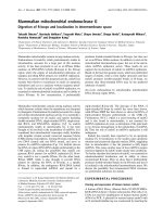
Figure 1
Intra-articular glucocorticoids do not increase synovial tissue apoptosis levels in rheumatoid arthritisIntra-articular glucocorticoids do not increase synovial tissue apoptosis levels in rheumatoid arthritis. Frozen sections of rheumatoid arthritis
synovial biopsy tissues (n = 12) show aminoethylcarbazole staining (red) for active caspase-3 (hematoxylin-counterstained) before (a) and after (b)
treatment and diaminobenzidine staining (brown) for TUNEL (hematoxylin-counterstained) before (c) and after (d) treatment (original magnification ×
125). (e) Results from image analysis of synovial biopsy sections for active caspase-3 and TUNEL staining before and after intra-articular corticos-
teroid injection. Values represent the mean ± standard error of the mean. TUNEL, terminal deoxynucleotidyl transferase-mediated dUTP-biotin nick
end-labeling.
Available online />Page 5 of 9
(page number not for citation purposes)
pared with control), suggesting that soluble factor(s) rather
than cellular contact are primarily responsible for induction of
the apoptosis-resistant phenotype of the RA synovial T cells
(Figure 5).
Discussion
Intra-articular glucocorticoids are a powerful adjuvant therapy
for a variety of inflammatory joint diseases which efficiently
reduces local joint inflammation. We demonstrate here that, in
RA patients, this effect is mediated through the reduction of
the synovial T-cell population as previously suggested in a
cohort of patients with arthritis of different pathogenesis [18].
Furthermore, we provide evidence for the first time that RA-
derived synovial T cells are resistant to apoptosis induction by
glucocorticoids due to a soluble factor(s)-mediated interaction
with monocytes.
Our immunohistochemistry results demonstrate that local
administration of glucocorticoids decreases the number of
lymphocytes without changes in the monocyte/macrophage
population, evaluated as both CD68
+
and CD163
+
cells. The
T cell-specific effect of locally administrated glucocorticoids
might reside in the imbalance between the two alternatively
spliced transcripts of the GR that have been suggested to
have different functional characteristics. Exposure of cells to
proinflammatory stimuli such as TNF and IL-1 can lead to
induction of -isoform of human glucocorticoid receptor
(hGR) and suppression of hGR, resulting in diminished glu-
cocorticoid responsiveness [19]. Furthermore, within the
same tissues, the levels of hGR may vary considerably
between different types of cells [20]. Thus, the local proinflam-
matory milieu in an inflamed joint might contribute to the cell
type-specific effect of locally administrated glucocorticoids.
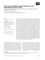
Figure 2
CD3
+
synovial T cells exhibit minimal levels of apoptosis in rheumatoid arthritis synovium both before and after intra-articular glucocorticoidsCD3
+
synovial T cells exhibit minimal levels of apoptosis in rheumatoid arthritis synovium both before and after intra-articular glucocorti-
coids. Photomicrographs illustrate fluorescent staining of CD3
+
cells (red, rhodamin red) before (a) and after therapy (b), TUNEL
+
cells (green, fluo-
rescein) before (c) and after therapy (d) (b, e), and superimposed stainings before (e) and after therapy (f) (original magnification × 320). TUNEL,
terminal deoxynucleotidyl transferase-mediated dUTP-biotin nick end-labeling.
Arthritis Research & Therapy Vol 10 No 6 Makrygiannakis et al.
Page 6 of 9
(page number not for citation purposes)
Our findings suggest a distinct effect of local as compared
with systemic administration of glucocorticoids which has
been shown to decrease both lymphocyte and macrophage
populations [21]. The difference might reside in the use of dis-
tinct synthetic glucocorticoid compounds for local versus sys-
temic administration (that is, triamcinolone versus
prednisolone/methylprednisolone). It has been suggested
that, at equivalent doses, the effects of triamcinolone and dex-
amethasone, but not of methylprednisolone, are suppressed
by overexpression of the hGR that acts as a natural dominant
negative inhibitor of the transactivation of glucocorticoid-
responsive genes [14]. However, when we tested the three
compounds at equivalent doses, we did not observe differ-
ences in the in vitro effect of any of the compounds in any cell
population studied. An alternative explanation is the apparently
specific effect of systemically as compared with locally admin-
istrated high-dose glucocorticoids to induce profound mono-
cytopenia in the peripheral blood [22] that would interfere with
local synovial accumulation of monocytes/macrophages.
The observed reduction in the number of synovial T cells might
be due either to a lower rate of recruitment or to a higher rate
of clearance at the site of inflammation. We have previously
demonstrated that intra-articular glucocorticoids decrease
synovial expression of ICAM-1 (intracellular adhesion mole-
cule-1), an adhesion molecule essential for leukocyte migra-
tion, despite minimal changes in the inflammatory phenotype
of the endothelial synovial cells [15]. Our current results show-
ing resistance of RA synovial T cells to glucocorticoid-induced
apoptosis provide further indirect support for decreased leu-
kocyte recruitment as the major mechanism responsible for
the decreased cellularity observed after treatment with intra-
articular glucocorticoids.
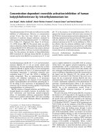
Figure 3
Rheumatoid arthritis (RA)-derived, but not psoriatic arthritis (PsA)-derived, synovial fluid (SF) T cells cocultured with monocytes are resistant to glu-cocorticoid-induced apoptosisRheumatoid arthritis (RA)-derived, but not psoriatic arthritis (PsA)-derived, synovial fluid (SF) T cells cocultured with monocytes are resist-
ant to glucocorticoid-induced apoptosis. Flow cytometric analysis shows that dexamethasone induces an increase in the number of SF CD3/
Annexin V double-positive T cells of PsA patients (n = 2) (a), whereas dexamethasone (n = 11) (b), triamcinolone (n = 4) (c), and metylprednisolone
(n = 4) (d) fail to induce similar changes in apoptosis in SF CD3
+
T cells of RA patients.
Available online />Page 7 of 9
(page number not for citation purposes)
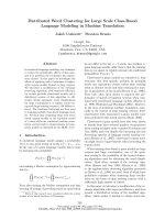
Figure 4
Rheumatoid arthritis (RA) synovial fluid (SF) T cells become susceptible to glucocorticoid-induced apoptosis upon separation from monocytesRheumatoid arthritis (RA) synovial fluid (SF) T cells become susceptible to glucocorticoid-induced apoptosis upon separation from mono-
cytes. Twenty-four-hour exposure to dexamethasone (a), triamcinolone (b), and metylprednisolone (c) of negatively isolated T cells from SF of RA
patients (n = 4) increases apoptosis evaluated as Annexin V
+
7-AAD
-
cells. Graphs demonstrate that all glucocorticoid compounds (d, e, f) induce
apoptosis in isolated T cells but not in nonisolated T cells (dashed line represents nonisolated T cells and continuous line represents isolated T
cells). Values are the mean ± standard error of the mean and are expressed as the ratio of Annexin V
+
cells in the experimental cultures to those in
the control cultures (fold). *P < 0.05. 7-AAD = 7-amino-actinomycin D; AnnV, Annexin V.
Arthritis Research & Therapy Vol 10 No 6 Makrygiannakis et al.
Page 8 of 9
(page number not for citation purposes)
In RA, synovial-derived T cells have a phenotype suggestive of
chronic immune activation but express low levels of cytokines
and show signs of anergy [2]. These cells are resistant to
apoptosis, partly due to their interaction with other cell popu-
lations present in the RA synovial inflamed milieu. It has been
previously demonstrated that synovial-derived isolated T cells
are rescued from spontaneous apoptosis through an integrin-
ligand interaction with stromal cells, an effect that was mim-
icked by the addition of several members of the IL-2R chain
cytokines, such as IL-15 [3]. Along the same line, coculture of
autologous synovial RA T cells with monocytes induces home-
ostatic proliferation of T cells which is dependent on the mem-
brane-bound TNF on monocytes [23]. We demonstrate that
not only spontaneous but also glucocorticoid-induced apopto-
sis is dependent on the complex cell-cell interaction in the
rheumatoid synovium. The essential factor in this situation
appears to be the T cell-monocyte interaction to the extent that
T-cell isolation renders the cells sensitive to apoptosis, while
coculture of T cells with monocytes in the absence of fibrob-
lasts prevented the effect of all tested glucocorticoid com-
pounds. Furthermore, we propose that the main mechanism by
which monocytes are able to rescue T cells is a soluble fac-
tor(s)-mediated interaction rather than cell-cell contact. It has
been demonstrated, for example, that monocytes isolated from
RA SF express IL-15 [24], a cytokine able to upregulate Bcl-2
expression [3] and to render activated T cells resistant to glu-
cocorticoid-mediated apoptosis [25]. The mechanism
appears to be RA-specific given that T-cell apoptosis induc-
tion was observed in cocultures of cells obtained from psori-
atic arthritis in the presence of dexamethasone at similar
doses.
Conclusion
We demonstrate that monocytes are essential in rescuing syn-
ovial T cells from glucocorticoid-induced apoptosis through a
soluble factor(s)-mediated mechanism, a feature that is spe-
cific for RA-derived synovial T cells. We propose that this
might be overcome by the combination of locally administrated
glucocorticoids with monocyte-targeted therapies rather than
T-cell apoptosis-inducing therapies.
Competing interests
The authors declare that they have no competing interests.
Authors' contributions
DM performed the immunohistochemistry and flow cytometry
experiments, participated in acquisition, analysis, and interpre-
tation of data, and drafted the manuscript. SR designed, per-
formed, and analyzed the transwell experiments and
participated in interpretation of the data and writing of the
manuscript. PN and EK recruited the patients for the study,
performed arthroscopies, and participated in acquisition and
interpretation of the data. OS participated in the flow cytome-
try experiments. CG participated in acquisition and analysis of
the data and drafting of the manuscript. AIC conceived the
study, participated in its design and coordination, analyzed the
data, and helped to draft the manuscript. All authors read and
approved the final manuscript.
Acknowledgements
This study was supported by grants from the Ulla and Gustaf af Ugglas
Foundation, Gustav den V:e Foundation, the Swedish Medical Research
Council, the Clas Groschinsky Foundation, and a EULAR young investi-
Figure 5
Coculturing of T cells in the presence of, but without direct contact with, monocytes rescues isolated T cells from glucocorticoid-induced apoptosisCoculturing of T cells in the presence of, but without direct contact with, monocytes rescues isolated T cells from glucocorticoid-induced
apoptosis. Graphs demonstrate that dexamethasone induces apoptosis in isolated T cells (a), an effect that disappears in the presence of mono-
cytes (b). Values are the mean ± standard error of the mean and are expressed as the ratio of Annexin V
+
cells in the experimental cultures to those
in the control cultures (fold). *P < 0.05.
Available online />Page 9 of 9
(page number not for citation purposes)
gator award. We thank Marianne Engstrom for excellent technical assist-
ance and help.
References
1. Pope RM: Apoptosis as a therapeutic tool in rheumatoid arthri-
tis. Nat Rev Immunol 2002, 2:527-535.
2. Cope AP: Studies of T-cell activation in chronic inflammation.
Arthritis Res 2002, 4(Suppl 3):S197-211.
3. Salmon M, Scheel-Toellner D, Huissoon AP, Pilling D, Shamsa-
deen N, Hyde H, D'Angeac AD, Bacon PA, Emery P, Akbar AN:
Inhibition of T cell apoptosis in the rheumatoid synovium. J
Clin Invest 1997, 99:439-446.
4. Firestein GS, Yeo M, Zvaifler NJ: Apoptosis in rheumatoid arthri-
tis synovium. J Clin Invest 1995, 96:1631-1638.
5. Isomaki P, Panesar M, Annenkov A, Clark JM, Foxwell BM, Cherna-
jovsky Y, Cope AP: Prolonged exposure of T cells to TNF down-
regulates TCR zeta and expression of the TCR/CD3 complex
at the cell surface. J Immunol 2001, 166:5495-5507.
6. Busteed S, Bennett MW, Molloy C, Houston A, Stone MA, Shana-
han F, Molloy MG, O'Connell J: Bcl-x(L) expression in vivo in
rheumatoid synovium. Clin Rheumatol 2006, 25:789-793.
7. Sugiyama M, Tsukazaki T, Yonekura A, Matsuzaki S, Yamashita S,
Iwasaki K: Localisation of apoptosis and expression of apopto-
sis related proteins in the synovium of patients with rheuma-
toid arthritis. Ann Rheum Dis 1996, 55:442-449.
8. Stahn C, Lowenberg M, Hommes DW, Buttgereit F: Molecular
mechanisms of glucocorticoid action and selective glucocorti-
coid receptor agonists. Mol Cell Endocrinol 2007, 275:71-78.
9. Kirsch AH, Mahmood AA, Endres J, Bohra L, Bonish B, Weber K,
Fox DA: Apoptosis of human T-cells: induction by glucocorti-
coids or surface receptor ligation in vitro and ex vivo. J Biol
Regul Homeost Agents 1999, 13:80-89.
10. Lanza L, Scudeletti M, Puppo F, Bosco O, Peirano L, Filaci G,
Fecarotta E, Vidali G, Indiveri F: Prednisone increases apoptosis
in in vitro activated human peripheral blood T lymphocytes.
Clin Exp Immunol 1996, 103:
482-490.
11. Herold MJ, McPherson KG, Reichardt HM: Glucocorticoids in T
cell apoptosis and function. Cell Mol Life Sci 2006, 63:60-72.
12. Sionov RV, Kfir S, Zafrir E, Cohen O, Zilberman Y, Yefenof E: Glu-
cocorticoid-induced apoptosis revisited: a novel role for glu-
cocorticoid receptor translocation to the mitochondria. Cell
Cycle 2006, 5:1017-1026.
13. Herr I, Gassler N, Friess H, Buchler MW: Regulation of differen-
tial pro- and anti-apoptotic signaling by glucocorticoids. Apop-
tosis 2007, 12:271-291.
14. Fruchter O, Kino T, Zoumakis E, Alesci S, De Martino M, Chrousos
G, Hochberg Z: The human glucocorticoid receptor (GR) iso-
form {beta} differentially suppresses GR{alpha}-induced
transactivation stimulated by synthetic glucocorticoids. J Clin
Endocrinol Metab 2005, 90:3505-3509.
15. af Klint E, Grundtman C, Engstrom M, Catrina AI, Makrygiannakis
D, Klareskog L, Andersson U, Ulfgren AK: Intraarticular glucocor-
ticoid treatment reduces inflammation in synovial cell infiltra-
tions more efficiently than in synovial blood vessels. Arthritis
Rheum 2005, 52:3880-3889.
16. Arnett FC, Edworthy SM, Bloch DA, McShane DJ, Fries JF, Cooper
NS, Healey LA, Kaplan SR, Liang MH, Luthra HS, Medsger TA Jr,
Mitchell DM, Neustadt DH, Pinals RS, Schaller JG, Sharp JT,
Wilder RL, Hunder GG: The American Rheumatism Association
1987 revised criteria for the classification of rheumatoid arthri-
tis. Arthritis Rheum 1988, 31:315-324.
17. Catrina AI, Trollmo C, af Klint E, Engstrom M, Lampa J, Hermans-
son Y, Klareskog L, Ulfgren AK: Evidence that anti-tumor necro-
sis factor therapy with both etanercept and infliximab induces
apoptosis in macrophages, but not lymphocytes, in rheuma-
toid arthritis joints: extended report. Arthritis Rheum 2005,
52:61-72.
18. Makrygiannakis D, af Klint E, Catrina SB, Botusan IR, Klareskog E,
Klareskog L, Ulfgren AK, Catrina AI: Intraarticular corticosteroids
decrease synovial RANKL expression in inflammatory arthritis.
Arthritis Rheum 2006, 54:1463-1472.
19. Webster JC, Oakley RH, Jewell CM, Cidlowski JA: Proinflamma-
tory cytokines regulate human glucocorticoid receptor gene
expression and lead to the accumulation of the dominant neg-
ative beta isoform: a mechanism for the generation of gluco-
corticoid resistance. Proc Natl Acad Sci USA 2001,
98:6865-6870.
20. Oakley RH, Webster JC, Sar M, Parker CR Jr, Cidlowski JA:
Expression and subcellular distribution of the beta-isoform of
the human glucocorticoid receptor. Endocrinology 1997,
138:5028-5038.
21. Gerlag DM, Haringman JJ, Smeets TJ, Zwinderman AH, Kraan MC,
Laud PJ, Morgan S, Nash AF, Tak PP: Effects of oral pred-
nisolone on biomarkers in synovial tissue and clinical
improvement in rheumatoid arthritis. Arthritis Rheum 2004,
50:3783-3791.
22. Fauci AS, Dale DC, Balow JE: Glucocorticosteroid therapy:
mechanisms of action and clinical considerations. Ann Intern
Med 1976, 84:304-315.
23. Wagner U, Pierer M, Wahle M, Moritz F, Kaltenhauser S,
Hantzschel H: Ex vivo homeostatic proliferation of CD4
+
T cells
in rheumatoid arthritis is dysregulated and driven by mem-
brane-anchored TNFalpha. J Immunol 2004, 173:2825-2833.
24. Miranda-Carus ME, Benito-Miguel M, Balsa A, Cobo-Ibanez T,
Perez de Ayala C, Pascual-Salcedo D, Martin-Mola E: Peripheral
blood T lymphocytes from patients with early rheumatoid
arthritis express RANKL and interleukin-15 on the cell surface
and promote osteoclastogenesis in autologous monocytes.
Arthritis Rheum 2006, 54:1151-1164.
25. Bulfone-Paus S, Ungureanu D, Pohl T, Lindner G, Paus R, Ruckert
R, Krause H, Kunzendorf U: Interleukin-15 protects from lethal
apoptosis in vivo. Nat Med 1997, 3:1124-1128.