Báo cáo y học: "Cartilage homeostasis in health and rheumatic diseases" pptx
Bạn đang xem bản rút gọn của tài liệu. Xem và tải ngay bản đầy đủ của tài liệu tại đây (453.47 KB, 16 trang )
Available online />Page 1 of 16
(page number not for citation purposes)
Abstract
As the cellular component of articular cartilage, chondrocytes are
responsible for maintaining in a low-turnover state the unique
composition and organization of the matrix that was determined
during embryonic and postnatal development. In joint diseases,
cartilage homeostasis is disrupted by mechanisms that are driven
by combinations of biological mediators that vary according to the
disease process, including contributions from other joint tissues. In
osteoarthritis (OA), biomechanical stimuli predominate with up-
regulation of both catabolic and anabolic cytokines and recapitula-
tion of developmental phenotypes, whereas in rheumatoid arthritis
(RA), inflammation and catabolism drive cartilage loss. In vitro
studies in chondrocytes have elucidated signaling pathways and
transcription factors that orchestrate specific functions that promote
cartilage damage in both OA and RA. Thus, understanding how the
adult articular chondrocyte functions within its unique environment
will aid in the development of rational strategies to protect cartilage
from damage resulting from joint disease. This review will cover
current knowledge about the specific cellular and biochemical
mechanisms that regulate cartilage homeostasis and pathology.
Introduction
Adult articular cartilage is an avascular tissue composed of a
specialized matrix of collagens, proteoglycans, and non-
collagen proteins, in which chondrocytes constitute the
unique cellular component. Although chondrocytes in this
context do not normally divide, they are assumed to maintain
the extracellular matrix (ECM) by low-turnover replacement of
certain matrix proteins. During aging and joint disease, this
equilibrium is disrupted and the rate of loss of collagens and
proteoglycans from the matrix may exceed the rate of
deposition of newly synthesized molecules. Originally con-
sidered an inert tissue, cartilage is now considered to
respond to extrinsic factors that regulate gene expression
and protein synthesis in chondrocytes. Numerous studies in
vitro and in vivo during the last two decades have confirmed
that articular chondrocytes are able to respond to mechanical
injury, joint instability due to genetic factors, and biological
stimuli such as cytokines and growth and differentiation
factors that contribute to structural changes in the surround-
ing cartilage matrix [1]. Mechanical influences on chondro-
cyte function are considered to be important in the patho-
genesis of osteoarthritis (OA), but chondrocyte responses to
molecular signals may vary in different regions, including the
calcified cartilage, and also occur at different stages over a
long time course (Figure 1). In rheumatoid arthritis (RA), the
inflamed synovium is the major source of cytokines and
proteinases that mediate cartilage destruction in areas
adjacent to the proliferating synovial pannus (Figure 2) [2].
However, the basic cellular mechanisms regulating chondro-
cyte responses are very different in OA and RA. Moreover,
mechanistic insights from in vitro studies ideally should be
interpreted in light of direct analysis of human cartilage and
other joint tissues and studies in experimental models, inclu-
Review
Cartilage homeostasis in health and rheumatic diseases
Mary B Goldring
1
and Kenneth B Marcu
2,3
1
Research Division, Hospital for Special Surgery, affiliated with Weill College of Medicine of Cornell University, Caspary Research Building,
535 E. 70th Street, New York, NY 10021, USA
2
Biochemistry and Cell Biology Department, Stony Brook University, Life Sciences Rm #330, Stony Brook, NY 11794, USA
3
Centro Ricerca Biomedica Applicata, S. Orsola-Malpighi University Hospital, University of Bologna, Via Massarenti 9, 40138 Bologna, Italy
Corresponding author: Mary B Goldring,
Published: 19 May 2009 Arthritis Research & Therapy 2009, 11:224 (doi:10.1186/ar2592)
This article is online at />© 2009 BioMed Central Ltd
ADAM = a disintegrin and metalloproteinase; ADAMTS = a disintegrin and metalloproteinase with thrombospondin-1 domains; AGE = advanced
glycation end product; CD-RAP = cartilage-derived retinoic acid-sensitive protein; COL2A1 = collagen, type II, alpha 1; COMP = cartilage
oligomeric matrix protein; COX-2 = cyclooxygenase 2; DDR-2 = discoidin domain receptor 2; DZC = deep zone chondrocyte; ECM = extracellular
matrix; ERK = extracellular signal-regulated kinase; FRZB = frizzled-related protein 3; GADD45β = growth arrest and DNA damage 45 beta; GLUT =
glucose transporter protein; HIF-1α = hypoxia-inducible factor-1-alpha; HMGB1 = high-mobility group protein 1; hTNFtg = human tumor necrosis
factor transgenic; IGF-1 = insulin-like growth factor 1; Ihh = Indian hedgehog; IKK = IκB kinase; IL = interleukin; JNK = c-jun N-terminal kinase;
MAPK = mitogen-activated protein kinase; MIA = melanoma inhibitory activity; MMP = matrix metalloproteinase; mPGES-1 = microsomal
prostaglandin E synthase 1; MSC = mesenchymal stem cell; MZC = middle zone chondrocyte; NF-κB = nuclear factor-kappa-B; NO = nitric oxide;
OA = osteoarthritis; PGE = prostaglandin E; PPAR = peroxisome proliferator-activated receptor; RA = rheumatoid arthritis; RAGE = receptor for
advanced glycation end products; RANK = receptor activator of nuclear factor-kappa-B; RANKL = receptor activator of nuclear factor-kappa-B
ligand; ROS = reactive oxygen species; SMAD = signal-transducing mothers against decapentaplegic; SOCS = suppressor of cytokine signaling;
SZC = superficial zone chondrocyte; TGF-β = transforming growth factor-beta; TLR = Toll-like receptor; TNF-α = tumor necrosis factor-alpha;
VEGF = vascular endothelial growth factor.
Arthritis Research & Therapy Vol 11 No 3 Goldring and Marcu
Page 2 of 16
(page number not for citation purposes)
ding knockout and transgenic mice [3,4]. The examination of
cartilage or chondrocytes from patients undergoing joint
replacement has yielded less information in RA patients, in
which cartilage damage is extensive, than studies of OA
patients. In both, the findings do not reflect early disease.
This review will cover current knowledge about the cellular
and biochemical mechanisms of cartilage in health and
disease derived from studies over the past 10 years.
Cartilage in health
Cartilage matrix in healthy articular cartilage
Articular cartilage is composed of four distinct regions: (a)
the superficial tangential (or gliding) zone, composed of thin
collagen fibrils in tangential array and associated with a high
concentration of decorin and a low concentration of aggre-
can, (b) the middle (or transitional) zone with radial bundles of
thicker collagen fibrils, (c) the deep (or radial) zone, in which
the collagen bundles are thickest and are arranged in a radial
fashion, and (d) the calcified cartilage zone, located
immediately below the tidemark and above the subchondral
bone [5,6]. The calcified zone persists after growth plate
closure as the ‘tidemark’ and serves as an important mecha-
nical buffer between the uncalcified articular cartilage and the
subchondral bone. From the superficial to the deep zone, cell
density progressively decreases, whereas cell volume and the
proportion of proteoglycan relative to collagen increase.
The interterritorial cartilage matrix, which is composed of a
fibrillar collagen network that bestows tensile strength, differs
from the territorial matrix closer to the cell, which contains
type VI collagen microfibrils but little or no fibrillar collagen.
The interterritorial collagen network consists primarily of type
II collagen fibrils with type XI collagen within the fibril and
type IX collagen integrated in the fibril surface with the non-
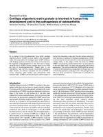
Figure 1
Cellular interactions in cartilage destruction in osteoarthritis. This scheme represents the destruction of the cartilage due to mechanical loading and
biological factors. The induction of stress-induced intracellular signals, catabolic cytokines, including interleukin-1 (IL-1) and tumor necrosis factor-
alpha (TNF-α), chemokines, and other inflammatory mediators produced by synovial cells and chondrocytes results in the upregulation of cartilage-
degrading enzymes of the matrix metalloproteinase (MMP) and ADAMTS families. Matrix degradation products can feedback regulate these cellular
events. Anabolic factors, including bone morphogenetic proteins (BMPs) and transforming growth factor-beta (TGF-β), may also be upregulated
and participate in osteophyte formation. In addition to matrix loss, evidence of earlier changes, such as chondrocyte proliferation and hypertrophy,
increased cartilage calcification with tidemark advancement, and microfractures with angiogenesis from the subchondral bone possibly mediated
by vascular endothelial growth factor (VEGF) can be observed in late osteoarthritis samples obtained from patients after total joint replacement.
ADAMTS, a disintegrin and metalloproteinase with thrombospondin-1 domains; C/EBP, CCAAT enhancer-binding protein; ESE1, epithelial-specific
ETS; ETS, E26 transformation specific; GADD45β, growth arrest and DNA damage 45 beta; HIF-1α, hypoxia-inducible factor-1-alpha; NF-κB,
nuclear factor-kappa-B; PA, plasminogen activator; TIMPs, tissue inhibitors of metalloproteinases.
collagen domain projecting outward, permitting association
with other matrix components and retention of proteoglycans
[7]. Collagen XXVII, a novel member of the fibrillar collagen
family, also contributes to the formation of a stable cartilage
matrix [8].
Compressive resistance is bestowed by the large aggre-
gating proteoglycan aggrecan, which is attached to
hyaluronic acid polymers via link protein. The half-life of
aggrecan core protein ranges from 3 to 24 years, and the
glycosaminoglycan components of aggrecan are synthesized
more readily under low-turnover conditions, with more rapid
matrix turnover in the pericellular regions. The proteoglycans
are essential for protecting the collagen network, which has a
half-life of more than 100 years if not subjected to inappro-
priate degradation. A large number of other noncollagen
molecules, including biglycan, decorin, fibromodulin, the
matrilins, and cartilage oligomeric matrix protein (COMP), are
also present in the matrix. COMP acts as a catalyst in
collagen fibrillogenesis [9], and interactions between type IX
collagen and COMP or matrilin-3 are essential for proper
formation and maintenance of the articular cartilage matrix
[10,11]. Perlecan enhances fibril formation [12], and collagen
VI microfibrils connect to collagen II and aggrecan via
complexes of matrilin-1 and biglycan or decorin [13].
Chondrocyte physiology and function in healthy
articular cartilage
Differences in the morphologies of zonal subpopulations of
chondrocytes may reflect matrix composition and are
ascribed largely to differences in the mechanical environment
[14]. The superficial zone chondrocytes (SZCs) are small and
flattened. The middle zone chondrocytes (MZCs) are rounded,
and the deep zone chondrocytes (DZCs) are grouped in
Available online />Page 3 of 16
(page number not for citation purposes)
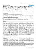
Figure 2
Cellular interactions in cartilage destruction in rheumatoid arthritis. This scheme represents the progressive destruction of the cartilage associated
with the invading synovial pannus in rheumatoid arthritis. As a result of immune cell interactions involving T and B lymphocytes,
monocytes/macrophages, and dendritic cells, a number of different cytokines are produced in the synovium due to the influx of inflammatory cells
from the circulation and synovial cell hyperplasia. The induction of proinflammatory cytokines produced primarily in the synovium, but also by
chondrocytes, results in the upregulation of cartilage-degrading enzymes at the cartilage-pannus junction. Chemokines, nitric oxide (NO), and
prostaglandins (PGE
2
) also contribute to the inflammation and tissue catabolism. ADAMTS, a disintegrin and metalloproteinase with
thrombospondin-1 domains; IFN-γ, interferon-gamma; IL, interleukin; MMP, matrix metalloproteinase; SDF-1, stromal derived factor 1; TGF-β,
transforming growth factor-beta; TNF-α, tumor necrosis factor-alpha; Treg, regulatory T (cell).
columns or clusters. In vitro studies with isolated SZCs and
DZCs indicate that differences in the expression of mole-
cules, such as lubricin (also known as superficial zone protein
or proteoglycan-4) and PTHrP by SZCs and Indian hedgehog
(Ihh) and Runx2 by DZCs, may determine the zonal differ-
ences in matrix composition and function [15-17].
How chondrocytes maintain their ECM under homeostatic
conditions has remained somewhat of a mystery since they
do not divide and the matrix isolates them from each other,
but gene expression and protein synthesis may be activated
by injury. Since the ECM normally shields chondrocytes, they
lack access to the vascular system and must rely on
facilitated glucose transport via constitutive glucose trans-
porter proteins, GLUT3 and GLUT8 [18], and active
membrane transport systems [19]. Chondrocytes exist at low
oxygen tension within the cartilage matrix, ranging from 10%
at the surface to less than 1% in the deep zones. In vitro,
chondrocytes adapt to low oxygen tensions by upregulating
hypoxia-inducible factor-1-alpha (HIF-1α), which can
stimulate expression of GLUTs [18], and angiogenic factors
such as vascular endothelial growth factor (VEGF) [20,21] as
well as a number of genes associated with cartilage anabo-
lism and chondrocyte differentiation [22]. One of our
laboratories has identified growth arrest and DNA damage 45
beta (GADD45β), which previously was implicated as an anti-
apoptotic factor during genotoxic stress and cell cycle arrest
in other cell types as a survival factor in healthy articular
chondrocytes [23]. Thus, by modulating the intracellular
expression of survival factors, including HIF-1α and
GADD45β, chondrocytes survive efficiently in the avascular
cartilage matrix and respond to environmental changes.
The aging process may affect the material properties of healthy
cartilage by altering the content, composition, and structural
organization of collagen and proteoglycan [24-26]. This has
been attributed to overall decreased anabolism and to the
accumulation of advanced glycation end products (AGEs) that
enhance collagen cross-linking [27]. Unless perturbed, healthy
chondrocytes remain in a postmitotic quiescent state
throughout life, with their decreasing proliferative potential
being attributed to replicative senescence associated with
erosion of telomere length [28]. The accumulation of cartilage
matrix proteins in the endoplasmic reticulum and Golgi of
chondrocytes, which have been modified by oxidative stress
during aging, may lead to decreased synthesis of cartilage
matrix proteins and diminished cell survival [29].
Cartilage in joint disease
The loss of balance between cartilage anabolism and
catabolism
Although the etiologies of OA and RA are different, both
diseases present states of inappropriate articular cartilage
destruction, which is largely the result of elevated expression
and activities of proteolytic enzymes. Whereas these enzymes
normally are involved in the formation, remodeling, and repair of
connective tissues, a shift in equilibrium between anabolic and
catabolic activities occurs in OA as a response to abnormal
mechanical loading in conjunction with genetic abnormalities or
injury to the cartilage and surrounding joint tissues. In RA, the
inflamed synovium is the major source of cytokine-induced
proteinases, although the episodic intra-articular inflammation
with synovitis indicates that the synovium may also be a source
of cytokines and cartilage-degrading proteinases in OA
[30,31]. However, in OA, these degradative enzymes are
produced primarily by chondrocytes due to inductive stimuli,
including mechanical stress, injury with attendant
destabilization, oxidative stress, cell-matrix interactions, and
changes in growth factor responses and matrix during aging.
Of the proteinases that degrade cartilage collagens and
proteoglycans in joint disease, matrix metalloproteinases
(MMPs) and aggrecanases have been given the greatest
attention because they degrade native collagens and proteo-
glycans [32-34]. These include the collagenases (MMP-1,
MMP-8, and MMP-13), the gelatinases (MMP-2 and MMP-9),
stromelysin-1 (MMP-3), and membrane type I (MT1) MMP
(MMP-14) [35]. MMP-10, similar to MMP-3, activates pro-
collagenases, is detectable in OA and RA synovial fluids and
joint tissues, and is produced in vitro by both the synovium
and chondrocytes in response to inflammatory cytokines [36].
MMP-14, produced principally by RA synovial tissue, is impor-
tant for synovial invasiveness [37], whereas the MMP-14
produced by OA chondrocytes activates pro-MMP-13, which
in turn cleaves pro-MMP-9 [38]. Other MMPs, including
MMP-16 and MMP-28 [32,39], and many members of the
reprolysin-related proteinases of the ADAM (a disintegrin and
metalloproteinase) family, including ADAM-17/TACE (tumor
necrosis factor-alpha [TNF-α]-converting enzyme), are
expressed in cartilage, but their specific roles in cartilage
damage in either OA or RA have yet to be defined [40-42].
Although several of the MMPs, including MMP-3, MMP-8,
and MMP-14, are capable of degrading proteoglycans,
ADAMTS (ADAM with thrombospondin-1 domains)-4 and
ADAMTS-5 are now regarded as the principal aggrecan-
degrading enzymes in cartilage [43,44]. Aggrecanase inhibi-
tors that target ADAMTS-5 have been developed and are
awaiting opportunities for clinical trials in OA [45].
OA and RA differ with respect to the sites as well as the
origins of disrupted matrix homeostasis. In OA, proteoglycan
loss and type II collagen cleavage initially occur at the
cartilage surface, with evidence of pericellular damage in
deeper zones as the lesion progresses [46]. In RA, intrinsic
chondrocyte-derived chondrolytic activity is present at the
cartilage-pannus junction, as well as in deeper zones of
cartilage matrix [47], although elevated levels of MMPs in RA
synovial fluids likely originate from the synovium. There are
also differences in matrix synthetic responses in OA and RA.
Whereas type II collagen synthesis is reduced in early RA
[48], there is evidence of compensatory increases in type II
collagen synthesis in deeper regions of OA cartilage [14].
Arthritis Research & Therapy Vol 11 No 3 Goldring and Marcu
Page 4 of 16
(page number not for citation purposes)
This is in agreement with findings of enhanced global syn-
thesis and gene expression of aggrecan and type II collagen
in human OA compared with healthy cartilage [49-51].
Importantly, microarray studies using full-thickness cartilage
have also shown that many collagen genes, including
collagen, type II, alpha 1 (COL2A1), are upregulated in late-
stage OA [23,51]. The latter applies mainly to MZCs and
DZCs, as revealed by laser capture microdissection, whereas
this anabolic phenotype is less obvious in the degenerated
areas of the upper regions [52].
Inflammation and cartilage destruction
In vivo and in vitro studies have shown that chondrocytes
produce a number of inflammatory mediators, such as inter-
leukin-1-beta (IL-1β) and TNF-α, which are present in RA or
OA joint tissues and fluids. Chondrocytes respond to these
proinflammatory cytokines by increasing the production of
proteinases, prostaglandins, and nitric oxide (NO) [2,25]. The
first recognition of IL-1 as a regulator of chondrocyte function
stems largely from work in in vitro culture models showing
that activities derived from synovium or monocyte macro-
phages induce the production of cartilage-degrading protein-
ases (reviewed in [2,53]).
IL-1, TNF-α, MMP-1, MMP-3, MMP-8, and MMP-13, and type
II collagen cleavage epitopes have been shown to colocalize
in matrix-depleted regions of RA cartilage [48,54] and OA
cartilage [46,55]. In addition, chondrocytes express several
chemokines as well as chemokine receptors that may
participate in cartilage catabolism [56,57]. IL-1β also induces
other proinflammatory cytokines such as IL-17, which has
similar effects on chondrocytes [58,59]. IL-32, a recently
discovered cytokine that induces TNF-α, IL-1β, IL-6, and
chemokines, is also expressed in the synovia of RA patients
and contributes to TNF-α-dependent inflammation and cartilage
proteoglycan loss [60]. The importance of synergisms
between IL-1 and TNF-α and with other cytokines, such as
IL-17, IL-6, and oncostatin M, in RA or OA joints has been
inferred primarily from culture models [61-63]. The up-
regulation of cyclooxygenase-2 (COX-2), MMP13, and NOS2
gene expression by IL-1β in chondrocytes and other cell
types is mediated by the induction and activation of a number
of transcription factors, including nuclear factor-kappa-B
(NF-κB), CCAAT enhancer-binding protein (C/EBP), activator
protein 1 (AP-1), and E26 transformation specific family
members, which regulate stress- and inflammation-induced
signaling [64]. IL-1β also uses these mechanisms to
suppress the expression of a number of genes associated
with the differentiated chondrocyte phenotype, including
COL2A1 and cartilage-derived retinoic acid-sensitive protein/
melanoma inhibitory activity (CD-RAP/MIA) [64-66]. The role
of epigenetics in regulating these cellular events in cartilage
is under current consideration [67].
The IL-1R/Toll-like receptor (TLR) superfamily of receptors,
which has a key role in innate immunity and inflammation,
has received recent attention with respect to cartilage
pathology. Human articular chondrocytes can express TLR1,
TLR2, and TLR4, and the activation of TLR2 by IL-1, TNF-α,
peptidoglycans, lipopolysaccharide, or fibronectin fragments
increases the production of MMPs, NO, prostaglandin E
(PGE), and VEGF [68-73]. In immune complex-mediated
arthritis, TLR4 regulates early-onset inflammation and
cartilage destruction by IL-10-mediated upregulation of Fcγ
receptor expression and enhanced cytokine production [74].
The IL-18 receptor shares homology with IL-1RI and has a
TLR signaling domain. IL-18 has effects similar to IL-1 in
human chondrocytes and stimulates chondrocyte apoptosis,
although studies do not suggest a pivotal role in cartilage
destruction in RA [75,76]. IL-33, an ST2-TLR ligand, is
associated with endothelial cells in RA synovium, but its role
in cartilage destruction has not been examined [77]. Of
recent interest are the suppressor of cytokine signaling
(SOCS) molecules, including SOCS3, which is induced by
IL-1 and acts as a negative feedback regulator during insulin-
like growth factor 1 (IGF-1) desensitization in the absence of
NO by inhibiting insulin receptor substrate 1 (IRS-1)
phosphorylation [78].
The increased production of prostaglandins by inflammatory
cytokines is mediated via induction of the expression of not
only COX-2 but also microsomal PGE synthase 1 (mPGES-1)
[79,80]. In addition to opposing the induction of COX-2,
inducible nitric oxide synthetase (iNOS), and MMPs and the
suppression of aggrecan synthesis by IL-1, activators of the
peroxisome proliferator-activated receptor gamma (PPARγ),
including the endogenous ligand 15-deoxy-Δ
12,14
prosta-
glandin J
2
(PGJ
2
), inhibit IL-1-induced expression of mPGES-
1 [81,82]. Recent evidence indicates that PPARα agonists
may protect chondrocytes against IL-1-induced responses by
increasing the expression of IL-1Ra [83].
White adipose tissue has been proposed as a major source
of both pro- and anti-inflammatory cytokines, including IL-1Ra
and IL-10 [84]. Roles for adipokines, identified originally as
products of adipocytes, have received recent attention, not
only because of their relationship to obesity, but also because
they can have pro- or anti-inflammatory effects in joint tissues
and may serve as a link between the neuroendocrine and
immune systems [85]. Leptin expression is enhanced during
acute inflammation, correlating negatively with inflammatory
markers in RA sera [86]. The expression of leptin is elevated
in OA cartilage and in osteophytes and it stimulates IGF-1
and transforming growth factor-beta-1 (TGF-β1) synthesis in
chondrocytes [87]. Leptin synergizes with IL-1 or interferon-
gamma to increase NO production in chondrocytes [88], and
leptin deficiency attenuates inflammatory processes in experi-
mental arthritis [89]. It has been proposed that the dys-
regulated balance between leptin and other adipokines, such
as adiponectin, promotes destructive inflammatory processes
[90]. Recent studies indicate that resistin plays a role in early
stages of trauma-induced OA and in RA at local sites of
Available online />Page 5 of 16
(page number not for citation purposes)
inflammation and that serum resistin reflects inflammation and
disease activity [91,92].
Effects of mechanical loading
In young individuals without genetic abnormalities, bio-
mechanical factors due to trauma are strongly implicated in
initiating the OA lesion. Mechanical disruption of cell-matrix
interactions may lead to aberrant chondrocyte behavior,
contributing to fibrillations, cell clusters, and changes in
quantity, distribution, or composition of matrix proteins
[93,94]. In the early stages of OA, transient increases in
chondrocyte proliferation and increased metabolic activity are
associated with a localized loss of proteoglycans at the
cartilage surface followed by cleavage of type II collagen
(reviewed in [95,96]). These events result in increased water
content and decreased tensile strength of the matrix as the
lesion progresses.
Chondrocytes can respond to direct biomechanical pertur-
bation by upregulating synthetic activity or by increasing the
production of inflammatory cytokines, which are also
produced by other joint tissues. In vitro mechanical loading
experiments have revealed that injurious static compression
stimulates proteoglycan loss, damages the collagen network,
and reduces synthesis of cartilage matrix proteins, whereas
dynamic compression increases matrix synthetic activity [97].
In response to traumatic injury, global gene expression is
activated, resulting in increased expression of inflammatory
mediators, cartilage-degrading proteinases, and stress
response factors [98,99]. Neuronal signaling molecules, such
as substance P and its receptor, NK1, and N-methyl-
D-
aspartic acid receptors (NMDARs), which require glutamate
and glycine binding for activation, have been implicated in
mechanotransduction in chondrocytes in a recent study [100].
Chondrocytes have receptors for responding to mechanical
stimulation, many of which are also receptors for ECM
components [101]. Among these are several of the integrins
that serve as receptors for fibronectin and type II collagen
fragments, which upon activation stimulate the production of
proteinases, cytokines, and chemokines [102]. Discoidin
domain receptor 2 (DDR-2), a receptor for native type II
collagen fibrils, is activated on chondrocytes via Ras/Raf/Mek
signaling and preferentially induces MMP-13 via p38
mitogen-activated protein kinase (MAPK); this is a universal
mechanism that occurs after loss of proteoglycans, not only
in genetic models, but also in surgical mouse OA and human
OA [103]. On the other hand, in RA the cell-cell adhesion
molecule, cadherin-11, is expressed at the interface between
the RA synovial pannus and cartilage and facilitates cartilage
invasion and erosion in mouse models in vivo and in human
RA tissues in vitro and ex vivo [104] in a TNF-α-dependent
manner [105]. Recent studies indicate that lubricin is an
important secreted product of chondrocytes, synovial cells,
and other joint tissues which is downregulated in OA and RA
and modulated by cytokines and growth factors [91,92].
Stress responses in cartilage
Injurious mechanical stress and cartilage matrix degradation
products are capable of stimulating the same signaling
pathways as those induced by inflammatory cytokines
[98,106-109]. Along with extracellular signal-regulated kinase
1/2 (ERK1/2), the key protein kinases in the c-jun N-terminal
kinase (JNK), p38 MAPK, and NF-κB signaling cascades are
activated, particularly in the upper zones of OA cartilage
[110]. Furthermore, the engagement of integrin receptors by
fibronectin or collagen fragments activates focal adhesion
kinase signaling and transmits signals intersecting with ERK,
JNK, and p38 pathways [111,112]. Cascades of multiple
protein kinases are involved in these responses, including
protein kinase Cζ, which is upregulated in OA cartilage and is
required for activation of NF-κB by IL-1 and TNF-α [113].
However, it remains controversial whether inflammatory cyto-
kines are primary or secondary effectors of cartilage damage
and defective repair mechanisms in OA since these same
pathways also induce or amplify the expression of cytokine
genes. Interestingly, physiological loading may protect
against cartilage loss by inhibiting IκB kinase-beta (IKKβ)
activity in the canonical NF-κB cascade and attenuating
NF-κB transcriptional activity [114] as well as by inhibiting
TAK1 (TGF-β-activated kinase 1) phosphorylation [115]. In
addition, genetic factors that cause disruption of chondrocyte
differentiation and function and influence the composition and
structure of the cartilage matrix may contribute to abnormal
biomechanics, independently of the influence of inflammation.
Reactive oxygen species (ROS) play a critical role in
chondrocyte homeostasis, but during aging, trauma, and OA,
partial oxygen variations and mechanical stress as well as
inflammation induce abnormal ROS production, which
exceeds the antioxidant capacity leading to oxidative stress.
ROS and attendant oxidative stress impair growth factor
responses, enhance senescence through telomere shorten-
ing, and impair mitochondrial function [28,116,117]. ROS
levels are also induced by activation of RAGE, the receptor
for AGEs, which regulates chondrocyte and synovial res-
ponses in OA [118]. In chondrocytes, interaction of RAGE
with S100A4, a member of the S100 family of calcium-
binding proteins, stimulates MMP-13 production via phos-
phorylation of Pyk2, MAPKs, and NF-κB signaling [119].
RAGE expression and S100A1 release are stimulated in
chondrocytes in vitro and increased in OA cartilage. Trans-
glutaminase 1, which is induced by inflammation and stress,
transforms S100A1 into a procatabolic cytokine that signals
through RAGE and the p38 MAPK pathway to induce
chondrocyte hypertrophy and aggrecan degradation [120]. In
experimental murine arthritis models, S100A8 and S100A9
are involved in the upregulation and activation of MMPs and
aggrecanases [121,122]. In addition, high-mobility group
protein 1 (HMGB1), another important RAGE ligand and also
a chromatin architectural protein, is produced by inflamed
synovium and thus acts as a RAGE-dependent proinflam-
matory cytokine in RA [123]. The differential regulation and
Arthritis Research & Therapy Vol 11 No 3 Goldring and Marcu
Page 6 of 16
(page number not for citation purposes)
expression of GLUT isoforms by hypoxia, growth factors, and
inflammatory cytokines may contribute to intracellular stress
responses [124]. COX-2 is also involved in the chondrocyte
response to high shear stress, associated with reduced
antioxidant capacity and increased apoptosis [125]. Modula-
tion of such intracellular stress response mechanisms may
provide strategies for novel therapies.
Biomarkers of cartilage pathology
The recent development of assays for specific biological
markers, which reflect quantitative and dynamic changes in
the synthetic and degradation products of cartilage and bone
matrix components, has provided a means of identifying
patients at risk for rapid joint damage and also for early
monitoring of the efficacy of disease-modifying therapies.
Molecules originating from the articular cartilage, including
aggrecan fragments, which contain chondroitin sulfate and
keratan sulfate, type II collagen fragments, and collagen
pyridinoline cross-links, are usually released as degradation
products as a result of catabolic processes. Specific
antibodies that detect either synthetic or cleavage epitopes
have been developed to study biological markers of cartilage
metabolism in synovial fluids, sera, and urine of patients with OA
or RA (reviewed in [126-129]). Aggrecan degradation
products are assayed using antibodies 846, 3B3(-), and 7D4
that detect chondroitin sulfate neoepitopes, 5D4 that detects
keratan sulfate epitopes, and the VIDIPEN and NITEGE
antibodies that recognize aggrecanase and MMP cleavage
sites, respectively, within the interglobular G1 domain of
aggrecan [33]. Similarly, the C2C antibody (previously known
as Col2-3/4C
Long mono
) has been used to detect specific
cleavage of the triple helix of type II collagen [48,129].
Increased ratios of C2C to the synthetic marker, CPII, are
associated with a greater likelihood of radiological
progression in OA patients [130]. Other markers included
COMP [131]; YKL-40/HC-gp39, or chitinase 3-like protein 1
(CH3L1), which is induced in chondrocytes by inflammatory
cytokines [132]; and CD-RAP, also known as MIA [133,134].
Such biomarker assays have been used as research tools
and are currently under evaluation for monitoring cartilage
degradation or repair in patient populations. C-reactive
protein, IL-6, and MMP-3 have also been identified as
potential biomarkers in both RA and OA patient populations.
A single marker has not proven to be sufficient, however, and
the major challenge will be to apply such biomarkers to the
diagnosis and monitoring of disease in individual patients and
to correlate them with structural changes in cartilage
identified by magnetic resonance imaging techniques [135].
The genetics of cartilage pathology
Results of epidemiological studies, analysis of patterns of
familial clustering, twin studies, and the characterization of
rare genetic disorders suggest that genetic abnormalities can
result in early onset of OA and increased susceptibility to RA.
For example, twin studies have shown that the influence of
genetic factors may approach 70% in OA that affects certain
joints. Candidate gene studies and genome-wide linkage
analyses have revealed polymorphisms or mutations in genes
encoding ECM and signaling molecules that may determine
OA susceptibility [136-138]. Gender differences have been
noted and gene defects may appear more prominently in
different joints [136,139]. Gene defects associated with
congenital cartilage dysplasias that affect the formation of
cartilage matrix and patterning of skeletal elements may
adversely affect joint alignment and congruity and thus
contribute to early onset of OA in these individuals [140].
Although whole-genome linkage analyses of RA patients have
not addressed cartilage specifically, this work has pointed to
immunological pathways and inflammatory signals that may
modulate cartilage destruction [141].
Genomic and proteomic analyses, which have been
performed in cytokine-treated chondrocytes, in cartilage from
patients with OA, and in rheumatoid synovium, have provided
some insights into novel mechanisms that might govern
chondrocyte responses in both OA and RA [57,63,102,142].
When coupled with biological analyses that address candi-
date genes, gene profiling studies of cartilage derived from
patients with OA have also begun to yield new information
about mediators and pathways [23,51,143,144]. Similarly,
microarray analysis of cocultures of synovial fibroblasts with
chondrocytes in alginate has identified markers of inflam-
mation and cartilage destruction associated with RA
pathogenesis [145].
Lessons from mouse models
Insight into cartilage pathology in RA has been gleaned from
the examination of type II collagen-induced arthritis and other
types of inflammatory arthritis in mice with transgenic over-
expression or knockout of genes encoding cytokines, their
receptors, or activators. These studies have led in part to the
conclusion that TNF-α drives acute inflammation whereas
IL-1 has a pivotal role in sustaining cartilage erosion [146]. In
support of this concept, crossing arthritic human TNF trans-
genic (hTNFtg) mice with IL-1α- and β-deficient strains
protected against cartilage erosion without affecting synovial
inflammation [147]. The success of anti-TNF-α therapy in
most but not all patients highlights the importance of
inflammation in joint destruction.
In vivo studies have also shown that alterations in cartilage
matrix molecules or in regulators of chondrocyte differen-
tiation can lead to OA pathology. The importance of the fine
protein network and ECM structural integrity in postnatal
cartilage health is well documented in studies of deficiencies
or mutations in cartilage matrix genes, including Col2a1,
Col9a1, Col11a1, aggrecan, matrilin-3, or fibromodulin alone
or together with biglycan, which lead to age-dependent
cartilage degeneration similar to that in OA patients
[140,148,149]. Deficiency of Timp3 (tissue inhibitor of metallo-
proteinases 3) or postnatal overexpression of constitutively
active Mmp13 also promotes OA-like pathology [150,151].
Available online />Page 7 of 16
(page number not for citation purposes)
Importantly, surgically induced OA disease models in mutant
mice have also implicated ADAMTS5 [152,153], DDR-2
[103], and Runx2 [154] as contributors to the onset and/or
severity of OA joint disease. Knockout of IL-1β is also
protective against OA induced by destabilization of the
medial meniscus [155]. Although single gene defects do not
model all aspects of human OA, the loss or mutation of a
gene that is involved in the synthesis or remodeling of the
cartilage matrix may lead to the disruption of other gene
functions in chondrocytes, thus resulting in joint instability
and OA-like pathology. Thus, novel mechanistic insights into
the initiation or progression of OA may be discovered by
identifying intracellular effectors of ECM homeostasis and
remodelling in vitro and evaluating their functions in animal
models of OA disease.
Chondrogenesis, chondrocyte hypertrophy, calcified
cartilage
,,
and bone in cartilage pathology
During skeletal development, the chondrocytes arise from
mesenchymal progenitors to synthesize the templates, or
cartilage anlagen, for the developing limbs in a process
known as chondrogenesis [156]. Following mesenchymal
condensation and chondroprogenitor cell differentiation,
chondrocytes undergo proliferation, terminal differentiation to
hypertrophy, and apoptosis, whereby hypertrophic cartilage is
replaced by bone in endochondral ossification. A number of
signaling pathways and transcription factors play stage-
specific roles in chondrogenesis and a similar sequence of
events occurs in the postnatal growth plate, leading to rapid
growth of the skeleton [64,156-158].
Chondrogenesis is orchestrated in part by Sox9 and Runx2,
two pivotal transcriptional regulators that determine the fate
of chondrocytes to remain within cartilage or undergo
hypertrophic maturation prior to ossification and is also
subject to complex regulation by interplay of the fibroblast
growth factor, TGF-β, BMP, and Wnt signaling pathways
[159-162]. Differential signaling during chondrocyte matura-
tion occurs via TGF-β-regulated signal-transducing mothers
against decapentaplegic (Smads) 2 and 3 that act to
maintain articular chondrocytes in an arrested state and
BMP-regulated Smads 1 and 5 that accelerate their differen-
tiation. Sox9, which is essential for type II collagen (COL2A1)
gene expression, is most highly expressed in proliferating
chondrocytes and has opposing positive and negative effects
on the early and late stages of chondrogenesis, respectively.
Sox9 cooperates with two related proteins, L-Sox5 and Sox6,
which are targets of Sox9 itself and function as architectural
HMG-like chromatin modifiers. Moreover, BMP signaling,
through the type I Bmpr1a and Bmpr1b receptors, redun-
dantly drives chondrogenesis via Sox9, Sox5, and Sox6. In
addition, Runx2, which drives the terminal phase of chondro-
genesis [163], is subject to direct inhibition by Sox9 [164]. In
cooperation with BMP-induced Smads, Runx2 also upregu-
lates GADD45β, a positive regulator of the terminal hyper-
trophic phase of chondrogenesis which drives the expression
of Mmp13 and Col10a1 in the mouse embryonic growth
plate [165]. More recently, the findings of our groups suggest
that GADD45β contributes to the homeostasis of healthy and
early OA articular chondrocytes as an effector of cell survival
and as one of the factors induced by NF-κB that contributes
to the imbalance in matrix remodelling in OA cartilage by
suppressing COL2A1 gene expression [23] and that the NF-
κB activating kinases, IKKα and IKKβ, differentially contribute
to OA pathology by also regulating matrix remodelling in
conjunction with chondrocyte differentiation [166].
Endochondral ossification, in which the hypertrophic
chondrocyte undergoes a stress response associated with
ECM remodelling, has been proposed as a ‘developmental
model’ to understand the contribution of exacerbated environ-
mental stresses to OA pathology [167-170]. Changes in the
mineral content and thickness of the calcified cartilage and
the associated tidemark advancement may be related to
recapitulation of the hypertrophic phenotype, including
COL10A1, MMP-13, and Runx2 gene expression, observed
in the deep zone of OA cartilage [167,171]. In addition to
COL10A1 and MMP-13, other chondrocyte terminal differen-
tiation-related genes, such as MMP-9 and Ihh, are detected
in the vicinity of early OA lesions along with decreased levels
of Sox9 mRNA [172]. However, Sox9 expression does not
always localize with COL2A1 mRNA in adult articular
cartilage [52,173]. Apoptosis is a rare event in OA cartilage
but may be a consequence of the chondrocyte stress res-
ponse associated with hypertrophy [174]. Interestingly, one
of our recent studies indicates that intracellular stress res-
ponse genes are upregulated in early OA, whereas a number
of genes encoding cartilage-specific and nonspecific
collagens and other matrix proteins are upregulated in late-
stage OA cartilage [23]. Moreover, articular chondrocytes in
micromass culture show ‘phenotypic plasticity’ comparable to
mesenchymal stem cells (MSCs) undergoing chondro-
genesis, by recapitulating processes akin to chondrocyte
hypertrophy [175], which one of our labs recently has shown
to be subject to differential control by canonical NF-κB
signaling and IKKα [166]. This process may also be
modulated by Src kinases [176,177].
Additional supporting evidence for dysregulation of endo-
chondral ossification as a factor in OA pathology comes from
genetic association studies identifying OA susceptibility
genes across different populations [138,170,178]. These
include the genes encoding asporin (ASPN), a TGF-β-
binding protein with biglycan and decorin sequence homology
[179], secreted frizzled-related protein 3 (FRZB), a WNT/β-
catenin signaling antagonist [180,181], and deiodinase 2
(DIO2), an enzyme that converts inactive thyroid hormone,
T4, to active T3 [182]. The activation of WNT/β-catenin in
mature postnatal growth plate chondrocytes stimulates
hypertrophy, matrix mineralization, and expression of VEGF,
ADAMTS5, MMP-13, and several other MMPs [183].
Findings from microarray analyses of bone from OA patients
Arthritis Research & Therapy Vol 11 No 3 Goldring and Marcu
Page 8 of 16
(page number not for citation purposes)
[184] and in Frzb knockout mice [185] also suggest that
signaling modifications in the calcified cartilage could
contribute to increased subchondral plate thickness accom-
panying tidemark advancement at the border with the
articular cartilage and the angiogenesis observed at the
osteochondral junction [186]. Moreover, endochondral
ossification also contributes to the formation of osteophytes
[187-189]. Interestingly, HMGB1 released by hypertrophic
cartilage, prior to the onset of programmed cell death,
contributes to endochondral ossification by acting as a
chemotactic factor for osteoclasts at the growth plate [190],
and HMGB1-induced NF-κB signaling is also required for
cellular chemotaxis in response to HMGB1-RAGE engage-
ment [191]. Thus, IKK-mediated NF-κB signaling not only
may intrinsically influence the differentiation of chondrocytes
toward a hypertrophy-like state [166], but also could
subsequently drive aspects of intercellular communication
culminating in endochondral ossification [190].
Changes in the periarticular and subchondral bone also
occur in both RA and OA and may contribute to cartilage
pathology. Receptor activator of NFκB (RANK), a member of
the TNF receptor family, RANK ligand (RANKL), and the
soluble receptor osteoprotegerin regulate osteoclast differen-
tiation and activity and are important mediators of bone
destruction in RA. IKKβ-mediated, but not IKKα-mediated,
NF-κB signaling is associated with inflammation-induced
bone loss [192] and is also critical for the survival of osteo-
clast precursors by suppressing JNK-dependent apoptosis in
response to RANKL signaling [193]. IL-17 induces RANKL,
inducing bone destruction independently of IL-1 and bypass-
ing the requirement for TNF in inflammatory arthritis [58].
Although RANK and RANKL are expressed in adult articular
chondrocytes, a direct action in cartilage has not been identi-
fied [194]. Since cartilage destruction is not blocked directly
by the inhibition of RANKL, at least in inflammatory models,
indirect effects may occur through protection of the bone
[195,196], as suggested by recent studies in experimental
models [197,198]. A link between RANKL and WNT has
been suggested by findings in hTNFtg mice and RA tissues,
in which decreased β-catenin and high DKK-1, a WNT
inhibitor, were demonstrated in synovium and in cartilage
adjacent to inflammatory tissue [199] (reviewed in [200]). In
contrast, increased β-catenin was observed in OA cartilage
and conditional overexpression in mouse cartilage leads to
premature chondrocyte differentiation and development of
OA-like phenotype [201]. Interestingly, Runx2-dependent
expression of RANKL occurs in hypertrophic chondrocytes at
the boundary next to the calcifying cartilage in the developing
growth plate [202].
Mesenchymal progenitor cells in cartilage and their use
in tissue engineering
MSCs from bone marrow and other adult tissues, including
muscle, adipose tissue, and synovium or other tissue sites,
which have the capacity to differentiate into cartilage, bone,
fat, and muscle cells, are under investigation as sources of
cartilage progenitor cells for cartilage tissue engineering
[203-206]. Studies in vitro indicate that the same growth and
differentiation factors that regulate different stages of
cartilage development may be able to promote cartilage
repair [207-209]. IGF-1 is a potent stimulator of proteoglycan
synthesis, particularly when combined with other anabolic
factors, including BMPs [210,211]. Moreover, ex vivo gene
transfer of anabolic factors such as BMPs, TGF-β, and IGF-1
has been explored as an approach to promote differentiation
of autologous chondrocytes or MSCs before implantation
[212,213]. Recently, endochondral ossification has been
achieved with murine embryonic stem cells in tissue-engi-
neered constructs implanted in cranial bone of rats [214].
BMP-2 and BMP-7 (osteogenic protein 1) are currently
approved for multiple indications in the area of bone fracture
repair and spinal fusion, but the capacity of BMPs and TGF-β
to induce chondrocyte hypertrophy in cartilage repair models
and to promote osteophyte formation may prevent controlled
repair of articular cartilage in vivo [207]. Since the injection of
free TGF-β or adenovirus-mediated delivery of TGF-β pro-
motes fibrosis and osteophyte formation, while stimulating
proteoglycan synthesis in cartilage, the local application of
molecules that block endogenous TGF-β signaling, such as
the soluble form of TGF-βRII, inhibitory SMADs, or the
physiological antagonist latency-associated peptide 1 (LAP-1),
has been proposed as a more effective strategy [188].
Additional strategies include gene transfer of Sox9, alone or
together with L-Sox5 and Sox6, into MSCs ex vivo or into
joint tissues in vivo to more directly promote the expression
of cartilage matrix genes [215,216]. Strategies to stably
express interfering RNAs in vivo could also provide a means
of blocking dysregulated ECM remodelling or inappropriate
endochondral ossification of articular chondrocytes.
Despite intensive investigation of cartilage repair strategies
and the increased understanding of the cellular mechanisms
involved, many issues remain to be resolved. These include
the fabrication and maintenance of the repair tissue in the
same zonal composition as the original cartilage, the recruit-
ment and maintenance of cells with an appropriate chondro-
cyte phenotype, and integration of the repair construct with
the surrounding cartilage matrix [217]. These issues are also
compounded when cartilage loss is severe or when chronic
inflammation exists, as in RA.
Conclusions
Laboratory investigations in vitro and in vivo regarding the
role of the chondrocyte in remodeling the cartilage matrix in
the RA and OA joint have identified novel molecules and
mechanisms and provided new understanding of the contri-
butions of known mediators. In RA, mediators involved in
immunomodulation and synovial cell function, including
cytokines, chemokines, and adhesion molecules, have primary
roles in the inflammatory and catabolic processes in the joint,
Available online />Page 9 of 16
(page number not for citation purposes)
but they may also, directly or indirectly, promote cartilage
damage. Despite our increasing knowledge of the mecha-
nisms regulating the responses of chondrocytes to anabolic
and catabolic factors involved in developing and adult
cartilage, the development of disease-modifying therapies for
OA patients has been elusive. In RA, in which significant
advances have been achieved in our understanding of the
cellular interactions in the RA joint involving macrophages, T
and B lymphocytes, and synovial fibroblasts, there is still a
need for therapeutic strategies that prevent the extensive
cartilage and bone loss, despite the clinical success of anti-
TNF therapy for RA. Further work using the principles of cell
and molecular biology, such as those described in this
review, will be necessary for uncovering new therapies for
targeting cartilage destruction in both degenerative and
inflammatory joint disease.
Competing interests
The authors declare that they have no competing interests.
Acknowledgments
Research relating to this review was supported by National Institutes of
Health (NIH) (Bethesda, MD, USA) grant AG022021 and by the Arthri-
tis Foundation. KBM greatly acknowledges his collaborators in the Lab-
oratorio di Immunologia e Genetica, Istituti Ortopedici Rizzoli (Bologna,
Italy), in particular Rosa Maria Borzi, Eleonora Olivotto, Stefania Pagani,
and Andrea Facchini. The research of KBM was supported in part by
the Rizzoli Institute, the Carisbo Foundation of Bologna, a Rientro dei
Cervelli award, the MAIN EU FPVI Network of Excellence, and NIH
grant GM066882.
References
1. Goldring MB, Goldring SR: Osteoarthritis. J Cell Physiol 2007,
213:626-634.
2. Dayer JM: The process of identifying and understanding
cytokines: from basic studies to treating rheumatic diseases.
Best Pract Res Clin Rheumatol 2004, 18:31-45.
3. van de Loo FA, Geurts J, van den Berg WB: Gene therapy works
in animal models of rheumatoid arthritis so what! Curr
Rheumatol Rep 2006, 8:386-393.
4. van den Berg WB: Lessons from animal models of osteoarthri-
tis. Curr Rheumatol Rep 2008, 10:26-29.
5. Poole AR: Cartilage in health and disease. In Arthritis and Allied
Conditions: A Textbook of Rheumatology. 15th edition. Edited by
Koopman WS. Philadelphia: Lippincott, Williams, and Wilkins;
2005:223-269.
6. Goldring MB: Chapter 3: cartilage and chondrocytes. In Kelley’s
Textbook of Rheumatology. 8th edition. Edited by Firestein GS,
Budd RC, McInnes IB, Sergent JS, Harris ED, Ruddy S. Philadel-
phia: WB Saunders, an imprint of Elsevier Inc.; 2008:37-69.
7. Eyre DR, Weis MA, Wu JJ: Articular cartilage collagen: an irre-
placeable framework? Eur Cell Mater 2006, 12:57-63.
8. Plumb DA, Dhir V, Mironov A, Ferrara L, Poulsom R, Kadler KE,
Thornton DJ, Briggs MD, Boot-Handford RP: Collagen XXVII is
developmentally regulated and forms thin fibrillar structures
distinct from those of classical vertebrate fibrillar collagens. J
Biol Chem 2007, 282:12791-12795.
9. Halasz K, Kassner A, Morgelin M, Heinegard D: COMP acts as a
catalyst in collagen fibrillogenesis. J Biol Chem 2007, 282:
31166-31173.
10. Leighton MP, Nundlall S, Starborg T, Meadows RS, Suleman F,
Knowles L, Wagener R, Thornton DJ, Kadler KE, Boot-Handford
RP, Briggs MD: Decreased chondrocyte proliferation and dys-
regulated apoptosis in the cartilage growth plate are key fea-
tures of a murine model of epiphyseal dysplasia caused by a
matn3 mutation. Hum Mol Genet 2007, 16:1728-1741.
11. Pirog-Garcia KA, Meadows RS, Knowles L, Heinegard D, Thorn-
ton DJ, Kadler KE, Boot-Handford RP, Briggs MD: Reduced cell
proliferation and increased apoptosis are significant patho-
logical mechanisms in a murine model of mild pseudoachon-
droplasia resulting from a mutation in the C-terminal domain
of COMP. Hum Mol Genet 2007, 16:2072-2088.
12. Kvist AJ, Johnson AE, Mörgelin M, Gustafsson E, Bengtsson E,
Lindblom K, Aszódi A, Fässler R, Sasaki T, Timpl R, Aspberg A:
Chondroitin sulfate perlecan enhances collagen fibril forma-
tion. Implications for perlecan chondrodysplasias. J Biol Chem
2006, 281:33127-33139.
13. Wiberg C, Klatt AR, Wagener R, Paulsson M, Bateman JF, Heine-
gard D, Morgelin M: Complexes of matrilin-1 and biglycan or
decorin connect collagen VI microfibrils to both collagen II
and aggrecan. J Biol Chem 2003, 278:37698-37704.
14. Poole AR, Guilak F, Abramson SB: Etiopathogenesis of
osteoarthritis. In Osteoarthritis: Diagnosis and Medical/Surgical
Management. 4th edition. Edited by Moskowitz RW, Altman RW,
Hochberg MC, Buckwalter JA, Goldberg VM. Philadelphia: Lippin-
cott, Williams, and Wilkins; 2007:27-49.
15. Cheng C, Conte E, Pleshko-Camacho N, Hidaka C: Differences
in matrix accumulation and hypertrophy in superficial and
deep zone chondrocytes are controlled by bone morpho-
genetic protein. Matrix Biol 2007, 26:541-553.
16. Eleswarapu SV, Leipzig ND, Athanasiou KA: Gene expression of
single articular chondrocytes. Cell Tissue Res 2007, 327:43-
54.
17. Chen X, Macica CM, Nasiri A, Broadus AE: Regulation of articu-
lar chondrocyte proliferation and differentiation by indian
hedgehog and parathyroid hormone-related protein in mice.
Arthritis Rheum 2008, 58:3788-3797.
18. Mobasheri A, Richardson S, Mobasheri R, Shakibaei M, Hoyland
JA: Hypoxia inducible factor-1 and facilitative glucose trans-
porters GLUT1 and GLUT3: putative molecular components of
the oxygen and glucose sensing apparatus in articular chon-
drocytes. Histol Histopathol 2005, 20:1327-1338.
19. Wilkins RJ, Browning JA, Ellory JC: Surviving in a matrix: mem-
brane transport in articular chondrocytes. J Membr Biol 2000,
177:95-108.
20. Lin C, McGough R, Aswad B, Block JA, Terek R: Hypoxia
induces HIF-1a and VEGF expression in chondrosarcoma
cells and chondrocytes. J Orthop Res 2004, 22:1175-1181.
21. Pufe T, Lemke A, Kurz B, Petersen W, Tillmann B, Grodzinsky AJ,
Mentlein R: Mechanical overload induces VEGF in cartilage
discs via hypoxia-inducible factor. Am J Pathol 2004, 164:185-
192.
22. Robins JC, Akeno N, Mukherjee A, Dalal RR, Aronow BJ,
Koopman P, Clemens TL: Hypoxia induces chondrocyte-spe-
cific gene expression in mesenchymal cells in association
with transcriptional activation of Sox9. Bone 2005, 37:313-
322.
23. Ijiri K, Zerbini LF, Peng H, Otu HH, Tsuchimochi K, Otero M,
Dragomir C, Walsh N, Bierbaum BE, Mattingly D, van Flandern G,
Komiya S, Aigner T, Libermann TA, Goldring MB: Differential
expression of GADD45b in normal and osteoarthritic carti-
lage: Potential role in homeostasis of articular chondrocytes.
Arthritis Rheum 2008, 58:2075-2087.
Arthritis Research & Therapy Vol 11 No 3 Goldring and Marcu
Page 10 of 16
(page number not for citation purposes)
This article is part of a special collection of reviews, The
Scientific Basis of Rheumatology: A Decade of
Progress, published to mark Arthritis Research &
Therapy’s 10th anniversary.
Other articles in this series can be found at:
/>The Scientific Basis
of Rheumatology:
A Decade of Progress
24. Dudhia J: Aggrecan, aging and assembly in articular cartilage.
Cell Mol Life Sci 2005, 62:2241-2256.
25. Loeser RF: Molecular mechanisms of cartilage destruction:
mechanics, inflammatory mediators, and aging collide. Arthri-
tis Rheum 2006, 54:1357-1360.
26. Aigner T, Haag J, Martin J, Buckwalter J: Osteoarthritis: aging of
matrix and cells—going for a remedy. Curr Drug Targets 2007,
8:325-331.
27. Verzijl N, Bank RA, TeKoppele JM, DeGroot J: AGEing and
osteoarthritis: a different perspective. Curr Opin Rheumatol
2003, 15:616-622.
28. Martin JA, Brown TD, Heiner AD, Buckwalter JA: Chondrocyte
senescence, joint loading and osteoarthritis. Clin Orthop Relat
Res 2004, (427 Suppl):S96-103.
29. Yang L, Carlson SG, McBurney D, Horton WE Jr.: Multiple
signals induce endoplasmic reticulum stress in both primary
and immortalized chondrocytes resulting in loss of differenti-
ation, impaired cell growth, and apoptosis. J Biol Chem 2005,
280:31156-31165.
30. Benito MJ, Veale DJ, FitzGerald O, van den Berg WB, Bresnihan
B: Synovial tissue inflammation in early and late osteoarthri-
tis. Ann Rheum Dis 2005, 64:1263-1267.
31. Pearle AD, Scanzello CR, George S, Mandl LA, DiCarlo EF, Peter-
son M, Sculco TP, Crow MK: Elevated high-sensitivity C-reac-
tive protein levels are associated with local inflammatory
findings in patients with osteoarthritis. Osteoarthritis Cartilage
2007, 15:516-523.
32. Cawston TE, Wilson AJ: Understanding the role of tissue
degrading enzymes and their inhibitors in development and
disease. Best Pract Res Clin Rheumatol 2006, 20:983-1002.
33. Sandy JD: A contentious issue finds some clarity: on the inde-
pendent and complementary roles of aggrecanase activity
and MMP activity in human joint aggrecanolysis. Osteoarthritis
Cartilage 2006, 14:95-100.
34. Rengel Y, Ospelt C, Gay S: Proteinases in the joint: clinical rel-
evance of proteinases in joint destruction. Arthritis Res Ther
2007, 9:221.
35. Murphy G, Nagase H: Reappraising metalloproteinases in
rheumatoid arthritis and osteoarthritis: destruction or repair?
Nat Clin Pract Rheumatol 2008, 4:128-135.
36. Barksby HE, Milner JM, Patterson AM, Peake NJ, Hui W, Robson
T, Lakey R, Middleton J, Cawston TE, Richards CD, Rowan AD:
Matrix metalloproteinase 10 promotion of collagenolysis via
procollagenase activation: implications for cartilage degrada-
tion in arthritis. Arthritis Rheum 2006, 54:3244-3253.
37. Rutkauskaite E, Volkmer D, Shigeyama Y, Schedel J, Pap G,
Muller-Ladner U, Meinecke I, Alexander D, Gay RE, Drynda S,
Neumann W, Michel BA, Aicher WK, Gay S, Pap T: Retroviral
gene transfer of an antisense construct against membrane
type 1 matrix metalloproteinase reduces the invasiveness of
rheumatoid arthritis synovial fibroblasts. Arthritis Rheum 2005,
52:2010-2014.
38. Dreier R, Grassel S, Fuchs S, Schaumburger J, Bruckner P: Pro-
MMP-9 is a specific macrophage product and is activated by
osteoarthritic chondrocytes via MMP-3 or a MT1-MMP/MMP-
13 cascade. Exp Cell Res 2004, 297:303-312.
39. Kevorkian L, Young DA, Darrah C, Donell ST, Shepstone L, Porter
S, Brockbank SM, Edwards DR, Parker AE, Clark IM: Expression
profiling of metalloproteinases and their inhibitors in carti-
lage. Arthritis Rheum 2004, 50:131-141.
40. Burrage PS, Huntington JT, Sporn MB, Brinckerhoff CE: Regula-
tion of matrix metalloproteinase gene expression by a
retinoid X receptor-specific ligand. Arthritis Rheum 2007, 56:
892-904.
41. Murphy G, Lee MH: What are the roles of metalloproteinases
in cartilage and bone damage? Ann Rheum Dis 2005, 64
Suppl 4:iv44-47.
42. Overall CM, Blobel CP: In search of partners: linking extracel-
lular proteases to substrates. Nat Rev Mol Cell Biol 2007, 8:
245-257.
43. Arner EC: Aggrecanase-mediated cartilage degradation. Curr
Opin Pharmacol 2002, 2:322-329.
44. Plaas A, Osborn B, Yoshihara Y, Bai Y, Bloom T, Nelson F, Mikecz
K, Sandy JD: Aggrecanolysis in human osteoarthritis: confocal
localization and biochemical characterization of ADAMTS5-
hyaluronan complexes in articular cartilages. Osteoarthritis
Cartilage 2007, 15:719-734.
45. Gilbert AM, Bursavich MG, Lombardi S, Georgiadis KE, Reifen-
berg E, Flannery CR, Morris EA: N-((8-hydroxy-5-substituted-
quinolin-7-yl)(phenyl)methyl)-2-phenyloxy/amin o-acetamide
inhibitors of ADAMTS-5 (Aggrecanase-2). Bioorg Med Chem
Lett 2008, 18:6454-6457.
46. Wu W, Billinghurst RC, Pidoux I, Antoniou J, Zukor D, Tanzer M,
Poole AR: Sites of collagenase cleavage and denaturation of
type II collagen in aging and osteoarthritic articular cartilage
and their relationship to the distribution of matrix metallopro-
teinase 1 and matrix metalloproteinase 13. Arthritis Rheum
2002, 46:2087-2094.
47. Kane D, Jensen LE, Grehan S, Whitehead AS, Bresnihan B,
Fitzgerald O: Quantitation of metalloproteinase gene expres-
sion in rheumatoid and psoriatic arthritis synovial tissue distal
and proximal to the cartilage-pannus junction. J Rheumatol
2004, 31:1274-1280.
48. Fraser A, Fearon U, Billinghurst RC, Ionescu M, Reece R, Barwick
T, Emery P, Poole AR, Veale DJ: Turnover of type II collagen
and aggrecan in cartilage matrix at the onset of inflammatory
arthritis in humans: relationship to mediators of systemic and
local inflammation. Arthritis Rheum 2003, 48:3085-3095.
49. Bau B, Gebhard PM, Haag J, Knorr T, Bartnik E, Aigner T: Rela-
tive messenger RNA expression profiling of collagenases and
aggrecanases in human articular chondrocytes in vivo and in
vitro. Arthritis Rheum 2002, 46:2648-2657.
50. Hermansson M, Sawaji Y, Bolton M, Alexander S, Wallace A,
Begum S, Wait R, Saklatvala J: Proteomic analysis of articular
cartilage shows increased type II collagen synthesis in
osteoarthritis and expression of inhibin bA (activin A), a regu-
latory molecule for chondrocytes. J Biol Chem 2004, 279:
43514-43521.
51. Aigner T, Fundel K, Saas J, Gebhard PM, Haag J, Weiss T, Zien A,
Obermayr F, Zimmer R, Bartnik E: Large-scale gene expression
profiling reveals major pathogenetic pathways of cartilage
degeneration in osteoarthritis. Arthritis Rheum 2006, 54:3533-
3544.
52. Fukui N, Ikeda Y, Ohnuki T, Tanaka N, Hikita A, Mitomi H, Mori T,
Juji T, Katsuragawa Y, Yamamoto S, Sawabe M, Yamane S,
Suzuki R, Sandell LJ, Ochi T: Regional differences in chondro-
cyte metabolism in osteoarthritis: a detailed analysis by laser
capture microdissection. Arthritis Rheum 2008, 58:154-163.
53. Arend WP, Goldring MB: The development of anticytokine
therapeutics for rheumatic diseases. Arthritis Rheum 2008, 58:
S102-109.
54. Tetlow LC, Woolley DE: Comparative immunolocalization
studies of collagenase 1 and collagenase 3 production in the
rheumatoid lesion, and by human chondrocytes and synovio-
cytes in vitro. Br J Rheumatol 1998, 37:64-70.
55. Tetlow LC, Adlam DJ, Woolley DE: Matrix metalloproteinase
and proinflammatory cytokine production by chondrocytes of
human osteoarthritic cartilage. Arthritis Rheum 2001, 44:585-
594.
56. Borzi RM, Mazzetti I, Marcu KB, Facchini A: Chemokines in carti-
lage degradation. Clin Orthop Relat Res 2004, (427 Suppl):
S53-61.
57. Sandell LJ, Xing X, Franz C, Davies S, Chang LW, Patra D: Exu-
berant expression of chemokine genes by adult human artic-
ular chondrocytes in response to IL-1b. Osteoarthritis Cartilage
2008, 16:1560-1571.
58. Lubberts E, Koenders MI, van den Berg WB: The role of T cell
interleukin-17 in conducting destructive arthritis: lessons
from animal models. Arthritis Res Ther 2005, 7:29-37.
59. Koenders MI, Joosten LA, van den Berg WB: Potential new
targets in arthritis therapy: interleukin (IL)-17 and its relation
to tumour necrosis factor and IL-1 in experimental arthritis.
Ann Rheum Dis 2006, 65 Suppl 3:iii29-33.
60. Joosten LA, Netea MG, Kim SH, Yoon DY, Oppers-Walgreen B,
Radstake TR, Barrera P, van de Loo FA, Dinarello CA, van den
Berg WB: IL-32, a proinflammatory cytokine in rheumatoid
arthritis. Proc Natl Acad Sci U S A 2006, 103:3298-3303.
61. Rowan AD, Koshy PJ, Shingleton WD, Degnan BA, Heath JK, Ver-
nallis AB, Spaull JR, Life PF, Hudson K, Cawston TE: Synergistic
effects of glycoprotein 130 binding cytokines in combination
with interleukin-1 on cartilage collagen breakdown. Arthritis
Rheum 2001, 44:1620-1632.
62. Koshy PJ, Henderson N, Logan C, Life PF, Cawston TE, Rowan
AD: Interleukin 17 induces cartilage collagen breakdown:
Available online />Page 11 of 16
(page number not for citation purposes)
novel synergistic effects in combination with proinflammatory
cytokines. Ann Rheum Dis 2002, 61:704-713.
63. Barksby HE, Hui W, Wappler I, Peters HH, Milner JM, Richards
CD, Cawston TE, Rowan AD: Interleukin-1 in combination with
oncostatin M up-regulates multiple genes in chondrocytes:
implications for cartilage destruction and repair. Arthritis
Rheum 2006, 54:540-550.
64. Goldring MB, Sandell LJ: Transcriptional control of chondrocyte
gene expression. In OA, Inflammation and Degradation: A Con-
tinuum. Edited by Buckwalter J, Lotz M, Stoltz JF. Amsterdam:
IOS Press; 2007:118-142.
65. Imamura T, Imamura C, Iwamoto Y, Sandell LJ: Transcriptional
Co-activators CREB-binding protein/p300 increase chondro-
cyte Cd-rap gene expression by multiple mechanisms includ-
ing sequestration of the repressor CCAAT/enhancer-binding
protein. J Biol Chem 2005, 280:16625-16634.
66. Peng H, Tan L, Osaki M, Zhan Y, Ijiri K, Tsuchimochi K, Otero M,
Wang H, Choy BK, Grall FT, Gu X, Libermann TA, Oettgen P,
Goldring MB: ESE-1 is a potent repressor of type II collagen
gene (COL2A1) transcription in human chondrocytes. J Cell
Physiol 2008, 215:562-573.
67. Roach HI, Yamada N, Cheung KS, Tilley S, Clarke NM, Oreffo
RO, Kokubun S, Bronner F: Association between the abnormal
expression of matrix-degrading enzymes by human
osteoarthritic chondrocytes and demethylation of specific
CpG sites in the promoter regions. Arthritis Rheum 2005, 52:
3110-3124.
68. Kim HA, Cho ML, Choi HY, Yoon CS, Jhun JY, Oh HJ, Kim HY:
The catabolic pathway mediated by Toll-like receptors in
human osteoarthritic chondrocytes. Arthritis Rheum 2006, 54:
2152-2163.
69. Su SL, Tsai CD, Lee CH, Salter DM, Lee HS: Expression and
regulation of Toll-like receptor 2 by IL-1b and fibronectin frag-
ments in human articular chondrocytes. Osteoarthritis Carti-
lage 2005, 13:879-886.
70. Varoga D, Paulsen F, Mentlein R, Fay J, Kurz B, Schutz R, Wruck
C, Goldring MB, Pufe T: TLR-2-mediated induction of vascular
endothelial growth factor (VEGF) in cartilage in septic joint
disease. J Pathol 2006, 210:315-324.
71. Bobacz K, Sunk IG, Hofstaetter JG, Amoyo L, Toma CD, Akira S,
Weichhart T, Saemann M, Smolen JS: Toll-like receptors and
chondrocytes: the lipopolysaccharide-induced decrease in
cartilage matrix synthesis is dependent on the presence of
toll-like receptor 4 and antagonized by bone morphogenetic
protein 7. Arthritis Rheum 2007, 56:1880-1893.
72. Haglund L, Bernier SM, Onnerfjord P, Recklies AD: Proteomic
analysis of the LPS-induced stress response in rat chondro-
cytes reveals induction of innate immune response compo-
nents in articular cartilage. Matrix Biol 2008, 27:107-118.
73. Zhang Q, Hui W, Litherland GJ, Barter MJ, Davidson R, Darrah C,
Donell ST, Clark IM, Cawston TE, Robinson JH, Rowan AD,
Young DA: Differential Toll-like receptor-dependent collage-
nase expression in chondrocytes. Ann Rheum Dis 2008, 67:
1633-1641.
74. van Lent PL, Blom AB, Grevers L, Sloetjes A, van den Berg WB:
Toll-like receptor 4 induced FcgR expression potentiates early
onset of joint inflammation and cartilage destruction during
immune complex arthritis: Toll-like receptor 4 largely regu-
lates FcgR expression by interleukin 10. Ann Rheum Dis 2007,
66:334-340.
75. Dai SM, Shan ZZ, Nishioka K, Yudoh K: Implication of inter-
leukin 18 in production of matrix metalloproteinases in articu-
lar chondrocytes in arthritis: direct effect on chondrocytes
may not be pivotal. Ann Rheum Dis 2005, 64:735-742.
76. John T, Kohl B, Mobasheri A, Ertel W, Shakibaei M: Interleukin-
18 induces apoptosis in human articular chondrocytes. Histol
Histopathol 2007, 22:469-482.
77. Barksby HE, Lea SR, Preshaw PM, Taylor JJ: The expanding
family of interleukin-1 cytokines and their role in destructive
inflammatory disorders. Clin Exp Immunol 2007, 149:217-225.
78. Smeets RL, Veenbergen S, Arntz OJ, Bennink MB, Joosten LA,
van den Berg WB, van de Loo FA: A novel role for suppressor
of cytokine signaling 3 in cartilage destruction via induction of
chondrocyte desensitization toward insulin-like growth factor.
Arthritis Rheum 2006, 54:1518-1528.
79. Masuko-Hongo K, Berenbaum F, Humbert L, Salvat C, Goldring
MB, Thirion S: Up-regulation of microsomal prostaglandin E
synthase 1 in osteoarthritic human cartilage: critical roles of
the ERK-1/2 and p38 signaling pathways. Arthritis Rheum
2004, 50:2829-2838.
80. Whiteman M, Spencer JP, Zhu YZ, Armstrong JS, Schantz JT:
Peroxynitrite-modified collagen-II induces p38/ERK and NF-
kB-dependent synthesis of prostaglandin E2 and nitric oxide
in chondrogenically differentiated mesenchymal progenitor
cells. Osteoarthritis Cartilage 2006, 14:460-470.
81. Cheng S, Afif H, Martel-Pelletier J, Pelletier JP, Li X, Farrajota K,
Lavigne M, Fahmi H: Activation of peroxisome proliferator-acti-
vated receptor g inhibits interleukin-1b-induced membrane-
associated prostaglandin E2 synthase-1 expression in human
synovial fibroblasts by interfering with Egr-1. J Biol Chem
2004, 279:22057-22065.
82. Li X, Afif H, Cheng S, Martel-Pelletier J, Pelletier JP, Ranger P,
Fahmi H: Expression and regulation of microsomal
prostaglandin E synthase-1 in human osteoarthritic cartilage
and chondrocytes. J Rheumatol 2005, 32:887-895.
83. Francois M, Richette P, Tsagris L, Fitting C, Lemay C, Benallaoua
M, Tahiri K, Corvol MT: Activation of the peroxisome prolifera-
tor-activated receptor a pathway potentiates interleukin-1
receptor antagonist production in cytokine-treated chondro-
cytes. Arthritis Rheum 2006, 54:1233-1245.
84. Dayer JM, Chicheportiche R, Juge-Aubry C, Meier C: Adipose
tissue has anti-inflammatory properties: focus on IL-1 recep-
tor antagonist (IL-1Ra). Ann N Y Acad Sci 2006, 1069:444-
453.
85. Otero M, Lago R, Gomez R, Dieguez C, Lago F, Gomez-Reino J,
Gualillo O: Towards a pro-inflammatory and immunomodula-
tory emerging role of leptin. Rheumatology (Oxford) 2006, 45:
944-950.
86. Popa C, Netea MG, Radstake TR, van Riel PL, Barrera P, van der
Meer JW: Markers of inflammation are negatively correlated
with serum leptin in rheumatoid arthritis. Ann Rheum Dis
2005, 64:1195-1198.
87. Dumond H, Presle N, Terlain B, Mainard D, Loeuille D, Netter P,
Pottie P: Evidence for a key role of leptin in osteoarthritis.
Arthritis Rheum 2003, 48:3118-3129.
88. Otero M, Lago R, Lago F, Reino JJ, Gualillo O: Signalling
pathway involved in nitric oxide synthase type II activation in
chondrocytes: synergistic effect of leptin with interleukin-1.
Arthritis Res Ther 2005, 7:R581-591.
89. Palmer G, Aurrand-Lions M, Contassot E, Talabot-Ayer D,
Ducrest-Gay D, Vesin C, Chobaz-Peclat V, Busso N, Gabay C:
Indirect effects of leptin receptor deficiency on lymphocyte
populations and immune response in db/db mice. J Immunol
2006, 177:2899-2907.
90. Lago F, Dieguez C, Gómez-Reino J, Gualillo O: The emerging
role of adipokines as mediators of inflammation and immune
responses. Cytokine Growth Factor Rev 2007, 18:313-325.
91. Lee JH, Ort T, Ma K, Picha K, Carton J, Marsters PA, Lohmander
LS, Baribaud F, Song XY, Blake S: Resistin is elevated follow-
ing traumatic joint injury and causes matrix degradation and
release of inflammatory cytokines from articular cartilage in
vitro. Osteoarthritis Cartilage 2008, Nov 1. [Epub ahead of print].
92. Senolt L, Housa D, Vernerova Z, Jirasek T, Svobodova R, Veigl D,
Anderlova K, Muller-Ladner U, Pavelka K, Haluzik M: Resistin in
rheumatoid arthritis synovial tissue, synovial fluid and serum.
Ann Rheum Dis 2007, 66:458-463.
93. Lee JH, Fitzgerald JB, Dimicco MA, Grodzinsky AJ: Mechanical
injury of cartilage explants causes specific time-dependent
changes in chondrocyte gene expression. Arthritis Rheum
2005, 52:2386-2395.
94. Alexopoulos LG, Williams GM, Upton ML, Setton LA, Guilak F:
Osteoarthritic changes in the biphasic mechanical properties
of the chondrocyte pericellular matrix in articular cartilage. J
Biomech 2005, 38:509-517.
95. Roach HI, Aigner T, Soder S, Haag J, Welkerling H: Pathobiology
of osteoarthritis: pathomechanisms and potential therapeutic
targets. Curr Drug Targets 2007, 8:271-282.
96. Sandell LJ: Anabolic factors in degenerative joint disease. Curr
Drug Targets 2007, 8:359-365.
97. Guilak F, Fermor B, Keefe FJ, Kraus VB, Olson SA, Pisetsky DS,
Setton LA, Weinberg JB: The role of biomechanics and inflam-
mation in cartilage injury and repair. Clin Orthop Relat Res
2004, (423):17-26.
98. Fitzgerald JB, Jin M, Dean D, Wood DJ, Zheng MH, Grodzinsky
Arthritis Research & Therapy Vol 11 No 3 Goldring and Marcu
Page 12 of 16
(page number not for citation purposes)
AJ: Mechanical compression of cartilage explants induces
multiple time-dependent gene expression patterns and
involves intracellular calcium and cyclic AMP. J Biol Chem
2004, 279:19502-19511.
99. Kurz B, Lemke AK, Fay J, Pufe T, Grodzinsky AJ, Schunke M:
Pathomechanisms of cartilage destruction by mechanical
injury. Ann Anat 2005, 187:473-485.
100. Ramage L, Martel MA, Hardingham GE, Salter DM: NMDA recep-
tor expression and activity in osteoarthritic human articular
chondrocytes. Osteoarthritis Cartilage 2008, 16:1576-1584.
101. Millward-Sadler SJ, Salter DM: Integrin-dependent signal cas-
cades in chondrocyte mechanotransduction. Ann Biomed Eng
2004, 32:435-446.
102. Pulai JI, Chen H, Im HJ, Kumar S, Hanning C, Hegde PS, Loeser
RF: NF-k B mediates the stimulation of cytokine and
chemokine expression by human articular chondrocytes in
response to fibronectin fragments. J Immunol 2005, 174:5781-
5788.
103. Xu L, Peng H, Glasson S, Lee PL, Hu K, Ijiri K, Olsen BR,
Goldring MB, Li Y: Increased expression of the collagen recep-
tor discoidin domain receptor 2 in articular cartilage as a key
event in the pathogenesis of osteoarthritis. Arthritis Rheum
2007, 56:2663-2673.
104. Lee DM, Kiener HP, Agarwal SK, Noss EH, Watts GF, Chisaka O,
Takeichi M, Brenner MB: Cadherin-11 in synovial lining forma-
tion and pathology in arthritis. Science 2007, 315:1006-1010.
105. Vandooren B, Cantaert T, ter Borg M, Noordenbos T, Kuhlman R,
Gerlag D, Bongartz T, Reedquist K, Tak PP, Baeten D: Tumor
necrosis factor a drives cadherin 11 expression in rheumatoid
inflammation. Arthritis Rheum 2008, 58:3051-3062.
106. Fanning PJ, Emkey G, Smith RJ, Grodzinsky AJ, Szasz N, Trippel
SB: Mechanical regulation of mitogen-activated protein
kinase signaling in articular cartilage. J Biol Chem 2003, 278:
50940-50948.
107. Deschner J, Hofman CR, Piesco NP, Agarwal S: Signal trans-
duction by mechanical strain in chondrocytes. Curr Opin Clin
Nutr Metab Care 2003, 6:289-293.
108. Zhou Y, Millward-Sadler SJ, Lin H, Robinson H, Goldring M, Salter
DM, Nuki G: Evidence for JNK-dependent up-regulation of
proteoglycan synthesis and for activation of JNK1 following
cyclical mechanical stimulation in a human chondrocyte
culture model. Osteoarthritis Cartilage 2007, 15:884-893.
109. Knobloch TJ, Madhavan S, Nam J, Agarwal S Jr., Agarwal S: Reg-
ulation of chondrocytic gene expression by biomechanical
signals. Crit Rev Eukaryot Gene Expr 2008, 18:139-150.
110. Fan Z, Soder S, Oehler S, Fundel K, Aigner T: Activation of inter-
leukin-1 signaling cascades in normal and osteoarthritic artic-
ular cartilage. Am J Pathol 2007, 171:938-946.
111. Ronziere MC, Aubert-Foucher E, Gouttenoire J, Bernaud J,
Herbage D, Mallein-Gerin F: Integrin a1b1 mediates collagen
induction of MMP-13 expression in MC615 chondrocytes.
Biochim Biophys Acta 2005, 1746:55-64.
112. Loeser RF, Forsyth CB, Samarel AM, Im HJ: Fibronectin frag-
ment activation of proline-rich tyrosine kinase PYK2 mediates
integrin signals regulating collagenase-3 expression by
human chondrocytes through a protein kinase C-dependent
pathway. J Biol Chem 2003, 278:24577-24585.
113. LaVallie ER, Chockalingam PS, Collins-Racie LA, Freeman BA,
Keohan CC, Leitges M, Dorner AJ, Morris EA, Majumdar MK, Arai
M: Protein kinase Cz is up-regulated in osteoarthritic cartilage
and is required for activation of NF-kB by tumor necrosis
factor and interleukin-1 in articular chondrocytes. J Biol Chem
2006, 281:24124-24137.
114. Dossumbekova A, Anghelina M, Madhavan S, He L, Quan N,
Knobloch T, Agarwal S: Biomechanical signals inhibit IKK
activity to attenuate NF-kB transcription activity in inflamed
chondrocytes. Arthritis Rheum 2007, 56:3284-3296.
115. Madhavan S, Anghelina M, Sjostrom D, Dossumbekova A, Gut-
tridge DC, Agarwal S: Biomechanical signals suppress TAK1
activation to inhibit NF-kB transcriptional activation in fibro-
chondrocytes. J Immunol 2007, 179:6246-6254.
116. Grishko VI, Ho R, Wilson GL, Pearsall AW 4th: Diminished mito-
chondrial DNA integrity and repair capacity in OA chondro-
cytes. Osteoarthritis Cartilage 2009, 17:107-113.
117. Ruiz-Romero C, Calamia V, Mateos J, Carreira V, Martínez-
Gomariz M, Fernández M, Blanco FJ: Mitochondrial dysregula-
tion of osteoarthritic human articular chondrocytes analyzed
by proteomics: a decrease in mitochondrial superoxide dis-
mutase points to a redox imbalance. Mol Cell Proteomics
2009, 8:172-189.
118. Steenvoorden MM, Huizinga TW, Verzijl N, Bank RA, Ronday HK,
Luning HA, Lafeber FP, Toes RE, DeGroot J: Activation of recep-
tor for advanced glycation end products in osteoarthritis
leads to increased stimulation of chondrocytes and synovio-
cytes. Arthritis Rheum 2006, 54:253-263.
119. Yammani RR, Carlson CS, Bresnick AR, Loeser RF: Increase in
production of matrix metalloproteinase 13 by human articular
chondrocytes due to stimulation with S100A4: role of the
receptor for advanced glycation end products. Arthritis Rheum
2006, 54:2901-2911.
120. Cecil DL, Terkeltaub R: Transamidation by transglutaminase 2
transforms S100A11 calgranulin into a procatabolic cytokine
for chondrocytes. J Immunol 2008, 180:8378-8385.
121. van Lent PL, Grevers L, Blom AB, Sloetjes A, Mort JS, Vogl T,
Nacken W, van den Berg WB, Roth J: Myeloid-related proteins
S100A8/S100A9 regulate joint inflammation and cartilage
destruction during antigen-induced arthritis. Ann Rheum Dis
2008, 67:1750-1758.
122. van Lent PL, Grevers LC, Blom AB, Arntz OJ, van de Loo FA, van
der Kraan P, Abdollahi-Roodsaz S, Srikrishna G, Freeze H, Sloet-
jes A, Nacken W, Vogl T, Roth J, van den Berg WB: Stimulation
of chondrocyte-mediated cartilage destruction by S100A8 in
experimental murine arthritis. Arthritis Rheum 2008, 58:3776-
3787.
123. Taniguchi N, Kawahara K, Yone K, Hashiguchi T, Yamakuchi M,
Goto M, Inoue K, Yamada S, Ijiri K, Matsunaga S, Nakajima T,
Komiya S, Maruyama I: High mobility group box chromosomal
protein 1 plays a role in the pathogenesis of rheumatoid
arthritis as a novel cytokine. Arthritis Rheum 2003, 48:971-981.
124. Mobasheri A, Bondy CA, Moley K, Mendes AF, Rosa SC, Richard-
son SM, Hoyland JA, Barrett-Jolley R, Shakibaei M: Facilitative
glucose transporters in articular chondrocytes. Expression,
distribution and functional regulation of GLUT isoforms by
hypoxia, hypoxia mimetics, growth factors and pro-inflamma-
tory cytokines. Adv Anat Embryol Cell Biol 2008, 200:1 p fol-
lowing vi, 1-84.
125. Healy ZR, Lee NH, Gao X, Goldring MB, Talalay P, Kensler TW,
Konstantopoulos K: Divergent responses of chondrocytes and
endothelial cells to shear stress: cross-talk among COX-2, the
phase 2 response, and apoptosis. Proc Natl Acad Sci U S A
2005, 102:14010-14015.
126. Charni-Ben Tabassi N, Garnero P: Monitoring cartilage
turnover. Curr Rheumatol Rep 2007, 9:16-24.
127. Rousseau JC, Delmas PD: Biological markers in osteoarthritis.
Nat Clin Pract Rheumatol 2007, 3:346-356.
128. Dayer E, Dayer JM, Roux-Lombard P: Primer: the practical use of
biological markers of rheumatic and systemic inflammatory
diseases. Nat Clin Pract Rheumatol 2007, 3:512-520.
129. Henrotin Y, Addison S, Kraus V, Deberg M: Type II collagen
markers in osteoarthritis: what do they indicate? Curr Opin
Rheumatol 2007, 19:444-450.
130. Cahue S, Sharma L, Dunlop D, Ionescu M, Song J, Lobanok T,
King L, Poole AR: The ratio of type II collagen breakdown to
synthesis and its relationship with the progression of knee
osteoarthritis. Osteoarthritis Cartilage 2007, 15:819-823.
131. Lindqvist E, Eberhardt K, Bendtzen K, Heinegard D, Saxne T:
Prognostic laboratory markers of joint damage in rheumatoid
arthritis. Ann Rheum Dis 2005, 64:196-201.
132. Baeten D, Steenbakkers PG, Rijnders AM, Boots AM, Veys EM,
De Keyser F: Detection of major histocompatibility complex/
human cartilage gp-39 complexes in rheumatoid arthritis syn-
ovitis as a specific and independent histologic marker. Arthri-
tis Rheum 2004, 50:444-451.
133. Saito S, Kondo S, Mishima S, Ishiguro N, Hasegawa Y, Sandell
LJ, Iwata H: Analysis of cartilage-derived retinoic-acid-sensi-
tive protein (CD-RAP) in synovial fluid from patients with
osteoarthritis and rheumatoid arthritis. J Bone Joint Surg Br
2002, 84:1066-1069.
134. Vandooren B, Cantaert T, van Lierop MJ, Bos E, De Rycke L, Veys
EM, De Keyser F, Bresnihan B, Luyten FP, Verdonk PC, Tak PP,
Boots AH, Baeten D: Melanoma inhibitory activity, a biomarker
related to chondrocyte anabolism, is reversibly suppressed by
proinflammatory cytokines in rheumatoid arthritis. Ann Rheum
Dis 2008, Jul 16. [Epub ahead of print].
Available online />Page 13 of 16
(page number not for citation purposes)
135. Hunter DJ, Li J, LaValley M, Bauer DC, Nevitt M, DeGroot J, Poole
R, Eyre D, Guermazi A, Gale D, Felson DT: Cartilage markers
and their association with cartilage loss on magnetic reso-
nance imaging in knee osteoarthritis: the Boston Osteoarthri-
tis Knee Study. Arthritis Res Ther 2007, 9:R108.
136. Valdes AM, Loughlin J, Oene MV, Chapman K, Surdulescu GL,
Doherty M, Spector TD: Sex and ethnic differences in the asso-
ciation of ASPN, CALM1, COL2A1, COMP, and FRZB with
genetic susceptibility to osteoarthritis of the knee. Arthritis
Rheum 2007, 56:137-146.
137. Valdes AM, Van Oene M, Hart DJ, Surdulescu GL, Loughlin J,
Doherty M, Spector TD: Reproducible genetic associations
between candidate genes and clinical knee osteoarthritis in
men and women. Arthritis Rheum 2006, 54:533-539.
138. Loughlin J: Polymorphism in signal transduction is a major
route through which osteoarthritis susceptibility is acting.
Curr Opin Rheumatol 2005, 17:629-633.
139. Bukulmez H, Matthews AL, Sullivan CM, Chen C, Kraay MJ, Elston
RC, Moskowitz RW, Goldberg VM, Warman ML: Hip joint
replacement surgery for idiopathic osteoarthritis aggregates
in families. Arthritis Res Ther 2006, 8:R25.
140. Li Y, Xu L, Olsen BR: Lessons from genetic forms of osteo-
arthritis for the pathogenesis of the disease. Osteoarthritis
Cartilage 2007, 15:1101-1105.
141. Gregersen PK, Behrens TW: Genetics of autoimmune diseases—
disorders of immune homeostasis. Nat Rev Genet 2006, 7:
917-928.
142. Vincenti MP, Brinckerhoff CE: Early response genes induced in
chondrocytes stimulated with the inflammatory cytokine inter-
leukin-1b. Arthritis Res 2001, 3:381-388.
143. Attur MG, Dave MN, Akamatsu M, Katoh M, Amin AR: A system
biology approach to bioinformatics and functional genomics
in complex human diseases: arthritis. Curr Issues Mol Biol
2002, 4:129-146.
144. Sato T, Konomi K, Yamasaki S, Aratani S, Tsuchimochi K, Yok-
ouchi M, Masuko-Hongo K, Yagishita N, Nakamura H, Komiya S,
Beppu M, Aoki H, Nishioka K, Nakajima T: Comparative analysis
of gene expression profiles in intact and damaged regions of
human osteoarthritic cartilage. Arthritis Rheum 2006, 54:808-
817.
145. Andreas K, Lubke C, Haupl T, Dehne T, Morawietz L, Ringe J,
Kaps C, Sittinger M: Key regulatory molecules of cartilage
destruction in rheumatoid arthritis: an in vitro study. Arthritis
Res Ther 2008, 10:R9.
146. van den Berg WB, van Riel PL: Uncoupling of inflammation and
destruction in rheumatoid arthritis: myth or reality? Arthritis
Rheum 2005, 52:995-999.
147. Zwerina J, Redlich K, Polzer K, Joosten L, Krönke G, Distler J,
Hess A, Pundt N, Pap T, Hoffmann O, Gasser J, Scheinecker C,
Smolen JS, van den Berg W, Schett G: TNF-induced structural
joint damage is mediated by IL-1. Proc Natl Acad Sci U S A
2007, 104:11742-11747.
148. Helminen HJ, Saamanen AM, Salminen H, Hyttinen MM: Trans-
genic mouse models for studying the role of cartilage macro-
molecules in osteoarthritis. Rheumatology (Oxford) 2002, 41:
848-856.
149. Ameye LG, Young MF: Animal models of osteoarthritis:
lessons learned while seeking the ‘Holy Grail’. Curr Opin
Rheumatol 2006, 18:537-547.
150. Sahebjam S, Khokha R, Mort JS: Increased collagen and aggre-
can degradation with age in the joints of Timp3(-/-) mice.
Arthritis Rheum 2007, 56:905-909.
151. Neuhold LA, Killar L, Zhao W, Sung ML, Warner L, Kulik J, Turner
J, Wu W, Billinghurst C, Meijers T, Poole AR, Babij P, DeGennaro
LJ: Postnatal expression in hyaline cartilage of constitutively
active human collagenase-3 (MMP-13) induces osteoarthritis
in mice. J Clin Invest 2001, 107:35-44.
152. Glasson SS, Askew R, Sheppard B, Carito B, Blanchet T, Ma HL,
Flannery CR, Peluso D, Kanki K, Yang Z, Majumdar MK, Morris
EA: Deletion of active ADAMTS5 prevents cartilage degrada-
tion in a murine model of osteoarthritis. Nature 2005, 434:644-
648.
153. Stanton H, Rogerson FM, East CJ, Golub SB, Lawlor KE, Meeker
CT, Little CB, Last K, Farmer PJ, Campbell IK, Fourie AM, Fosang
AJ: ADAMTS5 is the major aggrecanase in mouse cartilage in
vivo and in vitro. Nature 2005, 434:648-652.
154. Kamekura S, Kawasaki Y, Hoshi K, Shimoaka T, Chikuda H,
Maruyama Z, Komori T, Sato S, Takeda S, Karsenty G, Nakamura
K, Chung UI, Kawaguchi H: Contribution of runt-related tran-
scription factor 2 to the pathogenesis of osteoarthritis in mice
after induction of knee joint instability. Arthritis Rheum 2006,
54:2462-2470.
155. Glasson SS, Blanchet TJ, Morris EA: The surgical destabiliza-
tion of the medial meniscus (DMM) model of osteoarthritis in
the 129/SvEv mouse. Osteoarthritis Cartilage 2007, 15:1061-
1069.
156. Goldring MB, Tsuchimochi K, Ijiri K: The control of chondrogen-
esis. J Cell Biochem 2006, 97:33-44.
157. Lefebvre V, Smits P: Transcriptional control of chondrocyte fate
and differentiation. Birth Defects Res C Embryo Today 2005,
75:200-212.
158. Hidaka C, Goldring MB: Regulatory mechanisms of chondro-
genesis and implications for understanding articular cartilage
homeostasis. Curr Rheumatol Rev 2008, 4:136-147.
159. Haque T, Nakada S, Hamdy RC: A review of FGF18: its expres-
sion, signaling pathways and possible functions during
embryogenesis and post-natal development. Histol
Histopathol 2007, 22:97-105.
160. Wu X, Shi W, Cao X: Multiplicity of BMP signaling in skeletal
development. Ann N Y Acad Sci 2007, 1116:29-49.
161. Yoon BS, Pogue R, Ovchinnikov DA, Yoshii I, Mishina Y,
Behringer RR, Lyons KM: BMPs regulate multiple aspects of
growth-plate chondrogenesis through opposing actions on
FGF pathways. Development 2006, 133:4667-4678.
162. Zhong N, Gersch RP, Hadjiargyrou M: Wnt signaling activation
during bone regeneration and the role of Dishevelled in chon-
drocyte proliferation and differentiation. Bone 2006, 39:5-16.
163. Dong YF, Soung do Y, Schwarz EM, O’Keefe RJ, Drissi H: Wnt
induction of chondrocyte hypertrophy through the Runx2 tran-
scription factor. J Cell Physiol 2006, 208:77-86.
164. Zhou G, Zheng Q, Engin F, Munivez E, Chen Y, Sebald E, Krakow
D, Lee B: Dominance of SOX9 function over RUNX2 during
skeletogenesis. Proc Natl Acad Sci U S A 2006, 103:19004-
19009.
165. Ijiri K, Zerbini LF, Peng H, Correa RG, Lu B, Walsh N, Zhao Y,
Taniguchi N, Huang XL, Otu H, Wang H, Wang JF, Komiya S,
Ducy P, Rahman MU, Flavell RA, Gravallese EM, Oettgen P, Liber-
mann TA, Goldring MB: A novel role for GADD45b as a media-
tor of MMP-13 gene expression during chondrocyte terminal
differentiation. J Biol Chem 2005, 280:38544-38555.
166. Olivotto E, Borzi RM, Vitellozzi R, Pagani S, Facchini A, Battistelli
M, Penzo M, Li X, Flamigni F, Li J, Falcieri E, Facchini A, Marcu
KB: Differential requirements for IKKa and IKKb in the differ-
entiation of primary human osteoarthritic chondrocytes. Arthri-
tis Rheum 2008, 58:227-239.
167. Aigner T, Bartnik E, Sohler F, Zimmer R: Functional genomics of
osteoarthritis: on the way to evaluate disease hypotheses.
Clin Orthop Relat Res 2004, (427 Suppl):S138-143.
168. Drissi H, Zuscik M, Rosier R, O’Keefe R: Transcriptional regula-
tion of chondrocyte maturation: potential involvement of tran-
scription factors in OA pathogenesis. Mol Aspects Med 2005,
26:169-179.
169. Terkeltaub RA: Aging, inflammation, and altered chondrocyte
differentiation in articular cartilage calcification and
osteoarthritis. Amsterdam: IOS Press; 2007.
170. Bos SD, Slagboom PE, Meulenbelt I: New insights into osteo-
arthritis: early developmental features of an ageing-related
disease. Curr Opin Rheumatol 2008, 20:553-559.
171. Wang X, Manner PA, Horner A, Shum L, Tuan RS, Nuckolls GH:
Regulation of MMP-13 expression by RUNX2 and FGF2 in
osteoarthritic cartilage. Osteoarthritis Cartilage 2004, 12:963-
973.
172. Tchetina EV, Squires G, Poole AR: Increased type II collagen
degradation and very early focal cartilage degeneration is
associated with upregulation of chondrocyte differentiation
related genes in early human articular cartilage lesions. J
Rheumatol 2005, 32:876-886.
173. Aigner T, Gebhard PM, Schmid E, Bau B, Harley V, Poschl E:
SOX9 expression does not correlate with type II collagen
expression in adult articular chondrocytes. Matrix Biol 2003,
22:363-372.
174. Aigner T, Kim HA, Roach HI: Apoptosis in osteoarthritis. Rheum
Dis Clin North Am 2004, 30:639-653, xi.
175. Tallheden T, Dennis JE, Lennon DP, Sjogren-Jansson E, Caplan
Arthritis Research & Therapy Vol 11 No 3 Goldring and Marcu
Page 14 of 16
(page number not for citation purposes)
AI, Lindahl A: Phenotypic plasticity of human articular chondro-
cytes. J Bone Joint Surg Am 2003, 85-A Suppl 2:93-100.
176. Bursell L, Woods A, James CG, Pala D, Leask A, Beier F: Src
kinase inhibition promotes the chondrocyte phenotype. Arthri-
tis Res Ther 2007, 9:R105.
177. Pala D, Kapoor M, Woods A, Kennedy L, Liu S, Chen S, Bursell L,
Lyons KM, Carter DE, Beier F, Leask A: Focal adhesion kinase/
Src suppresses early chondrogenesis: central role of CCN2. J
Biol Chem 2008, 283:9239-9247.
178. Valdes AM, Spector TD: The contribution of genes to
osteoarthritis. Rheum Dis Clin North Am 2008, 34:581-603.
179. Kizawa H, Kou I, Iida A, Sudo A, Miyamoto Y, Fukuda A, Mabuchi
A, Kotani A, Kawakami A, Yamamoto S, Uchida A, Nakamura K,
Notoya K, Nakamura Y, Ikegawa S: An aspartic acid repeat poly-
morphism in asporin inhibits chondrogenesis and increases
susceptibility to osteoarthritis. Nat Genet 2005, 37:138-144.
180. Lane NE, Lian K, Nevitt MC, Zmuda JM, Lui L, Li J, Wang J,
Fontecha M, Umblas N, Rosenbach M, de Leon P, Corr M: Friz-
zled-related protein variants are risk factors for hip
osteoarthritis. Arthritis Rheum 2006, 54:1246-1254.
181. Loughlin J, Dowling B, Chapman K, Marcelline L, Mustafa Z,
Southam L, Ferreira A, Ciesielski C, Carson DA, Corr M: Func-
tional variants within the secreted frizzled-related protein 3
gene are associated with hip osteoarthritis in females. Proc
Natl Acad Sci U S A 2004, 101:9757-9762.
182. Meulenbelt I, Min JL, Bos S, Riyazi N, Houwing-Duistermaat JJ,
van der Wijk HJ, Kroon HM, Nakajima M, Ikegawa S, Uitterlinden
AG, van Meurs JB, van der Deure WM, Visser TJ, Seymour AB,
Lakenberg N, van der Breggen R, Kremer D, van Duijn CM, Klop-
penburg M, Loughlin J, Slagboom PE: Identification of DIO2 as a
new susceptibility locus for symptomatic osteoarthritis. Hum
Mol Genet 2008, 17:1867-1875.
183. Tamamura Y, Otani T, Kanatani N, Koyama E, Kitagaki J, Komori T,
Yamada Y, Costantini F, Wakisaka S, Pacifici M, Iwamoto M,
Enomoto-Iwamoto M: Developmental regulation of Wnt/b-
catenin signals is required for growth plate assembly, carti-
lage integrity, and endochondral ossification. J Biol Chem
2005, 280:19185-19195.
184. Hopwood B, Tsykin A, Findlay DM, Fazzalari NL: Microarray gene
expression profiling of osteoarthritic bone suggests altered
bone remodelling, WNT and transforming growth factor-b/
bone morphogenic protein signalling. Arthritis Res Ther 2007,
9:R100.
185. Lories RJ, Peeters J, Bakker A, Tylzanowski P, Derese I, Schrooten
J, Thomas JT, Luyten FP: Articular cartilage and biomechanical
properties of the long bones in Frzb-knockout mice. Arthritis
Rheum 2007, 56:4095-4103.
186. Walsh DA, Bonnet CS, Turner EL, Wilson D, Situ M, McWilliams
DF: Angiogenesis in the synovium and at the osteochondral
junction in osteoarthritis. Osteoarthritis Cartilage 2007, 15:743-
751.
187. Burr DB: Anatomy and physiology of the mineralized tissues:
role in the pathogenesis of osteoarthrosis.
Osteoarthritis Carti-
lage 2004, 12 Suppl A:S20-30.
188. Blaney Davidson EN, van der Kraan PM, van den Berg WB: TGF-
b and osteoarthritis. Osteoarthritis Cartilage 2007, 15:597-604.
189. van der Kraan PM, van den Berg WB: Osteophytes: relevance
and biology. Osteoarthritis Cartilage 2007, 15:237-244.
190. Taniguchi N, Yoshida K, Ito T, Tsuda M, Mishima Y, Furumatsu T,
Ronfani L, Abeyama K, Kawahara K, Komiya S, Maruyama I, Lotz
M, Bianchi ME, Asahara H: Stage-specific secretion of HMGB1
in cartilage regulates endochondral ossification. Mol Cell Biol
2007, 27:5650-5663.
191. Palumbo R, Galvez BG, Pusterla T, De Marchis F, Cossu G,
Marcu KB, Bianchi ME: Cells migrating to sites of tissue
damage in response to the danger signal HMGB1 require NF-
kB activation. J Cell Biol 2007, 179:33-40.
192. Ruocco MG, Maeda S, Park JM, Lawrence T, Hsu LC, Cao Y,
Schett G, Wagner EF, Karin M: I{k}B kinase (IKK){b}, but not
IKK{a}, is a critical mediator of osteoclast survival and is
required for inflammation-induced bone loss. J Exp Med 2005,
201:1677-1687.
193. Otero JE, Dai S, Foglia D, Alhawagri M, Vacher J, Pasparakis M,
Abu-Amer Y: Defective osteoclastogenesis by IKKb-null pre-
cursors is a result of receptor activator of NF-kB ligand
(RANKL)-induced JNK-dependent apoptosis and impaired dif-
ferentiation. J Biol Chem 2008, 283:24546-24553.
194. Komuro H, Olee T, Kuhn K, Quach J, Brinson DC, Shikhman A,
Valbracht J, Creighton-Achermann L, Lotz M: The osteoprote-
gerin/receptor activator of nuclear factor kB/receptor activa-
tor of nuclear factor kB ligand system in cartilage. Arthritis
Rheum 2001, 44:2768-2776.
195. Pettit AR, Ji H, von Stechow D, Muller R, Goldring SR, Choi Y,
Benoist C, Gravallese EM: TRANCE/RANKL knockout mice are
protected from bone erosion in a serum transfer model of
arthritis. Am J Pathol 2001, 159:1689-1699.
196. Schett G, Hayer S, Zwerina J, Redlich K, Smolen JS: Mecha-
nisms of Disease: the link between RANKL and arthritic bone
disease. Nat Clin Pract Rheumatol 2005, 1:47-54.
197. Kadri A, Ea HK, Bazille C, Hannouche D, Liote F, Cohen-Solal ME:
Osteoprotegerin inhibits cartilage degradation through an
effect on trabecular bone in murine experimental osteoarthri-
tis. Arthritis Rheum 2008, 58:2379-2386.
198. Kwan Tat S, Pelletier JP, Lajeunesse D, Fahmi H, Lavigne M,
Martel-Pelletier J: The differential expression of osteoprote-
gerin (OPG) and receptor activator of nuclear factor kB ligand
(RANKL) in human osteoarthritic subchondral bone
osteoblasts is an indicator of the metabolic state of these
disease cells. Clin Exp Rheumatol 2008, 26:295-304.
199. Diarra D, Stolina M, Polzer K, Zwerina J, Ominsky MS, Dwyer D,
Korb A, Smolen J, Hoffmann M, Scheinecker C, van der Heide D,
Landewe R, Lacey D, Richards WG, Schett G: Dickkopf-1 is a
master regulator of joint remodeling. Nat Med 2007, 13:156-
163.
200. Goldring SR, Goldring MB: Eating bone or adding it: the Wnt
pathway decides.
Nat Med 2007, 13:133-134.
201. Zhu M, Tang D, Wu Q, Hao S, Chen M, Xie C, Rosier R, O’Keefe
R, Zuscik M, Chen D: Activation of beta-catenin signaling in
articular chondrocytes leads to osteoarthritis-like phenotype
in adult beta-catenin conditional activation mice. J Bone Miner
Res 2009, 24:12-21.
202. Kishimoto K, Kitazawa R, Kurosaka M, Maeda S, Kitazawa S:
Expression profile of genes related to osteoclastogenesis in
mouse growth plate and articular cartilage. Histochem Cell
Biol 2006, 125:593-602.
203. Barry FP, Murphy JM: Mesenchymal stem cells: clinical applica-
tions and biological characterization. Int J Biochem Cell Biol
2004, 36:568-584.
204. Caplan AI: Review: mesenchymal stem cells: cell-based
reconstructive therapy in orthopedics. Tissue Eng 2005, 11:
1198-1211.
205. Huang JI, Kazmi N, Durbhakula MM, Hering TM, Yoo JU, John-
stone B: Chondrogenic potential of progenitor cells derived
from human bone marrow and adipose tissue: A patient-
matched comparison. J Orthop Res 2005, 23:1383-1389.
206. Palmer GD, Steinert A, Pascher A, Gouze E, Gouze JN, Betz O,
Johnstone B, Evans CH, Ghivizzani SC: Gene-induced chondro-
genesis of primary mesenchymal stem cells in vitro. Mol Ther
2005, 12:219-228.
207. Goldring MB: Are bone morphogenetic proteins effective
inducers of cartilage repair? Ex vivo transduction of muscle-
derived stem cells. Arthritis Rheum 2006, 54:387-389.
208. Kuroda R, Usas A, Kubo S, Corsi K, Peng H, Rose T, Cummins J,
Fu FH, Huard J: Cartilage repair using bone morphogenetic
protein 4 and muscle-derived stem cells. Arthritis Rheum
2006, 54:433-442.
209. Magne D, Vinatier C, Julien M, Weiss P, Guicheux J: Mesenchy-
mal stem cell therapy to rebuild cartilage. Trends Mol Med
2005, 11:519-526.
210. Loeser RF, Chubinskaya S, Pacione C, Im HJ: Basic fibroblast
growth factor inhibits the anabolic activity of insulin-like
growth factor 1 and osteogenic protein 1 in adult human artic-
ular chondrocytes. Arthritis Rheum 2005, 52:3910-3917.
211. Goodrich LR, Hidaka C, Robbins PD, Evans CH, Nixon AJ:
Genetic modification of chondrocytes with insulin-like growth
factor-1 enhances cartilage healing in an equine model. J
Bone Joint Surg Br 2007, 89:672-685.
212. Evans CH: Novel biological approaches to the intra-articular
treatment of osteoarthritis. BioDrugs 2005, 19:355-362.
213. Lories RJ, Luyten FP: Bone morphogenetic protein signaling in
joint homeostasis and disease. Cytokine Growth Factor Rev
2005, 16:287-298.
214. Jukes JM, Both SK, Leusink A, Sterk LMT, van Blitterswijk CA, de
Boer J: Endochondral bone tissue engineering using embry-
Available online />Page 15 of 16
(page number not for citation purposes)
onic stem cells. Proc Natl Acad Sci USA 2008, 105:6840-6845.
215. Ikeda T, Kamekura S, Mabuchi A, Kou I, Seki S, Takato T, Naka-
mura K, Kawaguchi H, Ikegawa S, Chung UI: The combination of
SOX5, SOX6, and SOX9 (the SOX trio) provides signals suffi-
cient for induction of permanent cartilage. Arthritis Rheum
2004, 50:3561-3573.
216. Tew SR, Li Y, Pothacharoen P, Tweats LM, Hawkins RE, Harding-
ham TE: Retroviral transduction with SOX9 enhances re-
expression of the chondrocyte phenotype in passaged
osteoarthritic human articular chondrocytes. Osteoarthritis
Cartilage 2005, 13:80-89.
217. Kuo CK, Li WJ, Mauck RL, Tuan RS: Cartilage tissue engineer-
ing: its potential and uses. Curr Opin Rheumatol 2006, 18:64-
73.
Arthritis Research & Therapy Vol 11 No 3 Goldring and Marcu
Page 16 of 16
(page number not for citation purposes)