Báo cáo y học: "Heavy metal exposure reverses genetic resistance to Chlamydia-induced arthritis" ppsx
Bạn đang xem bản rút gọn của tài liệu. Xem và tải ngay bản đầy đủ của tài liệu tại đây (2.68 MB, 10 trang )
Open Access
Available online />Page 1 of 10
(page number not for citation purposes)
Vol 11 No 1
Research article
Heavy metal exposure reverses genetic resistance to
Chlamydia-induced arthritis
Robert D Inman
1,2
and Basil Chiu
1,2
1
Division of Genes and Development, Toronto Western Research Institute, 399 Bathurst Street, Toronto, ON, M5T 2S8, Canada
2
Department of Medicine, University of Toronto, 190 Elizabeth Street, Toronto, ON, M5G 2C4, Canada
Corresponding author: Robert D Inman,
Received: 9 Aug 2008 Revisions requested: 10 Sep 2008 Revisions received: 19 Dec 2008 Accepted: 9 Feb 2009 Published: 9 Feb 2009
Arthritis Research & Therapy 2009, 11:R19 (doi:10.1186/ar2610)
This article is online at: />© 2009 Inman and Chiu; licensee BioMed Central Ltd.
This is an open access article distributed under the terms of the Creative Commons Attribution License ( />),
which permits unrestricted use, distribution, and reproduction in any medium, provided the original work is properly cited.
Abstract
Introduction We have previously observed that Brown Norway
(BN) rats display a relative resistance to experimental
Chlamydia-induced arthritis. In the present study, we examine
an environmental toxin, mercuric chloride (HgCl
2
), as a
modulator of this innate resistance to arthritis.
Methods To assess the effect of the heavy metal exposure, one
group of rats received two subcutaneous injections of HgCl
2
(1
mg/kg) 48 hours apart. Seven days later, the animals received
the intra-articular injection of synoviocyte-packaged Chlamydia.
Results Histopathology revealed that BN rats receiving only
Chlamydia had a minimal cellular infiltration in the joint, which
was predominantly mononuclear in character. In contrast,
mercury-exposed rats had a marked exacerbation of the
histopathological severity of the arthritis, and the infiltration was
predominantly neutrophilic. Mercury exposure was also
associated with marked enhancement in IgE levels and an
alteration in IgG2a/IgG1 ratio, reflecting a Th2 shift. The local
cytokine profile in the joint was markedly altered after mercury
exposure, with a suppression of tumour necrosis factor-alpha
and interferon-gamma but an enhancement of vascular
endothelial growth factor. This was associated with decreased
host clearance capacity reflected in enhanced bacterial load in
both the spleen and the joint and was accompanied by
enhanced detection of microbial antigens in the synovial tissues
by immunohistological staining.
Conclusions Genetically defined cytokine production in the
joint defines the severity of reactive arthritis by dictating the local
clearance of the pathogen. This interplay can be altered
dramatically by heavy metal exposure, which results in
suppression of protective cytokines in the microenvironment of
the joint.
Introduction
Many rheumatic diseases are thought to reflect an interplay of
genetic susceptibility and environmental triggers, but there are
few examples in which strong clues for the nature of these
interacting elements are known. One such example is reactive
arthritis (ReA), in which susceptible individuals develop an
aseptic arthritis following an extra-articular infection. Yet dis-
secting the immune mechanisms in ReA has proven to be dif-
ficult in the clinical setting. Experimental Chlamydia
trachomatis-induced arthritis (CtIA) affords the opportunity to
systematically address the factors that define outcomes after
exposure to this arthritogenic pathogen [1]. We have recently
observed that the Brown Norway (BN) rat is resistant to CtIA
[2], with minimal transient inflammation in the joint. The infiltrat-
ing cell population in the joint is primarily mononuclear in
nature and joint damage is minimal. This contrasts with an
aggressive neutrophilic infiltration with associated joint injury
that is seen in susceptible strains.
In contrast to their inherent resistance to this arthritis, BN rats
are uniquely susceptible to the development of a variety of
autoimmune conditions mediated mainly through Th2 mecha-
nisms [3]. One such condition is the development of autoim-
munity subsequent to an exposure of mercury [4]. The
hallmarks of this condition are the increase of circulating IgE,
the overproduction of interleukin-4 (IL-4), and the activation of
BN: Brown Norway; CFU: colony-forming units; CRP: C-reactive protein; Ct: Chlamydia trachomatis; CtIA: Chlamydia trachomatis-induced arthritis;
ELISA: enzyme-linked immunosorbent assay; Hg: mercury; HgCl
2
: mercuric chloride; hsp60: heat shock protein 60; IFN-γ: interferon-gamma; IL: inter-
leukin; NO: nitric oxide; PBS: phosphate-buffered saline; ReA: reactive arthritis; SF: synovial fibroblast; SpA: spondyloarthropathy; TNF-α: tumour
necrosis factor-alpha; VEGF: vascular endothelial growth factor.
Arthritis Research & Therapy Vol 11 No 1 Inman and Chiu
Page 2 of 10
(page number not for citation purposes)
Th2 cells [5,6]. The normal CD4/CD8 balance and their asso-
ciated cytokines are markedly disrupted [7]. This is accompa-
nied by the appearance of a range of autoantibodies [5,8,9].
Early pivotal studies examining the effects of mercuric chloride
(HgCl
2
) in rats documented a significant increase in total IgE
and this was observed in BN rats but not in Lewis rats [10].
Subsequent studies revealed that HgCl
2
induction of IL-4 was
accompanied by a decrease in CD23 expression on B cells
and that IL-2, IL-6, and IL-10 were upregulated as well as IL-4
following HgCl
2
exposure in BN rats [11,12]. Because the BN
rat displays a dichotomy of resistance to infection-triggered
arthritis but susceptibility to mercury-induced immune disrup-
tion, we addressed how these factors would interact if they
were temporally related in the rat.
Materials and methods
Animals
Eight week-old male BN rats were purchased from Harlan Lab-
oratories, Inc. (Indianapolis, IN, USA). The animals were main-
tained in microisolators under specific pathogen-free
conditions in the animal care facility of the Toronto Western
Hospital. All animals studied were less than 12 weeks of age.
The studies were conducted with the approval of the Animal
Care Committee of the University Health Network.
Induction of arthritis
Chlamydia trachomatis-induced arthritis
Arthritis was induced in the rats by intra-articular injection of
synoviocyte-packaged Chlamydia as described [2]. Briefly, C.
trachomatis serotype L2 was inoculated onto monolayers of
rat synovial fibroblasts (SFs) in tissue culture. These stable SF
lines were developed as described [1]. After overnight incuba-
tion, cells were harvested and adjusted to 5 × 10 PP
5PP
/mL.
Rats were anaesthetized with isoflurane (Pharmaceutical Part-
ners of Canada, Richmond Hill, ON, Canada), and 0.2 mL of
the infected cells containing 2 × 10 PP
5PP
colony-forming
units (CFU) of Chlamydia was injected into the knee joint. Joint
swelling was measured with a caliper and recorded in millime-
tres. Animals were euthanized at day 7 post-injection. Mock
injections on non-infected synoviocytes had previously been
shown to induce only a transient inflammation in the joint.
Protocol and injection schedule for HgCl
2
Several different dose schedules have been used previously
by investigators to address response to HgCl
2
[5,6,8,9]. Roos
and colleagues [13,14] used a two-injection abbreviated
schedule to investigate the short-term effects of HgCl
2
expo-
sure. This protocol was associated with less morbidity than
other protocols using five injections over a 10-day period.
These investigators observed that distinctive immunological
changes were rapid and were observed as early as 4 days
after the initial HgCl
2
exposure, with minimal toxicity. We there-
fore adopted the two-injection protocol of Roos and col-
leagues [13,14]. HgCl
2
(Sigma-Aldrich, St. Louis, MO, USA)
was dissolved in water at 1 mg/mL and then filter-sterilized.
The rats were anaesthetized by inhalation using isoflurane,
weighed, and injected with the HgCl
2
solution subcutaneously
at a dosage of 1 mg/kg of body weight. Two days later, the
injection procedure was repeated. The animals tolerated this
procedure well, with no local or systemic adverse effects
noted. There was no joint swelling or joint discomfort noted fol-
lowing the two injections of HgCl
2
. Seven days after the sec-
ond HgCl
2
injection, the rats were anaesthetized again and
synoviocyte-packaged Chlamydia (2 × 10
5
CFU) was deliv-
ered by intra-articular injection into the knee joint in accord-
ance with our established protocol [2]. Control rats without
antecedent exposure to HgCl
2
received a similar intra-articular
injection of synviocyte-packaged Chlamydia. One week after
the injection of the joint, the animals were sacrificed by anaes-
thetic overdose. Eight animals were used for each compara-
tive experimental condition.
Processing and pathology
The knee joints were removed and fixed in formalin. The lateral
width of each joint was measured with a caliper. After meas-
urement, the joints were decalcified and processed for his-
topathological scoring as described [2]. Immunopathology
studies used a primary antibody specific for C. trachomatis
(AbD Serotec, Oxford, Oxfordshire, UK) that was developed
with a peroxidase-conjugated anti-mouse antibody.
Chlamydia quantitation
The Dako IDEIA™ PCE Chlamydia Kit (Dako, Ely, Cambridge-
shire, UK) was used for determining the clearance of the path-
ogen. Seven days after the intra-articular injection of
Chlamydia, the HgCl
2
-exposed and non-exposed rats were
sacrificed. Spleens were removed and ground over a Falcon
Cell Strainer (BD Biosciences 2280 Argentia Road, Missis-
sauga, ON L5N 6H8, Canada) using the rubber end of a
syringe in a Petri dish containing 10 mL of phosphate-buffered
saline (PBS). The tissue homogenate suspensions were fro-
zen at -70°C until tested. For analysis of synovial tissue, the
injected joints were dissected and the tissues were removed.
Tissues were stored in 1 mL of the transport buffer provided
with the enzyme-linked immunosorbent assay (ELISA) kit and
then were frozen as above.
Serology
Blood was obtained from the rats at sacrifice by cardiac punc-
ture. Sera were separated and frozen at -70°C until used.
Results reflect serological changes 14 days after HgCl
2
expo-
sure and 7 days after onset of CtIA.
Total serum IgE
Elevation in serum IgE is the hallmark of Hg-induced autoim-
munity in the BN rat. The ELISA for total serum IgE was per-
formed using an antibody pair kit from AbD Serotec in
accordance with the protocol of the manufacturer. A mouse
anti-rat IgE monoclonal antibody was used to coat the ELISA
plate. All sera were diluted 1:100 and incubated in the wells
Available online />Page 3 of 10
(page number not for citation purposes)
for 90 minutes. This was followed by the second antibody, per-
oxidase-conjugated anti-rat kappa/lambda, which was used at
a 1:2,000 dilution for a second 90-minute incubation. O-phe-
nylenediamine (Sigma-Aldrich) was used for development,
and the plates were read at 490 nm.
Anti-Chlamydia antibodies
The anti-Chlamydia antibody ELISA followed our published
methods [1]. ELISA plates were coated with a 10-μg/mL prep-
aration of Chlamydia in a pH 9.6 carbonate-bicarbonate buffer
overnight at 4°C. A single batch of plates was prepared and
kept frozen. Rat sera were diluted 1:200 in PBS containing
1% bovine serum albumin (BSA) as blocking agent, and 0.2
mL was added to each well in triplicate. After 90 minutes of
incubation at 37°C, plates were washed and peroxidase-con-
jugated antibodies were added. Peroxidase-conjugated goat
anti-rat IgG1 and IgG2a secondary antibodies were obtained
from Bethyl Laboratories, Inc. (Montgomery, TX, USA) and
used at a 1:10,000 dilution. After a further 90-minute incuba-
tion at 37°C, the plates were washed and colour-developed
with o-phenylenediamine (0.4 mg/mL) as substrate. Plates
were read at 490 nm with an ELISA plate reader.
Anti-collagen
(II)
antibodies
The ELISA kit for the rat anti-type II collagen antibodies was
from Chondrex (Redmond, WA, USA) and followed the proto-
col of the manufacturer. Rat serum was run at a 1:200 dilution
in the assay.
Cytokines
Serum and synovial tissue cytokine levels in the Hg-exposed
and non-exposed BN rats were assayed as previously
described [2]. Rat interferon-gamma (IFN-γ), tumour necrosis
factor-alpha (TNF-α), IL-4, and IL-10 were assayed by ELISA
kits purchased from Pierce Endogen (Thermo Fisher Scientific
Inc., Rockford, IL, USA). The ELISA for rat vascular endothelial
growth factor (VEGF) was purchased from Bender MedSys-
tems (Vienna, Austria).
Nitric oxide
The kit to determine total nitric oxide (NO) concentrations was
from R&D Systems (Minneapolis, MN, USA) (KGE001). This is
a colorimetric assay using the Griess reaction with the entire
procedure performed on an ELISA plate. The serum samples
are filtered through 10,000-molecular weight cutoff spin filter
units (Millipore Microcon YM-10; Millipore Corporation, Biller-
ica, MA, USA) prior to the assay. Serum samples (0.5 mL in
size) were loaded on top of the filter units and then centrifuged
in Eppendorf centrifuge at a speed of 14,000 rotations per
minute for 30 minutes at room temperature.
Statistical analysis
The Student t test was used to compare statistical differences
between groups.
Results
Acute inflammatory response
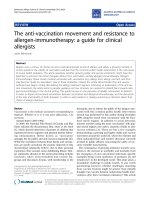
After induction of CtIA, a significant difference was observed
between the HgCl
2
-exposed rats compared with controls. The
rats with a prior exposure to HgCl
2
demonstrated significantly
more joint swelling compared with the normally mild swelling
of naïve BN rats using the same injection protocol. The mean
joint width in the HgCl
2
-exposed rats (n = 8) was 13.7 ± 0.53
mm in comparison with a control joint mean size (n = 8) of 9.6
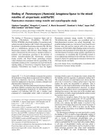
± 0.86 mm (P < 0.0001) (Figure 1a).
Histologically, two systemic injections of HgCl
2
induced no
pathological changes in the joint (Figure 2). The CtIA in the
non-HgCl
2
-exposed control rats was accompanied by only
modest synovial hypertrophy and hyperplasia (Figure 2). Syn-
ovial tissue cellular infiltration was pleomorphic, with mononu-
clear cells comprising the dominant population. Pannus
formation was minimal and there were only mild changes in
Figure 1
Joint swelling and histopathology scores demonstrating exacerbation of Chlamydia-induced arthritis following a prior systemic exposure to mercuric chloride (HgCl
2
) (n = 8 in each group)Joint swelling and histopathology scores demonstrating exacerbation of Chlamydia-induced arthritis following a prior systemic exposure to mercuric
chloride (HgCl
2
) (n = 8 in each group). The differences after HgCl
2
exposure are significant (P < 0.005) in both joint swelling (a) and histopatholog-
ical severity scores (b). Hg, mercury. Ct, Chlamydia trachomatis.
Arthritis Research & Therapy Vol 11 No 1 Inman and Chiu
Page 4 of 10
(page number not for citation purposes)
bone and cartilage. This is the pattern observed in BN rats in
our previous studies [2]. In contrast, the joints of the HgCl
2
-
exposed rats demonstrated more marked synovial hypertrophy
and hyperplasia (Figure 2). This was accompanied by massive
infiltration of the synovial tissues with a predominance of neu-
trophils. There were areas of necrosis and bone and cartilage
destruction by aggressive pannus formation. The invading
pannus could be seen invading subchondral bone. Thus, the
HgCl
2
exposure altered the mild arthritis characteristic of
resistant BN into the aggressive profile of the arthritis charac-
teristic of susceptible rats. When the histopathological scor-
ing system was applied, the joint scores of the HgCl
2
-exposed
test rats (n = 8) were significantly higher (average score of
99.28% ± 1.89% of maximum) than those of controls (average
score of 58.75% ± 7.44% of maximum) (P < 0.0001) (Figure
1b).
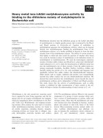
Figure 2
Histopathology (haematoxylin and eosin) in Chlamydia trachomatis-induced arthritis (CtIA) alone and after a prior exposure to mercuric chloride (HgCl
2
)Histopathology (haematoxylin and eosin) in Chlamydia trachomatis-induced arthritis (CtIA) alone and after a prior exposure to mercuric chloride
(HgCl
2
). CtIA in the non-HgCl
2
-exposed control rats is accompanied by only moderate synovial hypertrophy and hyperplasia and a mononuclear cell
infiltration (a-c). Eight animals were studied in each group. The joints of the HgCl
2
-exposed rats demonstrated more marked synovial hypertrophy
and hyperplasia and a marked infiltration of the synovial tissues, primarily by neutrophils. This is accompanied by bone and cartilage destruction and
by pannus formation, which invades subchondral bone (d-f). Panel (g) is representative of a rat that received the HgCl
2
injections alone and shows
no pathological change in the joint. Original magnifications × 200.
Available online />Page 5 of 10
(page number not for citation purposes)
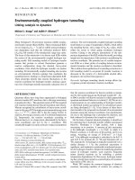
Serum IgE
There was a dramatic elevation of serum IgE levels (Figure 3)
in HgCl
2
-exposed animals (1.66 ± 0.05) compared with con-
trols (0.30 ± 0.07), representing a 4.5-fold increase with
HgCl
2
exposure (P < 0.0001) (n = 8 in each group).
Anti-Chlamydia antibodies
The HgCl
2
-exposed rats (n = 8) had significantly higher serum
IgG1 anti-Chlamydia antibodies (0.26 ± 0.05) than non-
exposed control rats (0.13 ± 0.03) (P < 0.001) (Figure 4a).
On the other hand, the control rats (n = 8) had slightly higher
(P = 0.12) IgG2a anti-Chlamydia antibodies (0.21 ± 0.02)
than the HgCl
2
-exposed rats (0.18 ± 0.03). The IgG2a to
IgG1 ratio was 1.57 for the control rats in contrast to 0.70 for
the HgCl
2
-exposed animals, consistent with a shift toward a
Th2 response after HgCl
2
exposure.
Anti-collagen
(II)
antibodies
The HgCl
2
-exposed BN rats (n = 8) had higher levels of IgG
antibodies to rat type II collagen than did controls: 0.108 ±
0.069 versus 0.032 ± 0.012 (P < 0.01). These serological
changes paralleled the enhanced severity of arthritis clinically
and histologically following HgCl
2
exposure (Figure 4b).
Cytokine profiles
After intra-articular injection with Chlamydia, there was an
increase in the serum levels of IFN-γ (n = 4) and TNF-α (n = 4)
(Figure 5a). These were significant increases compared with
normal rat serum for both IFN-γ (P < 0.005) and TNF-α (P <
0.001). There were no significant increases in serum IL-10 or
IL-4 after Chlamydia joint injection (n = 4). Prior HgCl
2
expo-
sure was associated with suppression of the expected rise in
serum IFN-γ and TNF-α seen with CtIA. The differences
between the HgCl
2
-exposed and non-exposed controls were
significant for IFN-γ (P = 0.002) and TNF-α (P = 0.014). No
comparable differences were detected for IL-4 or IL-10, but
the baseline levels were low in both cases. Inflamed synovial
tissues from Chlamydia-injected rats were harvested for
assays for tissue cytokine profiles using the dot-blot method
(Figure 5b). HgCl
2
exposure was accompanied by a decrease
in local cytokine production for IFN-γ (P < 0.005), TNF-α (P <
0.05), and IL-1 (P < 0.05). No significant difference was
observed for synovial IL-10.
Chlamydia clearance
HgCl
2
-exposed rats demonstrated a relative defect in host
clearance of the pathogen both locally and systemically (n = 5
in each group). The mean level of the splenic bacterial load
(measured in optical density units) for the HgCl
2
-exposed rats
was 0.092 ± 0.035 in contrast with 0.009 ± 0.003 for the con-
trols (Figure 6a). This clearance difference is statistically sig-
nificant (P < 0.005). In the joint, the average Chlamydia load
for the HgCl
2
-exposed rats was significantly higher (0.419 ±
0.264) than that of controls (0.028 ± 0.014) (P < 0.05) (Fig-
ure 6b). Immunohistology shows more intense staining with
anti-Chlamydia antibodies in the HgCl
2
-exposed rats (Figure
6c) than in controls (Figure 6d), reflecting the relative failure of
the HgCl
2
-exposed rat to achieve local clearance of the path-
ogen.
Nitric oxide and vascular endothelial growth factor
Serum NO was elevated (3.99 ± 0.83 μM) in CtIA compared
with serum from naïve rats (n = 4) (2.08 ± 0.47 μM). HgCl
2
alone had no impact on serum NO compared with naïve rats,
but there was an additive effect of Ct and HgCl
2
observed
(5.97 ± 1.58 μM; P < 0.001) (Figure 7a). Serum VEGF was
increased by exposure to HgCl
2
alone (56.67 ± 20.22 pg/mL)
in comparison with normal rat serum (9.21 ± 3.3 pg/mL), but
Ct alone did not elevate serum VEGF in comparison with nor-
mal rat serum (Figure 7b). In contrast, Ct and HgCl
2
had an
additive effect on the serum VEGF (110.56 ± 37.37 pg/mL; P
< 0.001 in comparison with normal rat serum). Four animals
were studied in each group. The elevation in NO and VEGF
was in contrast to some of the cytokine-suppressive effects of
HgCl
2
exposure, with serum levels of NO and VEGF rising
more with the combined exposure than to either exposure
alone. This corresponded to the most intense pathological
changes in the joints of animals with dual-exposure to both Ct
and HgCl
2
.
Discussion
The spondyloarthropathies (SpAs) refer to a group of diseases
that share several common features: association with HLA
class I genes, asymmetric oligoarthritis, axial involvement,
enthesitis, and characteristic extra-articular features such as
uveitis. The role of infection as a triggering factor is implicated
with varying degrees of certainty amongst the SpA subcatego-
Figure 3
The effect of prior exposure to mercuric chloride (HgCl
2
) on serum IgE levels in Chlamydia-induced arthritisThe effect of prior exposure to mercuric chloride (HgCl
2
) on serum IgE
levels in Chlamydia-induced arthritis. The serum IgE levels are signifi-
cantly increased compared with controls (P < 0.0001) (n = 8 in each
group). O.D., optical density.
Arthritis Research & Therapy Vol 11 No 1 Inman and Chiu
Page 6 of 10
(page number not for citation purposes)
ries, with ReA having the most clear evidence for a microbial
trigger [15]. The most direct causal evidence for microbial trig-
gers for ReA derives from a demonstration of microbial anti-
gens in the joint [16]. The case for intra-articular pathogens is
strongest for Chlamydia, and several investigators have used
polymerase chain reaction to demonstrate Chlamydia DNA or
RNA in the joints of patients with post-Chlamydia ReA [17-
19]. There is some evidence that these organisms are meta-
bolically altered and may have entered a quiescent phase
[20,21]. Several studies support the notion of a viable organ-
ism present, at least transiently, in the early stages of ReA. It is
speculated that this may reflect defective killing of the organ-
ism by failure either to internalize the pathogen or to effectively
initiate intracellular killing. There has been particular interest in
the Chlamydia heat shock proteins, notably heat shock protein
60 (hsp60), and recent studies have identified differential
expression of three C. trachomatis hsp60-encoding genes
that may differ in active versus persistent infections [22]. The
cellular response to Chlamydia infection has been studied by
microarray techniques, and 18 genes appeared to be selec-
tively upregulated following infection with C. trachomatis [23].
Profiling Chlamydia infection of U937 monocytic cells [24]
and human lung epithelial cells [25] has provided a profile of
induced gene expression and, in particular, which cytokines
are induced by this microbial challenge.
Analysis of cytokine profiles is a further method for studying
the link between infection and ReA. In a study of 11 patients
Figure 4
The effect of prior exposure to mercuric chloride (HgCl
2
) on serum anti-Chlamydia antibodies and anti-collagen antibodies in Chlamydia-induced arthritisThe effect of prior exposure to mercuric chloride (HgCl
2
) on serum anti-Chlamydia antibodies and anti-collagen antibodies in Chlamydia-induced
arthritis. (a) HgCl
2
-exposed rats had significantly higher serum IgG1 anti-Chlamydia antibodies than non-exposed control rats (P < 0.001). Control
rats had slightly higher IgG2a anti-Chlamydia antibodies than the HgCl
2
-exposed rats (P = 0.12). (b) HgCl
2
-exposed Brown Norway (BN) rats had
higher levels of IgG antibodies to rat type II collagen than controls (P < 0.01) (n = 8 in each group). O.D., optical density.
Figure 5
Serum and synovial cytokine profilesSerum and synovial cytokine profiles. (a) Serum cytokines in normal rats and in Chlamydia-induced arthritis alone or after prior exposure to mercuric
chloride (HgCl
2
). After the onset of Chlamydia trachomatis-induced arthritis (CtIA), there was a significant increase in the serum levels of interferon-
gamma (IFN-γ) (P < 0.005) and tumour necrosis factor-alpha (TNF-α) (P < 0.001). Prior HgCl
2
exposure was associated with significant suppres-
sion of IFN-γ (P = 0.002) and TNF-α (P = 0.014) seen with CtIA. (b) Synovial cytokines in Chlamydia-induced arthritis alone or after prior exposure
to HgCl
2
. HgCl
2
exposure was accompanied by a decrease in local cytokine production for IFN-γ (P < 0.005), TNF-α (P < 0.05), and interleukin (IL)-
1 (P < 0.05) compared with controls. No significant difference was observed for synovial IL-10 (n = 4 in each group).
Available online />Page 7 of 10
(page number not for citation purposes)
with ReA, it was observed that stimulation of SF mononuclear
cells resulted in secretion of low amounts of IFN-γ and TNF-α
but high amounts of IL-10 [26]. IL-10 was responsible for sup-
pression of IFN-γ and TNF-α as judged by the effect of adding
IL-10 or anti-IL-10 to the cells. The suppression of Th1-like
cytokines is likely mediated through suppression of IL-12 syn-
thesis. This IL-10/IL-12 balance, resulting in a predominance
of Th2 cytokines, may contribute to the persistence of bacteria
in the joint. In comparison with rheumatoid arthritis, SF levels
of TNF-α in ReA are lower despite comparable levels of IL-2
receptor, again implicating a relative deficiency of protective
antimicrobial cytokines in the local environment [27]. Analysis
of synovial fluid cytokines has been studied in patients with
Chlamydia-induced arthritis and it was found that B27
+
patients had lower SF IFN-γ levels and it was these patients
who had a more chronic course [28]. This suggests that dimin-
ished IFN-γ generation might account for the persistence of
the arthritis.
Our previous studies in experimental ReA have paralleled
these clinical findings. BN rats that are relatively resistant to
CtIA exhibit an enhanced IFN-γ and TNF-α expression in the
microenvironment of the joint. This is accompanied by
enhanced clearance of the pathogen and a more transient and
benign course of the arthritis. In the present study, we have
found that this inherent resistance to ReA can be overcome by
heavy metal exposure. Mercury exposure alters the cytokine
profile, host clearance capability, and histopathological out-
come from the resistant phenotype to the susceptible pheno-
type. Our findings contrast with the experience studying
murine collagen-induced arthritis, in which prior exposure to
HgCl
2
did not influence the outcome of the arthritis, but expo-
sure following the onset did have an aggravating effect on the
arthritis [29].
If HgCl
2
diminishes protective cytokines in the microenviron-
ment of the joint, what is driving the aggressive inflammation
and subsequent joint damage? Our study suggests that NO
and VEGF are two important candidates. We have previously
studied NO contribution to clearance of arthritogenic patho-
gens by synoviocytes and observed that in some instances
there was an IFN-γ-mediated suppression of NO in such cells,
Figure 6
Host clearance of Chlamydia from the spleen and the jointHost clearance of Chlamydia from the spleen and the joint. Tissues were harvested 5 days after the induction of Chlamydia trachomatis-induced
arthritis (CtIA). (a) The mean level of the splenic bacterial load for the mercuric chloride (HgCl
2
)-exposed rats was higher than that of controls (P <
0.005). (b) In the joint, the mean Chlamydia load for the HgCl
2
-exposed rats was significantly higher than that of controls (P < 0.05). Immunohistol-
ogy shows more intense staining with anti-Chlamydia antibodies in the HgCl
2
-exposed rats (c) than controls (d) (n = 5 in each group). Original mag-
nifications × 200. O.D., optical density.
Arthritis Research & Therapy Vol 11 No 1 Inman and Chiu
Page 8 of 10
(page number not for citation purposes)
suggesting a dynamic interaction between these two factors
[30]. This interaction may be specific to the cells under inves-
tigation since, in the case of J774 macrophages, IFN-γ medi-
ated an increase in NO production, which in turn reduced
viability of C. pneumoniae in the infected cells [31]. In our in
vivo model, the rise in NO was concurrent with an increase in
chlamydial load in the host, suggesting that the increase in NO
was not sufficient to contain an expanded replication profile of
the organism. In this regard, it is interesting to note that, in
murine cells, blockage of NO synthesis only partially rescues
chlamydial growth, suggesting that there are other important
IFN-γ-inducible antichlamydial mechanisms operative [32].
Prior studies on the effect of Hg on NO have yielded varying
results depending on the experimental conditions. Kim and
colleagues [33] found that Hg treatment of a macrophage cell
line results in a drop in NO production upon stimulation with
lipopolysaccharide. However, Huang and colleagues [34] dis-
covered that treatment of mice with Hg resulted in an increase
in serum NO levels. The latter results are very much in parallel
with our finding of an increase of NO following exposure to
HgCl
2
.
The pathological studies in our rats indicate that the exacerba-
tion of histological severity of the arthritis was accompanied by
a neovascularization process locally. It is known that VEGF
plays a crucial role in angiogenesis. Spondylarthritis is charac-
terized by enthesitis and synovitis, in which new blood vessels
participate in perpetuating the inflammation. Serum analysis
from SpA patients has documented that VEGF levels were sig-
nificantly higher in SpA patients than in controls. In SpA
patients, serum VEGF levels correlated with disease activity
indices as defined by the Bath Ankylosing Spondylitis Disease
Activity Index (BASDAI), erythrocyte sedimentation rate, or C-
reactive protein (CRP). These results suggest that VEGF and
therefore angiogenesis may play a role in SpA pathogenesis
and may serve as a disease activity marker in SpAs [35]. In
keeping with these findings, it has been observed that there is
a significant reduction in serum VEGF levels after the infliximab
treatment of SpA patients and that these changes correlate
with similar reductions in CRP and IL-6 [36]. Direct Chlamy-
dia-endothelial cell interactions are known to mediate induc-
tion of VEGF [37].
Mercury exists in elemental, inorganic, and organic forms. The
general population is exposed primarily to mercury vapour
from dental amalgam and to organic mercury from fish con-
sumption [38]. Mercury accumulates in the food chain such
that large fish such as tuna and swordfish have high concen-
trations of mercury in tissues [39]. Occupational exposure is
primarily to mercury vapour and occurs in dentistry, mining,
and the manufacture of electrical equipment. Mercury vapour
is well absorbed through the respiratory tract, but absorption
of elemental mercury is negligible orally whereas oral absorp-
tion of organic mercury is nearly complete [40]. There is recent
evidence from studies of human peripheral mononuclear cells
that low-dose exposure to mercury can polarize the immune
response toward Th2 [41].
The role of mercury in rheumatic diseases has received little
attention. One recent study implicated mercury exposure as a
risk factor for Wegener granulomatosis [42], but the mecha-
nism whereby mercury exposure could alter immune response
and set the stage for chronic inflammatory conditions has not
been resolved. Our experimental system demonstrates that
genetic susceptibility can be fundamentally altered by a heavy
Figure 7
Serum nitric oxide and VEGF profilesSerum nitric oxide and VEGF profiles. Serum nitric oxide (NO) levels (a) and vascular endothelial growth factor (VEGF) levels (b) with the local expo-
sure to Chlamydia trachomatis (Ct) or a systemic exposure to HgCl
2
(Hg) or both (Ct + Hg) in contrast to normal rat serum. For both NO and VEGF,
the increment seen with dual-exposure to Ct and HgCl
2
was statistically greater than that seen with either Ct or HgCl
2
alone (n = 4 in each group).
HgCl
2
, mercuric chloride.
Available online />Page 9 of 10
(page number not for citation purposes)
metal exposure. This suggests that several environmental fac-
tors may act in concert in the pathogenesis of ReA. The imme-
diate precipitating factor may be a bacterial infection, but this
occurs in the context of other factors in the environment which
influence the immune repertoire. The plasticity of the immune
response, even when encoded in its basic elements by herita-
ble factors, is highlighted in this genetic-environment interac-
tion. As significant advances in the genetic basis of rheumatic
diseases are being made, it will be a challenge to address the
environmental factors with equal rigour.
Conclusion
Genetically defined cytokine production in the joint defines the
severity of ReA by dictating the local clearance of the patho-
gen. This interplay can be dramatically altered by heavy metal
exposure, which results in suppression of protective cytokines
in the microenvironment of the joint.
Competing interests
The authors declare that they have no competing interests.
Authors' contributions
RDI and BC both contributed to the design and execution of
the study, to the data analysis, and to the writing of the manu-
script. Both authors read and approved the final manuscript.
Acknowledgements
The source of funding for this article was the Canadian Institutes of
Health Research.
References
1. Inman RD, Chiu B: Synoviocyte-packaged Chlamydia trachom-
atis induces a chronic aseptic arthritis. J Clin Invest 1998,
102:1776-1782.
2. Inman RD, Chiu B: Early cytokine profiles in the joint define
pathogen clearance and severity of arthritis in Chlamydia-
induced arthritis in rats. Arthritis Rheum 2006, 54:499-507.
3. Fournié GJ, Cautain B, Xystrakis E, Damoiseaux L, Mas M,
Lagrange D, Bernard I, Subra JF, Pelletier L, Druet P, Saoudi A:
Cellular and genetic factors involved in the difference between
Brown Norway and Lewis rats to develop respectively type-2
and type-1 immune-mediated diseases. Immunol Rev 2001,
184:145-160.
4. Fournie GJ, Saoudi A, Druet P, Pelletier L: Th2-type immun-
opathological manifestations induced by mercury chloride or
gold salts in the rat: signal transduction pathways, cellular
mechanisms and genetic control. Autoimmun Rev 2002,
1:205-212.
5. Gorrie MJ, Qasim FJ, Whittle CJ, Gillespie KM, Szeto CC, Nicoletti
F, Bolton EM, Bradley JA, Mathieson PW: Exogenous type-1
cytokine modulate mercury-induced hyper-IgE in the rat. Clin
Exp Immunol 2000, 121:17-22.
6. Kiely PDW, Thiru S, Oliveira DBG: Inflammatory polyarthritis
induced by mercuric chloride in the Brown Norway rat. Lab
Invest 1995, 73:284-293.
7. Qasim FJ, Thiru S, Mathieson PW, Oliveira DB: The time course
and characterization of mercuric chloride-induced immunopa-
thology in the Brown Norway rat. J Autoimmun 1995,
8:193-208.
8. Kosuda LL, Greiner DL, Bigazzi PE: Mercury induced renal
autoimmunity in BN to Lew.1N chimeric rats. Cell Immunol
1994, 155:77-94.
9. White KL, David DW, Butterworth LF, Klykken PC: Assessment
of autoimmunity-inducing potential using the brown Norway
rat challenge model. Toxicol Lett 2000, 12–113:443-451.
10. Prouvost-Danon A, Abadie A, Sapin C, Bazin H, Druet P: Induc-
tion of IgE synthesis and potentiation of anti-ovalbumin IgE
antibody response by HgCl2 in the rat. J Immunol 1981,
126:699-792.
11. Prigent P, Saoudi A, Pannetier C, Graber P, Bonnefoy JY, Druet P,
Hirsch F:
Mercuric chloride, a chemical responsible for T
helper cell (Th)2-mediated autoimmunity in brown Norway
rats, directly triggers T cells to produce interleukin-4. J Clin
Invest 1995, 96:1484-1489.
12. Gillespie KM, Saoudi A, Kuhn J, Whittle CJ, Druet P, Bellon B,
Mathieson PW: Th1/Th2 cytokine gene expression after mer-
curic chloride in susceptible and resistant rat strains. Eur J
Immunol 1996, 26:2388-2392.
13. Roos A, Claessen N, Weening JJ, Aten J: Enhanced T lym-
phocyte expression of LFA-1, ICAM-1, and the TNF receptor
family member OX40 in HgCl
2
-induced systemic autoimmu-
nity. Scand J Immunol 1996, 43:507-518.
14. Roos A, Claessen N, Schilder-Tol EJ, Weening JJ, Aten J: Differ-
ential expression of T-cell adhesion molecules and LFA-1
dependent intercellular adhesion in the HgCl2-induced
autoimmunity and immune suppression. Scand J Immunol
1998, 48:389-396.
15. Inman RD, Perl A: Infectious agents in chronic rheumatic dis-
eases. In Arthritis and Allied Conditions: A Textbook of Rheuma-
tology 15th edition. Edited by: Koopman WJ. Philadelphia:
Lippincott Williams & Wilkins; 2005:647-678.
16. Hannu H, Inman RD, Granfors K, Leirisalo-Repo M: Reactive
arthritis or postinfectious arthritis. Best Pract Res Clin Rheum
2006, 20:419-433.
17. Kuipers JG, Jurgen-Saathoff B, Bialowons A, Wollenhaupt J, Koh-
ler L, Zeidler H: Detection of Chlamydia trachomatis in periph-
eral blood lymphocytes of reactive arthritis patients by
polymerase chain reaction. Arthritis Rheum 1998,
41:1894-1895.
18. Branigan PJ, Gerard HC, Hudson AP, Schumacher HR, Pando J:
Comparison of synovial tissue and synovial fluid as the source
of nucleic acids for the detection of Chlamydia trachomatis by
polymerase chain reaction. Arthritis Rheum 1996,
39:1740-1746.
19. Bas S, Griffais R, Kvien TK, Glennas A, Melby K, Vischer TL:
Amplification of plasmid and chromosome Chlamydia DNA in
synovial fluids of patients with reactive arthritis an undifferen-
tiated seronegative spondyloarthropathies. Arthritis Rheum
1995, 38:1005-1013.
20. Gerard HC, Branigan PJ, Schumacher HR, Hudson AP: Synovial
Chlamydia trachomatis in patients with reactive arthritis/
Reiter's syndrome are viable but show aberrant gene expres-
sion. J Rheumatol 1998, 25:734-742.
21. Nanagara R, Li F, Beutler AM, Hudson A, Schumacher HR: Alter-
ation of Chlamydia trachomatis biological behavior in synovial
membranes: suppression of surface antigen production in
reactive arthritis. Arthritis Rheum 1995, 38:1410-1417.
22. Gerard HC, Whittum-Hudson JA, Schumacher HR, Hudson AP:
Differential expression of three Chlamydia trachomatis hsp60-
encoding genes in active vs persistent infections. Microb
Pathog 2004, 36:35-39.
23. Hess S, Rheinheimer C, Tidow F, Bartling G, Kaps C, Lauber J,
Buer J, Klos A: The reprogrammed host: Chlamydia trachoma-
tis-induced upregulation of glycoprotein 130 cytokines, tran-
scription factors and antiapoptotic genes. Arthritis Rheum
2001, 44:2392-2401.
24. Virok D, Loboda A, Kari L, Nebozhyn M, Chang C, Nichols C,
Endresz V, Gonczol E, Berencsi K, Showe MK, Showe LC: Infec-
tion of U937 monocytic cells with Chlamydia pneumoniae
induces extensive changes in host cell gene expression. J
Infect Dis 2003, 188:1310-1321.
25. Yang J, Hooper WC, Phillips DJ, Tondella ML, Talkington DF:
Induction of proinflammatory cytokines in human lung epithe-
lial cells during Chalmydia pneumoniae infection. Infect Immun
2003, 71:614-620.
26. Yin Z, Braun J, Neure L, Wu P, Liu L, Eggens U, Sieper J: Crucial
role of interleukin-10/interleukin-12 balance in the regulation
of the type 2 T helper cytokine response in reactive arthritis.
Arthritis Rheum 1997, 40:1788-1797.
27. Steiner G, Studnicka-Benke A, Witzmann G, Hofler E, Smolen J:
Soluble receptors for tumor necrosis factor and interleukin-2
in serum and synovial fluid of patients with rheumatoid arthri-
Arthritis Research & Therapy Vol 11 No 1 Inman and Chiu
Page 10 of 10
(page number not for citation purposes)
tis, reactive arthritis, and osteoarthritis. J Rheumatol 1995,
22:406-412.
28. Bas S, Kvien TK, Buchs N, Fulpius T, Gabay C: Lower level of
synovial fluid IFN-γ in HLA-B27-positive than in HLA-B27-neg-
ative patients with Chlamdyia trachomatis reactive arthritis.
Rheumatology (Oxford) 2003, 42:461-467.
29. Hansson M, Djerbi M, Rabbani H, Mellstedt H, Gharibdoost F,
Hassan M, Depierre JW, Abedi-Valugerdi M: Exposure to mercu-
ric chloride during the induction phase and after the onset of
collagen-induced arthritis enhances immune/autoimmune
responses and exacerbates the disease in DBA/1 mice.
Immunology 2005, 114:428-437.
30. Inman RD, Payne U: Determinants of synoviocyte clearance of
arthritogenic bacteria. J Rheumatol 2003, 30:1291-1297.
31. Carratelli CR, Rizzo A, Paolillo R, Catania MR, Catalanotti P, Ros-
sano F: Effect of nitric oxide on the growth of Chlamydophila
pneumoniae. Can J Microbiol 2005, 51:941-947.
32. Roshick C, Wood H, Caldwell HD, McClarty G: Comparison of
gamma interferon-mediated antichlamydial defense mecha-
nisms in human and mouse cells. Infect Immun 2006,
74:225-238.
33. Kim SH, Johnson VJ, Sharma RP: Mercury inhibits nitric oxide
production but activates proinflammatory cytokine expression
inmurine macrophage: differential modulation of NF-κB and
p38 MAPK signaling pathways. Nitric Oxide 2002, 7:67-74.
34. Huang CF, Liu SH, Lin-Shiau SY: Neurotoxicological effects of
cinnabar (HgS) in mice. Toxicol Appl Pharmacol 2007,
224:192-201.
35. Drouart M, Saas P, Billot M, Cedoz JP, Tiberghien P, Wendling D,
Toussirot E: High serum vascular endothelial growth factor
correlates with disease activity of spondylarthropathies. Clin
Exp Immunol 2003, 132:158-162.
36. Visvanathan S, Wagner C, Marini JC, Baker D, Gathany T, Han J,
van der Heijde D, Braun J: Inflammatory biomarkers, disease
activity, and spinal disease measures in patients with ankylos-
ing spondylitis after treatment with infliximab. Ann Rheum Dis
2008, 65:511-517.
37. Carratelli CR, Paolillo R, Rizzo A:
Chlamydia pneumoniae stimu-
lates the proliferation of HUVEC through the induction of
VEGF by THP-1. Int Immunopharmacol 2006, 7:287-294.
38. Brodkin E, Copes R, Mattman A, Kennedy J, Kling R, Yassi A: Lead
and mercury exposures: interpretation and action. CMAJ
2007, 176:59-63.
39. Bureau of Chemical Safety, Food Directorate, Health Products and
Food Branch (Government of Canada): Human Health Risk
Assessment of Mercury in Fish and Health Benefits of Fish Con-
sumption 2007:1-70 [ />merc_fish_poisson-eng.php]. Ottawa, ON: Health Canada
40. Mozaffarian D, Rimm EB: Fish intake, contaminants, and human
health: evaluating the risks and the benefits. JAMA 2006,
296:1885-1899.
41. Hemdan NY, Lehmann I, Wichmann G, Lehmann J, Emmrich F,
Sack U: Immunomodulation by mercuric chloride in vitro:
application of different cell activation pathways. Clin Exp
Immuno1 2007, 148:325-337.
42. Albert D, Clarkin C, Komoroski J, Brensinger CM, Berlin JA: Wege-
ner's granulomatosis: possible role of environmental agents in
its pathogenesis. Arthritis Rheum 2004, 51:656-664.