Báo cáo Y học: Dystrobrevin requires a dystrophin-binding domain to function in Caenorhabditis elegans doc
Bạn đang xem bản rút gọn của tài liệu. Xem và tải ngay bản đầy đủ của tài liệu tại đây (192.74 KB, 6 trang )
Dystrobrevin requires a dystrophin-binding domain to function
in
Caenorhabditis elegans
Karine Grisoni, Kathrin Gieseler and Laurent Se
´
galat
CGMC, CNRS-UMR, Universite
´
Lyon, Villeurbanne, France
Dystrobrevin is one of the intracellular components of the
transmembrane dystrophin–glycoprotein complex (DGC).
The functional role of th is complex in normal an d patho-
logical situations has not yet been clearly established.
Dystrobrevin disappears from t he muscle m embrane in
Duchenne muscular dystrophy (DMD), which results from
dystrophin mutations, as well as in limb girdle muscular
dystrophies (LGMD), which results f rom mutations affect-
ing other members of the DGC complex. These findings
therefore s uggest that dystrobrevin may play a pivotal role in
the progression of these clinically related diseases. In this
study, w e u sed t he Ca enorhabditis elegans mod el t o a ddress
the question of the relationship between dystrobrevin
binding to dystrophin and dystrobrevin function. Deletions
of the dystrobrevin protein w ere performed and the ability
of the mutated forms to bind t o dystrophin was tested both
in vitro and in a two-hybrid assay, as well as their ability t o
rescue dystrobrevin (dyb-1) mutations in C. elegans. The
deletions affecting the second helix of the D yb-1 coiled-coil
domain abolished the binding of dystrobrevin to dystrophin
both in vitro and in the two-hybrid assay. These deletions
also abolished t he rescuing activity of a functional transgene
in vivo. These r esults are consistent with a model according to
which dystrobrevin must bind to dystrophin to be able t o
function properly.
Keywords: dystrophin; dystrobrevin; nematode; Caeno-
rhabditis elegans.
Duchenne muscular dystrophy (DMD) is an inherited
muscular disease in which the patients’ muscles gradually
degenerate. So f ar, n o t reatment exists for DMD. The
disease i s c aused b y mutations affecting t he dystrophin gene,
which encodes a 3685-amino-acid protein (reviewed in [1]).
Dystrophin is a submembrane protein associated with a
transmembrane dystrophin–glycoprotein complex (DGC)
comprising dystroglycans, sarcoglycans, sarcospan, syntro-
phins and dystrobrevins [1–3]. DGC pro teins have attracted
an increasing amount of attention over the last few years,
because they might help to explain the physiopathology of
the disease, and may also provide therapeutic clues.
Dystrobrevins form a family of proteins that are uniqu e
in that they are both dystrophin-associated proteins, and
homologous to the C-terminal region of dystrophin. Alpha-
dystrobrevin was originally identified as a molecule that
copurifies with nicotinic acetylcholine receptors in sucrose
gradients [4,5]. I t was later recognized as one of the proteins,
which associates with dystrophin to form the dystrophin–
glycoprotein complex (DGC) [4,6,7]. A second dystro-
brevin, b-dystrobrevin, is mainly expressed in n erve tissues
[8,9]. Mice carrying a knockout mutation of the
a-dystrobrevin gene (adbn mice) suffer from a cardiac and
skeletal muscle myopathy reminiscent o f dystrophin (mdx )
mutations [10].
a-Dystrobrevin binds to dystrophin via a coiled-coiled
motif present in both proteins, and to the PDZ domain
containing syntrophins [11,12]. Indirect evidence suggests
that dystrobrevin may also bind to other members of the
DGC [13]. Although no enzymatic activity has yet been
assigned to dystrobrevins, there a re several i ndications that
they may play a role in signalling mechanisms. First,
dystrobrevins are tyrosine-phosphorylated proteins [5,14].
Secondly, in the absence of a-dystrobrevin, the signalling
molecule, neuronal nitric o xide synthase (nNO S) disappears
from the muscular membrane [10].
In addition, two lines of evidence suggest that dystro-
brevin may p lay a key role i n the muscle degeneration
observed in DMD and sarcoglycanopathies; first, dystro-
brevin immunostaining decreases greatly in DMD and in
several sarcoglycanopathies [15]. Secondly, although t he
DGC components (with the exception of NOS) are not
affected by the absence of dystrobrevin in adbn mice, musc le
degeneration occurs.
The nematode Caenorhabditis elegans has homologues of
most of the DGC proteins (L. Se
´
galat, unpublished results).
There is one dystrophin- and one dystrobrevin-like gene in
thegenomeofC. elegans (dys-1 and dyb-1, respectively)
[16,17]. C. elegans dystrophin and dystrobrevin are able t o
bind to each other in vitro [18] in the same way as their
mammalian counterparts [12], and they also bind to
syntrophin [18]. dys-1 and dyb-1 mutants d isplay a similar
behavioural phenotype c onsisting of hyperactivity, exagger-
ated bending of the head when moving forward, and a
tendency t o hypercontract [16,17]. In addition, progressive
muscle degeneration is observed when dys-1 or dyb-1
Correspondence t o L. Se
´
galat, CGMC, Universit e
´
Lyon1,43blddu11
Novembre, 69622 Villeurbanne cedex, France.
Fax: + 3 3 4 72 44 05 55, Tel.: + 33 4 72 43 29 51,
E-mail:
Abbreviations: DGC, dystrophin–glycoprotein complex;
DMD, Duchenne muscular dystrophy; LGMD, limb girdle muscular
dystrophy; nNOS, neuronal nitric oxide synthase; AD, activation
domain; DNA-BD, DNA binding domain; SD, synthetic dropout
medium; SBR, syntrophin binding region.
(Received 1 6 October 200 1, revised 1 0 January 2002 , accepted
11 January 2002)
Eur. J. Biochem. 269, 1607–1612 (2002) Ó FEBS 2002
mutations are introduced in a sensitized hlh-1(cc561)
genetic background that makes C. elegans muscles fragile
[19,20].
In this study, we addressed the questio n as to whether the
ability o f dystrobrevin to function properly may depend on
its association with dystrophin. First, we refined the
dystrophin-binding region on dystrobrevin (Dyb-1) by
performing deletion-mapping experiments in vitro.Wethen
tested the ability of t he truncated Dyb-1 proteins to bind to
dystrophin (Dys-1) in a yeast two-hybrid assay, as well as
their ability to rescue dyb-1 mutants.
EXPERIMENTAL PROCEDURES
Construction of deleted forms of Dyb-1
for
in vitro
binding experiments
Deletions were carried out on the dyb-1 coding sequence,
using clone AN450 [encoding Dyb-1 amino acids 3 90–543
fused in frame to the GST coding sequen ce; plasmid pGEX
3X (Pharmacia)] [18]. AN450 DNA (500 ng) was cut with
the restriction enzyme MfeI. The cut DNA was then
distributed among several tubes incubated with 0.05 lLof
BAL31 exonuclease for various times (typically 0–10 min).
The r eactions were stopped by adding EGTA to 4 m
M
and
heating a t 6 5 °C f or 10 min. DNA was purified on a Wizard
column (Promega) and the action of BAL31 was checke d by
loading an aliquot o f each tube onto a n agarose gel column.
The DNA corresponding to the deletions required was
treated b y applying T4 DNA polymerase in the presence of
nucleotides to create blunt ends, w hich were ligated and the
plasmids were transformed in Escherichia coli DH5. Clones
were picked randomly and analysed using sequencing
procedures. Any clones carrying a frame shift were rejected.
Construct 6¢4 was built using similar procedures, but using
the enzyme HindIII instead of MfeI. The amino acids
removed in the deletions were 489–499 (clone 2¢5), 487–513
(clone 5 ¢1), 489–528 ( clone 5 ¢2B), 471–517 (clone 5 ¢5B), 478–
543 ( clone 2 ¢1), and 391–450 (clone 6¢4). Clones 2¢1and6¢4
have been described previously [18], but clo ne 2¢1was
erroneously reported t o be deleted in amino a cids 478–521.
This correction make s no difference to the interpretation of
our previous data.
In vitro
interactions
Constructs were transformed into the E. coli strain BL21
DE3 and the fusion proteins were produced as follows.
After cell sonication and centrifugation, the supernatant
was loaded onto glutathione–Sepharose beads (Pharma-
cia). Approximately 20 lg of resin bound proteins were
washed in binding buffer [Hepes 20 m
M
pH 7.4, KOAc
110 m
M
,NaOAc5m
M
,Mg(OAc)
2
2.5 m
M
, NP40 0.05%,
dithiothreitol 1 m
M
, leupeptin 10 mgÆmL
)1
, aprotinin
10 mg ÆmL
)1
, pepstatin 10 mgÆmL
)1
, phenylmethanesulfo-
nyl fluoride (1 mM)]. The
35
S-labelled Dys-1 C-terminal
end was synthesized using a coupled in vitro transcription
and translation kit (Promega) with cDNA yk12c11 [16].
The p reparations were incub ated for 2 h at 4 °C. GST
controls were performed using 1–2 times the amount of
fusion protein. After five washes with binding buffer, the
labelled proteins were e luted by boiling t he preparation f or
3 min in gel loading buffer. Gels were dried, exposed
overnight and revealed using a radiographic analyser
(Fuji BAS-1500). Band intensity was quantitated using
the analyser software on at least three independent
experiments.
Constructs for the yeast two-hybrid assay
The C-terminal end of Dys -1 (amino a cids 2857–3674) was
fused t o t he DNA binding domain (DNA-BD) of t he Gal4
protein. For t his purpose, a 2,4 kb dys-1 cDNA fragment
(yk12c11) was cloned into the polylinker of pAS2-1
(Clontech) with respect to the reading frame.
Dyb-1 fragments and deletions were PCR a mplified using
clones AN 450, 2¢5, 5¢1, 5¢2B, 5 ¢5B and 6¢4inpGEX3Xas
templates (see b elow) and cloned into pACT2 (Clontech) in
frame w ith the activation domain ( AD) of the Gal4 protein
and the HA epitope. The resulting constructs were called
AD-AN450, AD-2¢5, AD-5¢1, AD-5¢2B, AD-5¢5B, and
AD-6¢4, respectively. All DNA constructs were checked by
performing DNA sequencing.
Yeast two-hybrid analysis
Construct DNA-BD-Dys-1 was transformed into the yeast
strain CG 1945 using the LiAc transformation procedure
(Clontech, Yeast protocols Handbook, PT 3024-1). Trans-
formants were selected on synthetic dropout (SD) media
(Clontech) minus tryptophan.
A DNA-BD-Dys-1 expressing yeast strain was selected
and transformed with plasmids AD-AN450, AD-2¢5,
AD-5¢1, AD-5¢2B, AD-5¢5B, or AD-6¢4. Transformants
were selected on SD media minus tryptophan and leucin.
Interactions between the DNA-BD-Dys-1 protein and t he
various forms of the AD-Dyb-1 fusion proteins were
analysed on the basis of transactivation of the HIS3
reporter gene a fter 3 days of growth on SD medium devoid
of tryptophan, leucin and histidin. A strain carrying both
the D NA-BD-Dys-1 p rote in a nd t he empty pAC T2 plasmid
was used as a negative control.
Western blots with yeast protein extracts
For Western blot analysis, yeast protein extracts were
prepared from strains carrying both DNA-BD-Dys-1
plasmids and AD-Dyb-1 plasmids (AN450, AD-2¢5,
AD-5¢1, AD-5¢2B, AD-5¢5B, or AD-6¢4). Overnight cul-
tures (5-mL) were prepared in SD media minus trypto-
phan and leucin. The next day, 1 mL of o vernight culture
was transferred into 10 mL of YPD medium. The diluted
culture w as i ncubated f or several hours at 30 °C
until D
600
¼ 0.3 for 1 mL. Cells (3 D
600
units) were
spun down and frozen at )70 °C for at least one hour.
The yeast pellet was resuspended in 60 lLofsample
buffer [21]. After boiling the mixture for 5 min, and
centrifuging f or 30 s a t 1 3 000 g,10lL of supernatant
was loaded onto each lane of a 0.1% SDS/10%
polyacrylamide gel. Proteins were transferred onto a
BA83 nitrocellulose membrane (Schleicher & Schuell) in
transfer buffer (Tris 25 m
M
, glycine 190 m
M
, SDS 0.01%,
ethanol 20%) for 1 h at 100 V. AD-Dyb-1 fusion proteins
were detected using a rabbit polyclonal anti-(Dyb-1) Ig
[19] at a dilution 1 : 500. Peroxidase-coupled anti-(rabbit
IgG) Ig (Biorad) was used at a dilution of 1 : 3000. Blots
1608 K. Grisoni et al. (Eur. J. Biochem. 269) Ó FEBS 2002
were revealed using the ECL+ kit (Amersham) as
recommended by the supplier.
Functional assay in
C. elegans
First, a dyb-1 functional construct was obtained by m odi-
fying a previously built dyb-1:gfp construct [19]. The
dyb-1:gfp construct, which has been previously described,
was shortened on the 5¢ end to leave 2.9 kb of upstream
sequence, and various restriction e nzyme s ites were removed
and a dded by performing synonymous point mutations to
yield the construct dyb-1:gfp VII, which h as single Age Iand
MluI sites at codons 390 and 543. This co nstruct encod es a
functional Dyb-1 gene as it can rescue dyb-1 muta tions ( data
not shown).
Secondly, Dyb-1 construct AN450 and the deletion
derivatives described above were transferred from pGEX
into dyb-1:gfp VII using PCR-amplifying procedures with
primers carrying AgeIandMluI sites, and cloned into the
single AgeIandMluI sites of dyb-1:gfp VII (Fig. 5).
Positives c lones were c hecked by determining their seq-
uence. dyb-1:gfp V II and constructs carrying e ither A N450
or the deletions were injected at a concentration of
1ngÆlL
)1
along with the transformation marker KP13
[22] using standard procedures [23] into worms carrying the
putative null allele dyb-1(cx36) [17]. Transgenic strains were
grown at 23 °C.
RESULTS
Mapping of the dystrophin-binding site on Dyb-1
The results of a previous study suggested that the Dys-1-
binding region on Dyb-1 w as located in the second helix of
the predicted coiled-coil domain [18]. We refined this
analysis by creating additional deletions by random muta-
genesis and testing their affinity for Dys-1. C lones 2¢5, 5¢1,
5¢2B, a nd 5¢5B were obtained by inducing exonuclease
digestion of the referen ce clone AN450, which encodes t he
amino acids 390–543 of Dyb-1 fused to the GST protein
[18]. These four clones contain various breakpoints within
the second helix of the predicted coiled-coil domain (H2)
(Fig. 1). The d eleted amino acids were 489–499 (clone 2¢5),
487–513 (clone 5¢1), 489–528 (clone 5¢2B) and 471–517
(clone 5¢5B). Clone 2¢1, lacking amino acids 478–543, was
used as a negative control [18]. Clone 6¢4, lacking amino
acids 391–450, was used as a second positive control [18].
The constructs were used to produce Dyb-1–GST chimeric
proteins in E. coli, which were affinity purified on gluthati-
one–Sepharose beads and subjected to in vitro binding with
35
S-labelled Dys-1. Clones 2¢5and5¢2B bound to Dys-1 at
levels that were not significantly different from those of the
positive controls AN450 (Fig. 2) and 6¢4 (gel not shown). In
contrast, the binding activity of clones 5¢1and5¢5B was
weaker (Fig. 2). The difference between clones 2¢5, 5¢1and
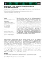
Fig. 1. Deletions used in this s tudy. The ‘WT’ line represents the amino-
acid sequence of the wild-type Dyb-1 protein in the predicted coiled-
coil domain region. The predicted helices forming the domain are
shown by hatched boxes. Numbers above the wild-type sequence
indicate the amino-acid coordinates of t he helices. Deletions are s hown
below the wild-type se quenc e. Numbers i ndicate the coordinates o f the
breakpoints. D eletions we re ge ne rated b y e xonuclease digestion . No te
that the 6¢4 de letion extends on the left side further tha n sh own on the
drawing. For in vitro binding experiments, the corresponding DNAs
were cloned into the pGEX vector to produce Dyb-1–GST fusion
proteins [18]. T he righ t column g ives t he binding affinity of the v arious
constructs to
35
S-labelled Dys-1 in arbitrary units (mean ± SD). One
unit is defined as t he autoradiogram intensity obtained with the neg-
ative control GST. Asterisks indicate values significantly different from
wild-type. Constructs 5¢1, 5¢5B and 2¢1 have significantly reduced
affinity to Dys-1.
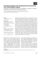
Fig. 2. In vitro binding of Dyb-1 (dystrobrevin) to Dys-1 (dystrophin).
Representative example of in vitro binding experiments. The same gel is
shown in Coomassie staining (top) and autoradiography (bottom).
The gel was loaded with various GST–Dyb-1 fusion proteins (and
GST alone) after inc ubation with equ al amounts of in vitro translated
35
S-labelled DYS-1. The signal intensity of the autoradiogram was
quantitated with a radiographic analyser (Biorad). MW, Molecular
mass markers. T, aliquot of the in vi tro translation product.
Ó FEBS 2002 Dystrophin–dystrobrevin interactions in C. elegans (Eur. J. Biochem. 269) 1609
5¢2B is of interest because these clones have left breakpoints
differing b y only two amino a cids. Although clones 2¢5and
5¢2B (cutting at position 489) display a wild-t ype p attern of
binding behaviour, clone 5¢1 (cutting at position 487) does
not. Therefore, the third heptad repeat of the second helix of
Dyb-1 (amino acids 484–490) seems to be critical for proper
Dys-1 binding to occur in vitro.
Dys-1/Dyb)1 interactions in the yeast two hybrid assay
Next, we t ested the ab ilit y of t he various forms of Dyb-1 t o
interact with Dys-1 in a two-hybrid assay. The Dyb-1
control and mutant clones w ere fused to the activation
domain of the Gal4 yeast transcription factor and were
tested against the entire C-terminal end of Dys-1 (amino
acids 2857–3674) fused t o the DNA-binding domain of
Gal4. The expression of wild-type and truncated proteins
was checked using the Western blotting procedure. This
confirmed that all the fusion proteins were correctly
expressed and that the experiment was not biased by any
differences in the protein expression levels (Fig. 3 ). Among
the six constructs tested , only the wild-type Dyb-1 fragment
and t he 6 ¢4 fragment resulted i n t he gr owth of yeasts on His -
plates, which can occur only if Dyb-1 binds to Dys-1
(Fig. 4). Similar r esults were obtained when a shorter Dys-1
fragment (amino acids 3402–3674) encompassing the syn-
trophin-binding domain and the coiled-coil domain (cor-
responding to BB810 in [18]) was used (not shown). These
results indicate that all the deletions affecting the second
helix of Dyb-1, including the shortest deletion (clone 2¢5),
greatly reduce t he interactions between Dys-1 and D yb-1 in
the yeast system.
Functional complementation of Dyb-1 deletions
in
C. elegans
The only functional assay available for dystrobrevin resides
in functional complementation. To test whether the dele-
tions of various parts of the coiled-coil domain had an effect
on the in vivo function of Dyb-1, we created transgenes
carrying the s ame deletions as those tested in v itro andinthe
yeast system. We transferred the deletions into the vector
dyb-1:gfp V II, w hich is a functional transgene consisting of
genomic Dyb-1 sequences (Fig. 5). Because the deletions
are derivatives of clone AN450, a cDNA fragment that
encompasses several exons, it was necessary first to check
whether removing introns 7 and 8 had any effect on the
rescuing activity of dyb-1:gfp VII. When the 1.2-kb AgeI–
MluI genomic fragment of dyb-1:gfp VII was replaced by
the 450 -bp A N450 cDNA fragment encoding the same
amino acids (Fig. 5), rescue of dyb-1(cx36) animals still
occurred in t wo out of thre e lines transgenic lines (Table 1),
which i ndicates that r emoving i ntrons 7 and 8 did not impair
the rescuing capacit y of dyb -1:gfp VII. The n we teste d the
various deletions. T hree out of the four lines obtained w ith
deletion 6¢4 showed consistent, a lthough only p artial, r escue
(Table 1). In these lines, the dyb-1 behavioural phenotype
(head bending and hyperlocomotion) was intermediate
between mutant and wild-type. This indicates that, although
deleting the syntrophin binding region (SBR) and the first
helix reduces the activity of the protein, it remains partly
functional. Five lines were obtained with deletion 2¢5(the
shortest deletion affecting the second helix); only one out of
the five lines tested show ed a w eak rescuing e ffect, which was
far less c onspicuous than that observed with construct 6¢4
(Table 1). Worms carrying the remaining constructs (5¢1,
5¢2B and 5 ¢5B) did not display any visible rescue. All in all,
these data indicate that deletions affecting t he second helix
of the Dyb-1 coiled-coil domain strongly decrease or abolish
the functional p roperties of D yb-1.
DISCUSSION
The aim of this study was twofold: first, to confirm and
refine th e localization of D ys-1 binding sites o n Dyb-1, and
secondly to test whether there may exist a correlation
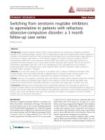
Fig. 3. Western blot of D yb-1 deletions produced i n yeast. Western blots
were prepared with protein extracts of yeast carrying DN A-BD-Dys-1
plasmids and empty pACT2 (lane 1), AD-AN450 (lane 2), AD-2¢5
(lane 3), AD-5¢1(lane4),AD-5¢2B (lane 5), AD-5¢5B (lan e 6) a nd
AD-6¢4 (lane 7). Th e blot was probed w ith mouse monoclonal
antibodies directed against the HA epitope situated between the
C-terminal end of the activation domain of the Gal4 protein and
the various Dyb-1 proteins. Molecular mass standards are shown on
the right.
Fig. 4. Yeast two-hybrid assay. A plate containing SD media minus
leucin, t ryp tophan and histidine was seeded with yeast c arrying DNA-
BD-Dys-1 and various AD-Dyb-1 p lasmids or empty pACT2 (as a
negative control), an d incubated at 30 °C for 3 days. Gro wth on m edia
devoid of histidine can occur only if D ys-1 and Dyb-1 interact and the
HIS3 reporter gene is transactivated. Only t he wild-type construct A D-
AN450 and the AD-6¢4 construct promoted growth in this assay. The
other constructs, all carrying deletions in the second h e lix of the Dyb-1
coiled-coil domain (H2), were unable t o p romote gr owth in this assay,
which indicates that the H2 helix is a critical prerequisite for Dys-1/
Dyb-1 interactions to b e possible.
1610 K. Grisoni et al. (Eur. J. Biochem. 269) Ó FEBS 2002
between th e binding of dystrobrevin to dystrophin and
functional activity of dystrobrevin. The C. elegans mode l
organism was particularly well suited for the latter part
because transgenic a nimals can be quickly obtained in this
species. As long as the catalytic, enzymatic, or other
functional activity of the dystrobrevin protein will remain
unknown, the only way of making functional investi-
gations will continue to be through complementation of
mutations.
A preliminary in vitro study on Dys-1–Dyb-1 interactions
pointed to the second helix (H2) of the predicted coiled-coil
domain of Dyb-1 [18]. Here, we refined this analysis by
studying additional d eletions that s ubdivide the H2 domain.
Our results confirm the previously published data and show
that H2 is involved in the interaction with Dys-1 in vitro.
Within this domain, the first half seems to be particularly
critical as deletions infringing on th is side lead to a d ecrease
in binding. Constructs 2¢5, 5¢2B and 5¢1 are of particular
interest; t he first two constructs break at amino acid 489 and
retain binding to Dys-1, whereas the third one breaks at
position 487 and its binding affinity is reduced two-fold.
Alternatively, this discrepancy might also be attributable to
differences in the three dimensional structure of the helix
imposed by amino a cids on the C -terminal side of the
breakpoint. The predicted three-dimensional structure of
the various constructs has not been investigated so far.
The two deletions of the H2 region that retained some
in vitro binding activity (clones 2¢5and5¢2B) were unable to
promote Dys-1–Dyb-1 interaction in the yeast two-hybrid
system, which indicates that the yeast assay i s more selective
than the in v itro assay. A possible explanation may r eside in
the d ifferences in protein concentrations. In the in vitro pull
down experiment, the GST–Dys-1 moiety is in great excess
to Dyb-1, whereas Dys-1 and D yb-1 are thought to be in the
same range of concentration in the yeast assay. In agree-
ment with the yeast results, these two constructs (2¢5and
Fig. 5. Drawing o f the c onstructs used for
in vivo complementation t es ts. To p, map of the
dyb-1:gfp VII construct. dyb-1:gfp V II is a
8-kb genomic fragment of the dyb-1 gene
cloned i nto a pGEM backbone, in which the
gfp coding sequence has been added termin-
ally to t he dyb-1 coding sequence. Bottom:
map of t he constructs used for t h e in vivo
experiments. The various de letions are deri-
vatives of the A N450 construct, a fragment of
dyb-1 cDNA cloned into the GST-containing
vector pGEX. Deleted regions are indica ted
in dark. T he deletions were transfered into
dyb-1:gfp VII by PCR using the unique Age I
and MluI restriction sites o f dyb-1:gfp VII. As
a result, introns 7 an d 8 of dyb-1:gfp VII we re
removed in t he se constructs. These construct
were injected in dyb-1(cx36) mutants to assay
their ability to re sc ue the mutant phenotype.
Table 1. Rescuing activity o f Dyb-1 deletions. Constructs were injec ted in dyb-1(cx36) animals a long with the t ransformation marker KP13 [22].
dyb-1(cx36) animals display a behavioral phenotype consisting of hyperactivity, exaggerated be nding of the head w h en moving forward , and a
tendency to hyp ercontract when m oving backwards. + + +, t ransgenic animals n ot distinguishable from wild-type animals; + +, transgenic
animals resemble w ild-type but remain slightly hyperactive and bend their h ead more than wild-type; +, transgenic animals remain hyperactive and
bend their h ead, but can be distingu ished from nontransgenic siblings in blind tests; ±, some t ransgenic animals show a slightly improved behavior
but transgenics cannot be reco gnized with certainty in b lind tests; –, no m odification of the phenotype c ou ld be observed.
Construct
Number of
transgenic lines
Number of
rescuing lines
Rescue
in best line(s)
dyb-1:gfp VII 3 2 + + +
AN450 in dyb-1:gfp VII 3 2 + +
2¢5 in dyb-1:gfp VII 5 1 ±
5¢1 in dyb-1:gfp VII 6 0 –
5¢2B in dyb-1:gfp VII 4 0 –
5¢5B in dyb-1:gfp VII 8 0 –
6¢4 in dyb-1:gfp VII 4 3 +
Ó FEBS 2002 Dystrophin–dystrobrevin interactions in C. elegans (Eur. J. Biochem. 269) 1611
5¢2B) had no or very limited functional a ctivity in t he worm.
Howev er, the 6¢4 construct, which removes the syntroph in-
binding re gion and most of t he fi rst helix, continued to s how
full binding activity. The results obtained in vivo by
transgenesis in C. elegans are in agreement with the yeast
results. Although t he 6¢4 deletion was still partly fu nctional,
only very w eak activity could b e observed with d eletion 2¢5.
In conclusion, the results presented in this paper show
that the s econd helix of the coiled-coil domain of Dyb-1 is
necessary for binding to Dys-1 both in vitro and in vivo.
These results are consistent with a model in which
dystrobrevin must bind to dystrophin t o be able to f unction
properly.
ACKNOWLEDGEMENTS
We thank L. Granger and P. Morales for their excellent technical
assistance. We w ish to thank R. Bourette, C . Bourgin and V. Corset for
helpful discussions, and A. Dumont, P. Me
´
rel, and S. Q ue
´
tat for their
participation i n this w ork during their training courses. K. G. and L. S.
were supported by a Rhoˆ ne-Alpes district grant and L. S. by an
Association Franc¸ a ise contre les M yopathies (A. F. M.) g rant.
REFERENCES
1. Ahn, A.H. & Kunkel, L.M. (1993) The structural and fu nctional
diversity of d y strophin. Nat. Genet. 3, 283– 291.
2. Matsumura, K. & Campbell, K.P. (1994) Dystrophin-glycopro-
tein complex: its role i n in the molecular p athogenesis o f muscular
dystroph ies . Muscle Nerve 17 , 2–15.
3. Michalak, M. & Opas, M . (1997) Functions of dystrophin and
dystrophin associated pr oteins. Curr . Opin. Ne urol. 10, 436–442.
4. Carr, C., Fischbach, G.D. & Cohen, J.B. (1989) A novel 87,000-
M
r
protein associated with acetylcholine receptors in Torpedo
electric organ and vertebrate skeletal muscle. J. Cell Biol. 109,
1753–1764.
5. Wagner, K.R., Cohen, J.B. & Huganir, R.L. (1993) The
87K postsynaptic membrane pro tein from torpedo is a protein-
tyrosine kinase substrate homologous to dystrophin. Neuron 10,
511–522.
6. Sadoulet-Puccio, H.M., Khurana, T.S., Cohen, J.B. & Kunkel,
L.M. (1996) Cloning and characterization of the human
homologue o f a dystrophin r elat ed phosphoprotein found at the
Torpedo electric o rgan post-synaptic membrane. Hum. Mol.
Genet. 5, 489–496.
7. Blake, D., Nawrotzki, R., Peters, M., Froehner, S. & Davies, K.
(1996) Isoform diversity of dystrobrevin, the murine 87-kDa
postsynaptic protein. J. Biol . Chem. 271, 7 802–7810.
8. Peters, M., Adams, M. & Froehner, S. (1997) Differential
association of syntrophin pairs with the dystrophin complex.
J. Cell Bio l. 138, 81–93.
9. Blake, D., Nawrotzki, R., Loh, N., Gorecki, D. & Davies, K.
(1998) b-Dystrobrevin, a member of the dystrophin-related
protein family. Proc. Natl A cad. Sci. USA 95, 241–246.
10. Grady, R.M., G range, R.W., Lau, K.S., Maimone, M.M., Nichol,
M.C., Stull, J .T. & Sanes, J.R. ( 1999) R ole f or a lpha-dystrobrevin
in the pathogenesis of dystrophin-dependen t muscular dystro-
phies. Nat. Cell Biol. 1, 215–220.
11. Peters, M., Sadoulet -Puccio, H., Grady, R., Kram arcy, N .,
Kunkel,L.,Sanes,J.,Sealock,R.&Froehner,S.(1998)
Differential membrane localization and intermolecular associa-
tions of a-dystrobrevin isoforms in skeletal muscle. J. Cell Biol.
142, 1269–1278.
12. Sadoulet-Puccio, H.M., Rajala, M. & Kunkel, L.M. ( 1997)
Dystrobrevin and dystrophin: an interaction through coiled-coil
motifs. Proc. N atl Acad. Sci. USA 94, 12413–12418.
13. Crawford, G.E., Faulkner, J .A., Crosbie, R.H ., Campbell, K .P.,
Froehner, S.C. & Chamberlain, J.S. (2000) Assembly of the
dystrophin-associated protein complex does not require the
dystrophin COOH-te rminal domain. J. Cell Biol. 150, 1399 –1410.
14. Balasubramanian, S., Fung, E.T. & Huganir, R.L. (1998)
Characterization of t he tyrosine p hosphorylation and distribution
of dystrobrevin isoforms. FEBS Let t. 432, 133–140.
15. Metzinger, L., Blake, D .J., Squier, M.V., Anderson, L.V.B.,
Deconinck, A.E., Nawrotzki, R., Hilto n-Jone s, D. & Davies, K.E.
(1997) Dystrobrevin deficiency at the sar colemma of patients with
muscular dystrophy. Hum. Mo l. Genet. 6, 1185–1191.
16. Bessou, C., Giugia, J B., Franks, C .J., Holden-Dye, L. & Se
´
galat,
L. (1998) Mutations in the Caenorhabditis elegans dystrophin-like
gene dys-1 lead to hyperactivity a nd suggest a link with cholinergic
transmission. Neurogenetics 2, 61–72.
17. Gieseler, K., Bessou, C. & Se
´
galat, L. (1999) Dystrobrevin- and
dystrophin-like mutants display similar phenotypes in the nema-
tode Caenorhabditis elegans. Neurogenetics 2, 87–90.
18. Gieseler, K., Abdel-Dayem, M. & Se
´
galat, L. (1999) In vitro
interacti ons of Caenorhabditis elegans dystrophin with
dystrobrevin and syntrophin. FEBS Lett. 461 , 59–62.
19. Gieseler,K.,Mariol,M.,Bessou,C.,Migaud,M.,Franks,C.,
Holden-Dye,L.&Se
´
galat, L. (2001) Molecular, genetic, and
physiological characterisation of dystrobrevin-like (dyb-1)
mutants of Caenorhabditis elegans. J. Mol. Biol. 307, 1 07–117.
20. Gieseler, K., Grisoni, K . & Se
´
galat, L. (2000 ) Genetic suppression
of phenotypes arisin g from m utations in d ystrophin -related genes
in Caenorhabditis elegans. Curr. Biol. 10 , 1092–1097.
21. Harlow,E.&Lane,D.(1988)Antibodies, a Laboratory Manual.
Cold Spring Harbor Laboratory Press, Cold Spri ng Har bor, N ew
York.
22. Se
´
galat, L., Elkes, D.A. & Kaplan, J.M. (1995) Modulation of
serotonin-controlled behaviors by Go in Caenorhabditis elegans.
Science 267, 1648–1651.
23. Mello, C. & Fire, A. (1995) DNA transformation. Methods C ell
Biol. 48, 451–482.
1612 K. Grisoni et al. (Eur. J. Biochem. 269) Ó FEBS 2002