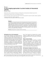
Báo cáo y học: " In vivo imaging approaches in animal models of rheumatoid arthritis" pot
Bạn đang xem bản rút gọn của tài liệu. Xem và tải ngay bản đầy đủ của tài liệu tại đây (64.53 KB, 7 trang )
165
CCD = charge-coupled device; FIAU = 2′-fluoro-2′-deoxy-1-beta-D-arabinofuranosyl-5-iodo-uracil; HSV-Tk = herpes simplex virus thymidine kinase;
MHC = major histocompatibility complex; MRI = magnetic resonance imaging; PET = positron emission tomography.
Available online />Introduction: the
in vivo
renaissance
The early phase of exploration of the lymphoid system gen-
erated a wealth of information about anatomy and in vivo
responses. Our ability to define molecular structures in the
context of the anatomy of the in vivo immune response, first
with antibodies and more recently with tools of molecular
genetics, has increased the ability to incisively test hypothe-
ses through in vivo experimentation. This is leading to a
renaissance in a variety of in vivo studies, mostly focused
around genetic manipulations. The molecular genetics tools
are also complemented by new technologies to image the
movements and interactions of cells in vivo.
The present review will focus on emerging technologies
that allow in vivo imaging of specific cells or molecules
using noninvasive methods or direct microscopic imaging
of single cells in the in vivo environment using minimally
invasive methods. Microscopic imaging has the advantage
of being able to study single cells in action. Invasiveness in
this case refers specifically to the need for surgical proce-
dures to expose tissues for high-resolution imaging of
cells or molecules of interest. The advantages and limita-
tions of each approach are discussed with a specific
emphasis on imaging in joints and on work directly rele-
vant to rheumatoid arthritis. This information is summarized
in Table 1.
Whole animal imaging
Imaging of events in intact live animals is a powerful
approach primarily because it allows studies over time
with minimal perturbation of the experiment. These
methods also couple in powerful ways with molecule
genetics technologies that allow in situ labeling of cell
populations expressing specific genes. The present review
will also discuss recent studies in this area with direct rel-
evance to animal models of rheumatoid arthritis.
Bioluminescence imaging in intact animals [1]
The expression of luciferase has for many years been a
powerful tool in gene expression studies. This is because
the substrates in the luciferase reaction generate no signal
(light) in the absence of luciferase. Instruments that detect
luminescent reactions can be optimized for sensitivity to
light without the necessity of rejecting any significant
Review
In vivo
imaging approaches in animal models of rheumatoid
arthritis
Michael L Dustin
Skirball Institute of Biomolecular Medicine and Department of Pathology, New York University School of Medicine, New York, USA
Corresponding author: Michael L Dustin (e-mail: )
Received: 14 Nov 2002 Revisions requested: 29 Nov 2002 Revisions received: 4 Apr 2003 Accepted: 10 Apr 2003 Published: 1 May 2003
Arthritis Res Ther 2003, 5:165-171 (DOI 10.1186/ar768)
© 2003 BioMed Central Ltd (Print ISSN 1478-6354; Online ISSN 1478-6362)
Abstract
The interaction of activated leukocytes with the rheumatoid synovial environment is a key process in
arthritis. Understanding this process will play an important role in designing effective treatments. In vivo
imaging approaches combined with molecular genetics in animal models provide important tools to
address these issues. The present review will focus on approaches to in vivo imaging, with particular
attention to approaches that are proving useful for, or have promise for, research on animal models of
rheumatoid arthritis. These approaches will probably shed light on the specific local mechanisms
involved in chronic inflammation and provide real time monitoring approaches to follow cellular and
molecular events related to disease development.
Keywords: arthritis, fluorescence, imaging, luminescence, microscopy
166
Arthritis Research & Therapy Vol 5 No 4 Dustin
background signals. Only in recent years have cameras
become sensitive enough to detect the faint light emis-
sions of the luciferase reaction from within intact animals.
The most useful detectors are back-illuminated, cooled,
charge-coupled device (CCD) cameras that have very low
background and very high ‘quantum efficiency’ (the propor-
tion of photons hitting the detector that are converted into
a usable signal). Back-illumination refers to a method of
preparing the CCD sensor so that the photons directly
strike the light-sensitive thinned back surface, in contrast to
conventional CCDs where photons pass through nonlight-
sensitive elements on the front of the CCD with a resulting
loss of efficiency. These systems also have very low noise,
and long exposures can therefore be used to integrate the
signal over time and to obtain a usable signal.
To apply this approach, the luciferase gene can be intro-
duced into an animal using transgenic or homologous
recombination technology to place luciferase expression
under the control of specific genetic elements. When tran-
scription of luciferase is activated, the cells or tissues
expressing the gene can metabolize injected substrates
(luciferin in the presence of endogenous ATP), which are
nontoxic. The substrate metabolism can generate a signal
detected by the external camera with the only requirement
that the animal is anesthetized so that it does not move
during the imaging period. Breathing causes movements in
the thoracic area, but these are not significant compared
with the general resolution. The drawback of this method is
that the light emitted from the luciferase reaction is
yellow–green, and thus is highly scattered as it passes
through tissues and exits the animal. The resolving power is
therefore low (millimeters). However, this is certainly ade-
quate to identify cell migration or gene expression within the
joint with a detection threshold in the order of 10–100 cells.
Lymphocytes for transfer studies could be prepared from
luciferase expressing transgenic mice. Luminescence
imaging has been applied to studies on cell transfer in the
murine autoimmune disease model experimental autoim-
mune encephalitis [2] and has been applied to examina-
tion of transcription factor nuclear factor-κB in inflamed
mouse joints [3]. This approach has also been used to
track antigen-specific T cells for gene therapy of collagen-
induced arthritis in mice [4]. Application of the lumines-
cence methodology to humans would be problematic due
to the greater thickness of human skin as a barrier to
photon escape and detection. Shifting the luminescent
emission to the red end of the spectrum might improve
these prospects [5]. Transcutaneous imaging of cells
expressing green fluorescent protein and other fluorescent
dyes has also been demonstrated with similar resolution to
the luminescence-based imaging, but with less sensitivity
owing to the greater background from autofluorescence
and scattered excitation light [6].
Radioactive tracer imaging in intact animals
Radioactive tracer studies offer greater penetration and
quantitative integrity compared with optical imaging
methods because the emissions from radioisotopes have
less interaction with tissues than does light. Of the avail-
able methods for radioisotope imaging, that with the best
resolution for small animal imaging is positron emission
tomography (PET).
PET imaging is based on isotopes such as
14
F and
64
Cu,
which decay by emitting positrons that, on collision with
an electron, emit γ-rays at 180° to each other. Arrays of
detectors surrounding an animal can simultaneously
detect these γ-emissions and then determine with great
precision the line along which the emission was localized.
From a number of such emissions, the PET method can
build an image in which the source can be localized with a
resolution of ~2 mm.
The limitation of PET imaging is that the positron-emitting
isotopes have short half-lives so they can only be used to
follow the cell or molecule in vivo for a day or two at most.
Within this time span, however, very important results can
Table 1
Summary of
in vivo
imaging methodologies
Imaging mode Invasiveness Sensitivity, resolution, time scale Advantages Disadvantages
Bioluminescence Anesthesia ~100 cells, 5 mm, minutes Noninvasive, sensitive, Resolution, penetration
quantitative
Micro positron emission Anesthesia 1000 cells, 2 mm, minutes Noninvasive, resolution Short half-life of isotopes
tomography/single photon
emission commuted
tomography
Magnetic resonance imaging Anesthesia 1000 cells, 0.1 mm, minutes Noninvasive, resolution Sensitivity, slow
Intravital microscopy Anesthesia/surgery 1 cell, 0.2 µm, seconds Highest resolution Invasive, penetration limited
167
be obtained. A striking recent example is a study on the
interaction of antibodies to glucose-6-phosphate iso-
merase, a ubiquitous enzyme [7]. These antibodies trans-
fer arthritis and are specifically produced in mice
transgenic for the KRN T-cell receptor on a nonobese dia-
betic mouse genetic background. A mystery in this
disease process is why antibodies to a generally
expressed enzyme would specifically induce a joint
disease. Anti-glucose-6-phosphate isomerase antibodies
were labeled with
64
Cu and injected into recipient mice,
which were then subjected to micro-PET analysis (a PET
scanner configured to produce high-resolution images of
small animals). It was found that the anti-glucose-6-phos-
phate isomerase antibody was rapidly concentrated in
distal joints (the targets of the disease), while control IgG
did not show this localization [7]. Therefore, an important
advance in understanding the pathological effects of
autoantibodies in a rheumatoid arthritis model was made
using PET imaging of molecules. PET imaging is per-
formed with human subjects where the short-lived isotopes
are considered to pose a small risk and much information is
gained, particularly regarding the metabolic status of tissue
[8]. In vivo studies on autoantibody involvement in human
rheumatoid arthritis are thus possible.
An alternative mode of imaging is the use of single photon
imaging of γ-emitting isotopes like
111
In or
99
Tc. Imaging of
γ-emitting isotopes is referred to as single photon emission
commuted tomography. This approach as been used to
follow isotope-labeled materials in joints of arthritis
patients. It has the advantage that the individual compo-
nents can be radiolabeled and followed in vivo, but has the
disadvantage that γ-emitters of sufficiently high activity also
have relatively short half-lives. Cells can be labeled prior to
transfer to animals or can be labeled in situ by injection of
monoclonal antibodies labeled with appropriate isotopes
[9,10]. This method has lower resolution than PET, but is
simpler and utilizes isotopes such as
111
In that are readily
incorporated into live cells. These isotopes can also be
detected with γ-cameras with similar resolution.
A drawback of both the PET and single photon emission
commuted tomography methods is that the isotopes have
short half-lives, making long-term tracking impractical. This
problem has been partially overcome for experimental
animal models through the expression of herpes simplex
virus thymidine kinase (HSV-Tk) in cells of animals and
then injecting the animals with 2′-fluoro-2′-deoxy-1-beta-
D-
arabinofuranosyl-5-iodo-uracil (FIAU), a compound that is
specifically accumulated in cells expressing the HSV-Tk
gene product [11]. Similar experiments have been per-
formed with rat myocardium using other tracer com-
pounds, but FIAU appears to be the best [12–14]. This
approach allows an elegant combination of molecular
genetics and noninvasive imaging: the presence of the
HSV-Tk gene can mark a specific cell population in a spe-
cific state of activation based on the activity of the pro-
moter controlling expression of the HSV-Tk gene. The
animals expressing tagged cells can then be labeled with
radionuclide-tagged FIAU (for either single photon emis-
sion commuted tomography or for PET imaging) on
repeated occasions over a long period of time. The
HSV-Tk cells can then be located as long as they are not
in organs like the bladder that accumulate FIAU as part of
normal metabolism and excretion of the FIAU.
MRI of transferred lymphocytes
A promising technology for tracking cells deep in animals
is the use of paramagnetic contrast agents taken into cells
using cell-penetrating peptides in conjunction with MRI
[15,16]. This method uses the HIV tat peptide, a highly
cationic peptide that has the ability to enter into cells
through the plasma membrane in an energy-independent
process and to bring along large cargo [17], linked to
superparamagnetic iron [18]. In vitro MRI imaging of bone
marrow material populated with a few cells that had taken
up the paramagnetic iron shows that single cells are
detected as ‘signal voids’. Because this is a dark signal on
a light and variable background, the actual sensitivity may
not reach the single-cell level in vivo. However, T-cell infil-
trates in nonobese diabetic mice were readily detected in
the pancreas [16]. This suggests that the sensitivity is suf-
ficient to be useful in tracking cells in inflammatory infil-
trates. This contrast agent allows the detection of cells in
the context of the normal high level of tissue contrast that
can be attained with MRI. This method is relatively new
and has not been extensively applied to autoimmune situa-
tions. One important issue will be the minimum number of
cells, which can be tracked.
Ultrasound imaging with microbubbles
A novel type of specific tracer for noninvasive cellular
imaging is the use of ultrasound to image cells specifically
tagged with stable microbubbles [19–22]. These studies
demonstrated that the microbubble contract agents of
various surface chemistries are readily phagocytosed by
leukocytes attached to inflamed blood vessels. These
phagocytosed microbubbles were more stable than extra-
cellular microbubbles and thus could be imaged with high
contrast. Microbubbles could also be specifically targeted
to inflamed endothelium with antibodies to P-selectin
(CD62P). The tendency of microbubbles to attach to
leukocytes in inflamed vessels may correlate with the utility
of these contrast agents in detecting active arthritis in the
knee [23]. The utility of ultrasound may be enhanced, and
the mechanism of contract agent accumulation is better
understood and specific targeting strategies for contrast
agents developed for clinical use.
Microscopy approaches
Microscopic approaches allow the resolution of cellular
and subcellular details with high numerical aperture objec-
Available online />168
tives. The general drawback of these methods is that they
do not allow this level of resolution transcutaneously, and
therefore require surgical exposure of the organ or tissue of
interest. These invasive methods must be approached with
great care since the surgical procedures are well known to
induce leukocyte adhesion to endothelial cells and other
effects, which may render the surgical preparations differ-
ent in some ways from intact tissues. Nonetheless,
microscopy is essential to address questions of single cell
and supramolecular dynamics in vivo.
Intravital microscopy
Fortunately for immunologists and rheumatologists inter-
ested in surgical procedures for in vivo imaging, there is a
rich arsenal of procedures for imaging within almost all
major organs of mice or rats. Almost all were developed
originally for microvascular research and then adapted for
inflammation research. A nonexhaustive list includes the
brain, the liver, the lungs, the muscle, the spleen, the
lymph nodes, the pancreas, the mesenteries and the skin
[24–28]. Each of these preparations has unique strengths
and caveats, and most show some effects of surgical
trauma that must be considered in interpreting the results.
For example, in the cremaster muscle preparation, the
abundant rolling leukocytes in the venules are due to
P-selectin upregulation on endothelial cells in response to
surgical trauma [25].
It is important to note that there is a recently developed
intravital preparation for mouse joint synovium [29]. The
synovium is exposed for imaging by partial resection of the
patella tendon. This preparation has been used to evaluate
the effects of anti-inflammatory drugs and nitric oxide inhi-
bition on leukocyte recruitment to rheumatoid synovium
[30–32]. The important results were that inducible nitric
oxide synthase was protective in acute joint inflammation
but had no influence on chronic synovial inflammation. The
nonconventional anti-inflammatory drug oxaceprol reduced
leukocyte adherence to synovial microvessels and gener-
ally reduced the signs of inflammation. The groundwork for
further studies on the dynamics of lymphocyte interactions
in the synovium has thus been established.
Most of the work in intravital imaging of leukocytes has
focused on the interaction of lymphocytes with endothelial
cells, and has only minimally addressed the issues of what
leukocytes do after they extravasate. While leukocytes in
blood vessels have high contrast, the extravasated leuko-
cytes in tissues generally lack contrast and can only be
tracked by fluorescence imaging of labeled cells. Those
workers studying leukocyte interactions with blood vessels
have also had a very clear hypothesis in the form of the
multistep paradigm, which argued for rolling, activation
and arrest steps executed by selectins, chemokines and
integrins [33,34]. This hypothesis created a clear frame-
work for many studies to identify these components, or
their absence, in different tissue sites for different leuko-
cyte subsets.
A hypothetical framework for migration of leukocytes and
lymphocytes in tissues is provided by the multistage guid-
ance of leukocytes by chemokines and bacterial products
[35], and by the concept that antigen receptor engagement
delivers a stop signal for lymphocytes [36]. While the move-
ment of leukocytes in blood vessels is fast and much data
can be collected in a couple of minutes of recording, the
migration of leukocytes in tissues is relatively slow and
requires many minutes of recording to track cells. This
longer imaging period requires greater stability of the prepa-
rations. A few studies have now documented that leukocyte
and lymphocyte migration in the parenchyma of tissues can
be followed in vivo by imaging in thin tissues like the mesen-
teries or by fluorescence intravital microscopy, but there has
been very little systematic analysis of this migration at this
point [37–39]. Werr and colleagues clearly established that
the collagen receptor VLA-2 has an important role in the
migration of leukocytes in the rat mesenteries [37]. At this
point, the adhesion systems used by lymphocytes for migra-
tion in tissues are not known.
An intermediate step between in vitro and in vivo studies
on tissue migration of lymphocytes is the use of organ
culture systems. A very useful experimental system is
based on thymic organ cultures in which positive and neg-
ative selection in thymocyte maturation can be recapitu-
lated in long-term culture models. Imaging of fluorescently
labeled thymocyte migration in thymic organ cultures
demonstrates both dynamic and stable interactions that
were dependent upon positive selecting MHC–peptide
complexes [40]. The power of this system is that imaging
results can be directly related to the functional maturation
of thymocytes in the culture system.
Lymph node organ cultures are not a traditional system in
immunology, yet imaging of lymph nodes from mice into
which a few million fluorescently labeled T cells or B cells
had been transferred was an informative experiment. The
lymph nodes were excised and immediately superperfused
with highly oxygenated media in an effort to maintain
oxygen levels within the intact mouse lymph node, which is
about 1 mm in diameter. Both T cells and B cells in the cul-
tured lymph nodes displayed dramatic motility, which was
restricted to the T-cell zones and the follicles, respectively,
but was otherwise random in direction [41]. While it was
not clear whether oxygen was a critical parameter for
these experiments, a clearly critical parameter was tem-
perature. The motility of T cells in the lymph node was criti-
cally dependent on the temperature being close to 37°C.
The motility dropped steeply below, and also above, this
level. The increased local temperature associated with
inflammation in tissues may therefore play a role in optimiz-
ing leukocyte migration in the site. It is probable that this
Arthritis Research & Therapy Vol 5 No 4 Dustin
169
rapid, random migration, which was not previously postu-
lated, is a critical element in the search of lymphocytes for
presenting cells with appropriate antigens. T-cell receptor
engagement appeared to deliver a stop signal in both
systems [41,42].
These organ culture experiments will probably serve as
stepping stones to in vivo observations now that it is clear
that there are interesting things to be learned from follow-
ing migration of labeled lymphocyte populations. It was
also demonstrated that a sufficiently high resolution can
be obtained for imaging the distribution of molecules
within individual cells, making it possible to approach
analysis of formation of the immunological synapse, a spe-
cific supramolecular pattern of receptors involved in
immune cell communication, in vivo [42,43].
Two-photon microscopy
One of the limitations of high-resolution optical imaging is
that it is very sensitive to light scattering by biological
tissues. This makes the effective imaging depth for con-
ventional high-resolution microscopy around 0–50 µm into
a tissue. Cells can still be detected for another 50 µm, but
all detail on the micrometer scale is lost.
Two-photon microscopy is a powerful method for imaging
deeper within tissues that takes advantage of the lower
light scattering with infrared light [44,45]. This is demon-
strated by the classic childhood experiment of holding a
flashlight to one’s hand and observing that the light that
penetrates is red. Two-photon excitation is based on the
excitation of fluorescence for typical visible excitation fluo-
rophores with two photons of low-energy infrared light.
The two photons have to be absorbed by the fluorophore
in rapid succession such that the instantaneous intensity
of light has to be millions of times brighter than that typi-
cally used for conventional fluorescence excitation. This
extreme brightness is accomplished using a mode-locked
titanium–sapphire laser, which emits light in fentosecond
pulses. While the average power is similar to that used in
conventional confocal microscopy, the peak power is 10
6
times higher. The beam is then expanded to fill the back
aperture of the objective and is focused to a diffraction-
limited spot in the tissue. Only at this focal point is the
density of photons sufficiently high to achieve multiphoton
fluorescence excitation, resulting in a very small volume of
0.2 µm wide × 0.5 µm high. The laser beam is scanned
through the specimen and all the light that is emitted is
collected by a photomultiplier mounted as close to the
back of the objective as possible. No pinhole is needed
since the excitation volume defines the image plane. The
emission can be highly scattered as it exits the tissue, but
only needs to hit the detector to count toward the signal.
The practical depth of imaging achieved with multiphoton
imaging depends on the objective used, on the tissue and
on the exact wavelength that is used for excitation. In the
brain, it is possible to image up to 300 µm with submicron
resolution. Lymph nodes appear to have more background
signal and scattering than the brain, but imaging over
100 µm deep is still readily achieved and cellular signals
can be identified up to 200 µm [46]. Advances in technol-
ogy such as gradient refractive index lenses may enable
much deeper high-resolution imaging in the future.
Future studies
A clear direction for future studies will be the direct exami-
nation of T-cell migration and cell–cell interactions in the
rheumatoid synovium. This process may be studied at
many levels, from cell populations by noninvasive methods
to single cells by direct microscopic observation after
simple surgical procedures to expose the synovium. Mice
expressing fluorescent proteins in specific tissues will be
valuable for these future studies.
There are a number of key questions about cell dynamics
in the synovium. Do T cells form stable immunological
synapses with antigen presenting cells in the synovium?
Is stable synapse formation related to the assembly of
ectopic secondary lymphoid tissues in the vicinity of the
synovium? How do T cells interact with different types of
synoviocytes — the macrophage-like type I cells and the
fibroblast-like type II cells? Do T cells interact in specific
ways with macrophage-like cells at sites of bone
erosion? How do autoantibodies interact with tissues
and immune cells, including mast cells, at the micro-
scopic level? These and other questions can be
addressed by combining molecule genetic methods with
new imaging modes.
We should know in the near future the general utility of
these approaches in evaluating therapeutics and disease
models. It is most probable that these approaches will
yield surprising results and will be highly informative in the
effort to cure arthritis.
Competing interests
None declared.
Acknowledgements
The author thanks his laboratory group for inspiring discussions and
the Irene Diamond Fund for generous support. The work is also sup-
ported by grants from the National Institutes of Health. MLD is a past
recipient of an Arthritis Foundation Research Grant, which supported
work on the TCR stop signal.
References
1. Contag CH, Bachmann MH: Advances in in vivo biolumines-
cence imaging of gene expression. Annu Rev Biomed Eng
2002, 4:235-260.
2. Costa GL, Sandora MR, Nakajima A, Nguyen EV, Taylor-Edwards
C, Slavin AJ, Contag CH, Fathman CG, Benson JM: Adoptive
immunotherapy of experimental autoimmune encephalo-
myelitis via T cell delivery of the IL-12 p40 subunit. J Immunol
2001, 167:2379-2387.
Available online />170
3. Carlsen H, Moskaug JO, Fromm SH, Blomhoff R: In vivo imaging
of NF-kappa B activity. J Immunol 2002, 168:1441-1446.
4. Nakajima A, Seroogy CM, Sandora MR, Tarner IH, Costa GL,
Taylor-Edwards C, Bachmann MH, Contag CH, Fathman CG:
Antigen-specific T cell-mediated gene therapy in collagen-
induced arthritis. J Clin Invest 2001, 107:1293-1301.
5. Branchini BR, Magyar RA, Murtiashaw MH, Anderson SM, Helger-
son LC, Zimmer M: Site-directed mutagenesis of firefly
luciferase active site amino acids: a proposed model for bio-
luminescence color. Biochemistry 1999, 38:13223-13230.
6. Mahmood U, Tung CH, Tang Y, Weissleder R: Feasibility of in
vivo multichannel optical imaging of gene expression: experi-
mental study in mice. Radiology 2002, 224:446-451.
7. Wipke BT, Wang Z, Kim J, McCarthy TJ, Allen PM: Dynamic visu-
alization of a joint-specific autoimmune response through
positron emission tomography. Nat Immunol 2002, 3:366-372.
8. Antonini A, Moresco RM, Gobbo C, De Notaris R, Panzacchi A,
Barone P, Calzetti S, Negrotti A, Pezzoli G, Fazio F: The status of
dopamine nerve terminals in Parkinson’s disease and essen-
tial tremor: a PET study with the tracer [11-C]FE-CIT. Neurol
Sci 2001, 22:47-48.
9. Jorgensen C, Apparailly F, Couret I, Canovas F, Jacquet C, Sany
J: Interleukin-4 and interleukin-10 are chondroprotective and
decrease mononuclear cell recruitment in human rheumatoid
synovium in vivo. Immunology 1998, 93:518-523.
10. Kipper SL, Rypins EB, Evans DG, Thakur ML, Smith TD, Rhodes
B: Neutrophil-specific 99mTc-labeled anti-CD15 monoclonal
antibody imaging for diagnosis of equivocal appendicitis.
J Nucl Med 2000, 41:449-455.
11. Tjuvajev JG, Finn R, Watanabe K, Joshi R, Oku T, Kennedy J,
Beattie B, Koutcher J, Larson S, Blasberg RG: Noninvasive
imaging of herpes virus thymidine kinase gene transfer and
expression: a potential method for monitoring clinical gene
therapy. Cancer Res 1996, 56:4087-4095.
12. Inubushi M, Wu JC, Gambhir SS, Sundaresan G, Satyamurthy N,
Namavari M, Yee S, Barrio JR, Stout D, Chatziioannou AF, Wu L,
Schelbert HR: Positron-emission tomography reporter gene
expression imaging in rat myocardium. Circulation 2003, 107:
326-332.
13. Brust P, Haubner R, Friedrich A, Scheunemann M, Anton M, Koufaki
ON, Hauses M, Noll S, Noll B, Haberkorn U, Schackert G, Schack-
ert HK, Avril N, Johannsen B: Comparison of [18F]FHPG and
[124/125I]FIAU for imaging herpes simplex virus type 1 thymi-
dine kinase gene expression. Eur J Nucl Med 2001, 28:721-729.
14. MacLaren DC, Toyokuni T, Cherry SR, Barrio JR, Phelps ME, Her-
schman HR, Gambhir SS: PET imaging of transgene expres-
sion. Biol Psychiatry 2000, 48:337-348.
15. Dodd CH, Hsu HC, Chu WJ, Yang P, Zhang HG, Mountz JD Jr,
Zinn K, Forder J, Josephson L, Weissleder R, Mountz JM, Mountz
JD: Normal T-cell response and in vivo magnetic resonance
imaging of T cells loaded with HIV transactivator-peptide-
derived superparamagnetic nanoparticles. J Immunol Methods
2001, 256:89-105.
16. Moore A, Sun PZ, Cory D, Hogemann D, Weissleder R, Lipes MA:
MRI of insulitis in autoimmune diabetes. Magn Reson Med
2002, 47:751-758.
17. Wender PA, Mitchell DJ, Pattabiraman K, Pelkey ET, Steinman L,
Rothbard JB: The design, synthesis, and evaluation of mole-
cules that enable or enhance cellular uptake: peptoid molecu-
lar transporters. Proc Natl Acad Sci USA 2000, 97:
13003-13008.
18. Lewin M, Carlesso N, Tung CH, Tang XW, Cory D, Scadden DT,
Weissleder R: Tat peptide-derivatized magnetic nanoparticles
allow in vivo tracking and recovery of progenitor cells. Nat
Biotechnol 2000, 18:410-414.
19. Lindner JR, Song J, Christiansen J, Klibanov AL, Xu F, Ley K:
Ultrasound assessment of inflammation and renal tissue
injury with microbubbles targeted to P-selectin. Circulation
2001, 104:2107-2112.
20. Lindner JR, Song J, Xu F, Klibanov AL, Singbartl K, Ley K, Kaul S:
Noninvasive ultrasound imaging of inflammation using
microbubbles targeted to activated leukocytes. Circulation
2000, 102:2745-2750.
21. Lindner JR, Dayton PA, Coggins MP, Ley K, Song J, Ferrara K,
Kaul S: Noninvasive imaging of inflammation by ultrasound
detection of phagocytosed microbubbles. Circulation 2000,
102:531-538.
22. Lindner JR, Coggins MP, Kaul S, Klibanov AL, Brandenburger GH,
Ley K: Microbubble persistence in the microcirculation during
ischemia/reperfusion and inflammation is caused by integrin-
and complement-mediated adherence to activated leuko-
cytes. Circulation 2000, 101:668-675.
23. Carotti M, Salaffi F, Manganelli P, Salera D, Simonetti B, Grassi
W: Power Doppler sonography in the assessment of synovial
tissue of the knee joint in rheumatoid arthritis: a preliminary
experience. Ann Rheum Dis 2002, 61:877-882.
24. Piccio L, Rossi B, Scarpini E, Laudanna C, Giagulli C, Issekutz
AC, Vestweber D, Butcher EC, Constantin G: Molecular mecha-
nisms involved in lymphocyte recruitment in inflamed brain
microvessels: critical roles for P-selectin glycoprotein ligand-
1 and heterotrimeric G(i)-linked receptors. J Immunol 2002,
168:1940-1949.
25. Kunkel EJ, Jung U, Bullard DC, Norman KE, Wolitzky BA, Vestwe-
ber D, Beaudet AL, Ley K: Absence of trauma-induced leuko-
cyte rolling in mice deficient in both P-selectin and
intercellular adhesion molecule 1. J Exp Med 1996, 183:57-
65.
26. Andonegui G, Goyert SM, Kubes P: Lipopolysaccharide-
induced leukocyte–endothelial cell interactions: a role for
CD14 versus toll-like receptor 4 within microvessels.
J Immunol 2002, 169:2111-2119.
27. Ley K, Linnemann G, Meinen M, Stoolman LM, Gaehtgens P:
Fucoidin, but not yeast polyphosphomannan PPME, inhibits
leukocyte rolling in venules of the rat mesentery. Blood 1993,
81:177-185.
28. von Andrian UH: Intravital microscopy of the peripheral lymph
node microcirculation in mice. Microcirculation 1996, 3:287-
300.
29. Veihelmann A, Harris AG, Krombach F, Schutze E, Refior HJ,
Messmer K: In vivo assessment of synovial microcirculation
and leukocyte–endothelial cell interaction in mouse antigen-
induced arthritis. Microcirculation 1999, 6:281-290.
30. Veihelmann A, Hofbauer A, Krombach F, Dorger M, Maier M,
Refior HJ, Messmer K: Differential function of nitric oxide in
murine antigen-induced arthritis. Rheumatology (Oxford) 2002,
41:509-517.
31. Veihelmann A, Landes J, Hofbauer A, Dorger M, Refior HJ,
Messmer K, Krombach F: Exacerbation of antigen-induced
arthritis in inducible nitric oxide synthase-deficient mice.
Arthritis Rheum 2001, 44:1420-1427.
32. Veihelmann A, Hofbauer A, Refior HJ, Messmer K: Oxaceprol, an
atypical inhibitor of inflammation, reduces leukocyte adher-
ence in mouse antigen-induced arthritis. Acta Orthop Scand
2001, 72:293-298.
33. Lawrence MB, Springer TA: Leukocytes roll on a selectin at
physiologic flow rates: distinction from and prerequisite for
adhesion through integrins. Cell 1991, 65:859-873.
34. von Andrian UH, Chambers JD, McEvoy LM, Bargatze RF, Arfors
KE, Butcher EC: Two-step model of leukocyte–endothelial cell
interaction in inflammation: distinct roles for LECAM-1 and the
leukocyte beta 2 integrins in vivo. Proc Natl Acad Sci USA
1991, 88:7538-7542.
35. Foxman EF, Campbell JJ, Butcher EC: Multistep navigation and
the combinatorial control of leukocyte chemotaxis. J Cell Biol
1997, 139:1349-1360.
36. Dustin ML, Bromley SK, Kan Z, Peterson DA, Unanue ER:
Antigen receptor engagement delivers a stop signal to
migrating T lymphocytes. Proc Natl Acad Sci USA 1997,
94:3909-3913.
37. Werr J, Johansson J, Eriksson EE, Hedqvist P, Ruoslahti E,
Lindbom L: Integrin alpha(2)beta(1) (VLA-2) is a principal
receptor used by neutrophils for locomotion in extravascular
tissue. Blood 2000, 95:1804-1809.
38. Miura S, Tsuzuki Y, Fukumura D, Serizawa H, Suematsu M,
Kurose I, Imaeda H, Kimura H, Nagata H, Tsuchiya M, et al.:
Intravital demonstration of sequential migration process of
lymphocyte subpopulations in rat Peyer’s patches. Gastroen-
terology 1995, 109:1113-1123.
39. Grayson MH, Chaplin DD, Karl IE, Hotchkiss RS: Confocal fluo-
rescent intravital microscopy of the murine spleen. J Immunol
Methods 2001, 256:55-63.
40. Bousso P, Bhakta NR, Lewis RS, Robey E: Dynamics of thymo-
cyte–stromal cell interactions visualized by two-photon
microscopy. Science 2002, 296:1876-1880.
Arthritis Research & Therapy Vol 5 No 4 Dustin
171
41. Miller MJ, Wei SH, Parker I, Cahalan MD: Two-photon imaging
of lymphocyte motility and antigen response in intact lymph
node. Science 2002, 296:1869-1873.
42. Stoll S, Delon J, Brotz TM, Germain RN: Dynamic imaging of T
cell–dendritic cell interactions in lymph nodes. Science 2002,
296:1873-1876.
43. Grakoui A, Bromley SK, Sumen C, Davis MM, Shaw AS, Allen PM,
Dustin ML: The immunological synapse: a molecular machine
controlling T cell activation. Science 1999, 285:221-227.
44. Denk W, Strickler JH, Webb WW: Two-photon laser scanning
fluorescence microscopy. Science 1990, 248:73-76.
45. Xu C, Zipfel W, Shear JB, Williams RM, Webb WW: Multiphoton
fluorescence excitation: new spectral windows for biological
nonlinear microscopy. Proc Natl Acad Sci USA 1996, 93:
10763-10768.
46. Miller MJ, Wei SH, Cahalan MD, Parker I: Autonomous T cell
trafficking examined in vivo with intravital two-photon
microscopy. Proc Natl Acad Sci USA 2003, 100:2604-2609.
Correspondence
Michael L Dustin, Program in Molecular Pathogenesis, Skirball Institute
of Biomolecular Medicine and Department of Pathology, New York
University School of Medicine, 540 First Avenue, New York, NY
10016, USA. Tel: +1 212 263 3207; fax: +1 212 263 5711;
e-mail:
Available online />