Báo cáo y học: "A review of anabolic therapies for osteoporosis" ppt
Bạn đang xem bản rút gọn của tài liệu. Xem và tải ngay bản đầy đủ của tài liệu tại đây (86.96 KB, 9 trang )
214
bFGF = basic FGF; BMD = bone mineral density; BMP = bone morphogenic protein; eNOS = endothelial nitric oxide synthase; FGF = fibroblast
growth factor; GH = growth hormone; HRT = hormone replacement therapy; IGF = insulin-like growth factor; IL-6 = interleukin-6; OP-1 =
osteogenic protein 1; PTH = parathyroid hormone; rhBMP = recombinant human BMP; rhPTH = recombinant human PTH; TGF = transforming
growth factor; VEGF = vascular endothelial growth factor.
Arthritis Research & Therapy Vol 5 No 5 Lane and Kelman
Introduction
Osteoporosis is a disease causing skeletal fragility due to
low bone mass or architectural changes in bone structure,
and results in fractures from low impact. It is also a
disease that increases with the age of the patient.
Throughout adult life, the skeleton turns over or remodels
to remove old bone tissue and lays down new bone tissue.
Bone remodeling is a tightly coupled process in which an
area of the bone undergoes osteoclastic bone resorption
and then the location of the bone resorption is filled in by
osteoblasts. This bone remodeling cycle is synchronized,
with resorption and formation being equal, until metabolic
or lifestyle changes occur that unbalance the system [1].
Events such as the menopause, taking glucocorticoids, or
aging are examples of situations in which bone resorption
is greater than bone formation, with a resulting loss of
bone mass and structure. In adults, most bone diseases
are in bone remodeling, while in children many bone dis-
eases result from remodeling defects [1].
Over the past 10 years, many patients with osteoporosis
have been treated with antiresorptive agents (estrogens,
bisphosphonates, calcitonin) that reduce osteoclast bone
resorption. These agents prevent bone from being broken
down, allow remodeling spaces to fill in, and improve bone
strength and reduce fracture risk. These agents intro-
duced both the prevention of and treatment of osteoporo-
sis [2–4].
Today, another type of bone-active agents is available in the
United States, recombinant human parathyroid hormone
(rhPTH) (1-34), which can increase bone mass and
strength, and treatment with these bone agents is referred
to as ‘anabolic therapy’. These anabolic bone-active agents
primarily work by stimulating new bone formation on quies-
cent bone surface that is not simultaneously undergoing
remodeling. In addition, these agents increase bone mass
to a greater degree than just filling in the bone remodeling
space. These new agents have the potential to restore bone
Review
A review of anabolic therapies for osteoporosis
Nancy E Lane and Ariella Kelman
Division of Rheumatology, University of California, San Francisco, San Francisco, CA, USA
Corresponding author: Nancy Lane (e-mail: )
Received: 26 Jun 2003 Accepted: 10 Jul 2003 Published: 5 Aug 2003
Arthritis Res Ther 2003, 5:214-222 (DOI 10.1186/ar797)
© 2003 BioMed Central Ltd (Print ISSN 1478-6354; Online ISSN 1478-6362)
Abstract
Osteoporosis results from a loss of bone mass and bone structure such that the bone becomes weak
and fractures with very little trauma. Until recently, the approved osteoporosis therapies prevented
more bone loss by altering osteoclast activity and lifespan. Recently, attention has turned away from
osteoclast inhibition to agents that can stimulate the osteoblast to form new bone, or anabolic agents.
This article reviews both approved and experimental anabolic agents that improve bone mass by
improving osteoblast activity, or increasing osteoblast number. The use of the anabolic agents to
improve bone mass and strength followed by agents that prevent the new bone mass from being lost
may offer the ability to cure osteoporosis and reduce bone fracture healing time.
Keywords: anabolic; bone morphogenic protein-2 (BMP-2); bone morphogenic protein-7 (BMP-7); parathyroid
hormone rhPTH (1-34); parathyroid hormone hPTH (1-84)
215
Available online />mass, bringing it back toward normal, and may reduce the
risk of osteoporotic fracture more than the currently avail-
able antiresorptive agents.
This article provides an overview of a number of anabolic
therapies, including parathyroid hormone (PTH), growth
hormone (GH), insulin-like growth factor (IGF) 1, strontium,
fluoride, bone morphogenetic protein (BMP)-2, BMP-7
(also called osteogenic protein-1 [OP-1]), basic fibroblast
growth factors (bFGFs) and vascular endothelial growth
factor (VEGF), as examples of approved anabolic thera-
pies and those currently under development. Since a
number of excellent reviews on anabolic agents have been
published in the past few years, we refer the reader to
additional reviews on some of these anabolic agents [5,6].
Parathyroid hormone
Proposed mechanisms of action
Hyperparathyroidism is associated with a continuously
high serum level of PTH, and bone loss occurs over time
[7,8]. However, when PTH is administered by a daily sub-
cutaneous injection, an increase in bone mass occurs in
both animals and humans [9–12]. In humans, the anabolic
effect of PTH is most pronounced in the trabecular bone.
However, histomorphometric studies of iliac crest biopsies
from clinical studies of PTH find both thickened trabeculae
and increased cortical cross-sectional diameter and
increased trabecular number and connections [12]. This
could result from PTH stimulating bone-forming cells on
the trabecular surface. In addition, by increasing the pro-
duction of FGF and IGF-1 in the localized bone environ-
ment, osteoprogenitor cells adjacent to the endocortical
bone surface are stimulated to differentiate into
osteoblasts and form osteoid, new bone spicules, and
connections [13,14]. Interestingly, PTH injections also
stimulate the osteoclast-stimulating cytokines (receptor
activator of nuclear factor κB ligand [RANKL] and IL-6),
thus increasing bone reasorption simultaneously with the
bone-formation actions [15–20]. However, both animal
and clinical studies show that PTH exerts major action on
bone formation on the trabecular bone surface, followed
by some periosteal and endocortical bone surfaces. The
bone resorption appears to be localized haversian remod-
eling within the cortical bone wall [5,12,21] (Fig. 1).
PTH treatment for postmenopausal osteoporosis
Recombinant human PTH (1-34) has now been approved
in the United States as monotherapy for the treatment of
postmenopausal women with osteoporosis and men with
low bone density and osteoporosis. Neer and colleagues
performed a large placebo-controlled trial using daily
rhPTH (1-34) of 20 or 40 µg, or placebo, for a median
follow-up of 21 months [11]. With both rhPTH (1-34)
doses, lumbar spine bone mineral density (BMD)
increased by 9 to 13%, femoral neck BMD increased by
up to 3%, and radial BMD decreased by 2 to 4% [11].
However, compared with placebo-treated subjects, the
risk of new vertebral fractures was reduced in both groups
given rhPTH (1-34) by about 65% and the risk of nonver-
tebral fractures was reduced by about 35%. Interestingly,
patients treated with rhPTH (1-34) had less back pain and
less height loss than placebo-treated patients. Adverse
side effects including headache, nausea, and hypercal-
cemia were reported in 3% of subjects in the 20-µg group
and 11% in the 40-µg group [5,11]. The daily dose of
rhPTH (1-34) approved by the US Food and Drug Admin-
istration is 20 µg a day by subcutaneous injection for up to
24 months [5,11,13].
PTH treatment in men with osteoporosis
Two randomized, placebo-controlled studies with PTH
were done in men with osteoporosis. Kurland and
colleagues randomized men to either PTH (1-34) or
placebo for 18 months. Lumbar spine BMD increased by
14% and femoral neck BMD increased by about 3% with
PTH in comparison with the placebo-treated group
[12,22]. The investigators also performed iliac crest biop-
sies on eight subjects before and after PTH treatment and
performed standard two-dimensional histomorphometry
and microcomputed tomography for a three-dimensional
assessment. The three-dimensional assessment of trabec-
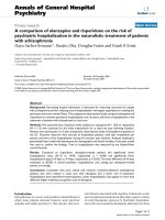
Figure 1
Cell differentiation from mesenchymal stem cells (MSCs) to
osteoblasts and osteocytes. Parathyroid hormone (PTH) promotes
osteoblast proliferation via several mechanisms. PTH stimulates
preosteoblasts (PreOBs) and osteoblasts to make growth factors
(GFs), which promote proliferation of MSCs to PreOBs. PTH
stimulates the conversion of bone-lining cells to osteoblasts, and it
prevents osteoblast and osteocyte apoptosis. BMP, bone
morphogenetic protein ; FGF, fibroblast growth factor; IGF-1, insulin-
like growth factor 1; TGF-β, transforming growth factor β; VEGF,
vascular endothelial growth factor. Adapted from Whitfield [13].
GFs
• VEGF
• FGF-2
• IGF-1
• TGF-βs
• BMPs
MSC
Imm.
PreOB
PreOB OB
Lining
Cell
Osteocyte
Apoptosis
+
+
PTH
PTH
–
–
216
Arthritis Research & Therapy Vol 5 No 5 Lane and Kelman
ular bone showed an increase in trabecular bone volume
and trabecular connections [12]. The histomorphometric
assessment showed bone formation on both the
periosteal and endocortical surface, with a suggestion of
less erosion surface. The investigators suggested that
PTH might be improving bone mass and bone strength by
producing a positive bone balance during remodeling
[5,12,22].
Orwoll and colleagues performed a large randomized,
placebo-controlled trial of PTH in 437 men with osteo-
porosis (either idiopathic or hypogonadal) [23]. The men
were randomized to placebo or 20 or 40 µg of daily sub-
cutaneous injections of rhPTH (1-34) for an average dura-
tion of 11 months. The BMD of the lumbar spine increased
in the treatment groups by 6 to 9% and the femoral neck
BMD by 1.5 to 3%, and the radial BMD decreased by
<1%. Study subjects followed up for 18 months after dis-
continuation of PTH had a nearly 50% reduction in the risk
of vertebral fracture [23].
PTH in combination with other antiresorptive agents
Previously, there was a concern that PTH treatment would
increase the trabecular bone mass at the expense of corti-
cal bone. To protect the skeleton from enlarged remodel-
ing space created by PTH treatment as well as to attempt
to obtain further gain in bone density and prevent any
decline, a number of investigators evaluated the use of
PTH in the presence of antiresorptive agents that would
prevent cortical bone remodeling and bone loss. Initial
combination studies were performed with hormone
replacement therapy (HRT), since bisphosphonates were
not yet available. Current combination studies are evaluat-
ing bisphosphonate treatment together with or after PTH
therapy [5].
Lindsay and colleagues performed the initial randomized,
controlled trial of estrogen with PTH (1-34) in post-
menopausal women with osteoporosis for 3 years [24].
PTH treatment resulted in BMD increases in the lumbar
spine of nearly 13% and in the total hip of about 4%. The
incident vertebral fracture risk was also reduced in the
PTH-treated group [5].
Roe and Arnaud and colleagues performed an random-
ized, controlled trial of PTH (1-34) at 40 µg per day with
HRT in postmenopausal women for 2 years [25]. After
2 years, the BMD of the lumbar spine as measured by
dual-energy x-ray absorptiometry increased by nearly 30%
and that of the femoral neck increased by about 8%, in
comparison with estrogen alone. Quantitative-computed-
tomography measurements of the lumbar spine for trabec-
ular bone volume increased by nearly 80% in the
PTH-treated group compared with the placebo group [25]
and three-dimensional quantitative computed tomography
of the hip showed significant increases in cortical bone
thickness directed centrally on the endocortical surface of
the femoral neck [26]. However, since the newly formed
bone on the endocortical surface was less mineralized
than the cortical bone in the hip, the real changes in hip
cortical bone were not well reflected by BMD, because it
is a ratio of bone mineral content to bone area.
Recently, Rittmaster and colleagues conducted a random-
ized, controlled trial with PTH (1-84) treatment for 1 year,
followed by alendronate (10 mg/day) for 1 to 2 years [27].
After the 2-year treatment period, the group given a high
dose of PTH had about a 14% increase in lumbar spine
BMD; however, the placebo group that was treated with
alendronate for the second part of the study had a gain of
about 6%. It appears that PTH treatment followed by a
bisphosphonate was additive in this study. One explana-
tion for the additional gains in bone mass after PTH
therapy is that PTH increased bone mass but also opened
up remodeling space, especially in the cortical bone com-
partment. Alendronate treatment allowed remodeling
space opened up by PTH to fill in, thereby allowing a sub-
stantial increase in bone mass. Whether this type of
sequential therapy of an anabolic agent followed by an
antiresorptive agent will reduce the risk of fracture is not
known. However, additional studies should now be per-
formed to assess whether fracture risk is reduced with this
type of sequential therapy [28–30].
Since PTH has been approved for the treatment of osteo-
porosis, a number of questions have arisen. At present, we
do not know if the combination of PTH plus a bisphospho-
nate will be additive or synergistic to the anabolic bone
response [28–30]. Also, we are not sure if patients who
have been treated for several years (> 3) with a bisphos-
phonate such as alendronate will have a good anabolic
response to PTH. Small pilot studies suggest that patients
who are treated for 3 years with a bisphosphonate, alen-
dronate, and are then treated with PTH have a delayed
response in biochemical markers of bone turnover and
increases in bone mass over the first year compared with
patients treated with raloxifene for 3 years prior to PTH
[31]. Additional research is needed to determine when
best to prescribe PTH in patients chronically treated with
a bisphosphonate. At this time, there is no contraindica-
tion to treating patients with PTH that have been treated
with a bisphosphonate; however, we have no data to
support the use of the PTH with a bisphosphonate.
The approval of rhPTH (1-34) was a dramatic step forward
in the treatment of osteoporosis. However, a number of
other PTH fragments are now being studied. Some are at
the preclinical stage and some have gone on to clinical
evaluation. Examples are listed but are not limited to
PTH (1-84), PTHrP, PTH (1-31), PTH (2-34), PTH (8-84),
and PTH (1-28), PTH (13-34), PTH (3-34) [13]. Interest-
ing results were reported from a small placebo-controlled
217
clinical trial of women with osteoporosis who were treated
for 3 months with PTHrP [32]. The study subjects had a
4.7% increase in lumbar spine mass in the PTHrP group,
associated with an increase of 60% above the baseline
level in the serum osteocalcin, a measurement of bone for-
mation. However, during this 3-month study, the bone
resorption markers serum N-telopeptide (NTX) crosslinks
and urine deoxypyridinoline (DPD) crosslinks did not
change from the baseline levels in the PTHrP or the
placebo group [32]. These results suggest that PTHrP,
unlike PTH (1-34), may be a more effective uncoupler of
bone turnover, as PTHrP did not increase bone resorption
at 3 months while all clinical studies of PTH (1-34) and
PTH (1-84) show bone resorption markedly increased by
3 months. Additional, large and longer-term studies are
needed to determine the durability of this finding [32].
Growth hormone and insulin-like growth
factor 1
GH is critical for the development and maintenance of
bone mass [33]. It exerts its bone effects via IGF-1. GH
secretion decreases with aging, and therefore so does
that of IGF-1. GH deficiency is associated with an
increased incidence of fracture in adults [34,35,5].
Studies have suggested that recombinant human GH may
improve muscle and bone mass in men over 60 years of
age [36], and recombinant human GH has been shown to
improve muscle and bone mass in patients with GH defi-
ciency, and has been approved by the Food and Drug
Administration for this use.
Mechanisms for the role of IGF-1 in bone metabolism have
yet to be clearly defined [37]. In the process of bone
remodeling, once bone resorption occurs, growth factors,
e.g. IGFs and transforming growth factors (TGFs), are
released from bone matrix and promote the recruitment of
osteoblasts and osteoclasts to the bone surface. Mice,
which lack the IGF-1 gene, have relatively low cortical
bone density. IGFs are present in the skeleton, as well as
circulation. Type I IGF receptors are present on both
osteoblasts and osteoclasts. Most skeletal IGF-1 is
derived from local osteoblasts and plays a role in cell dif-
ferentiation in the osteoblast lineage. Hormones known to
exert effects on bone turnover in part regulate IGF-1
expression. Specifically, PTH and estradiol have been
shown to enhance IGF-1 transcription in rats [5,37].
There has been concern about the safety of therapeutic
GH/IGF-1, because of epidemiologic studies suggesting
an association of normal to high serum IGF-1 levels with
breast, prostate, and colon cancer [38–40]. Also, use of
GH may result (theoretically) in direct metabolic side
effects such as diabetes mellitus.
However, GH has been used in osteoporosis studies.
Recently, Landin-Wilhelmsen and colleagues performed a
randomized, placebo-controlled trial of postmenopausal
women with osteoporosis [41]. The use of subcutaneous
recombinant human GH for 18 months in combination
with HRT, followed by HRT alone for 30 additional
months, resulted in a 14% increase in lumbar spine bone
mineral content at the 4-year follow-up versus HRT and
placebo. Interestingly, not only did the group given HRT +
GH experience an increase in the bone mineral content of
the spine and hip within the group and relative to the HRT-
only group, but also the lumbar spine and femoral neck
bone area was increased from baseline to year 4 in the
group given HRT + GH [41]. Therefore, these results
demonstrate that GH with HRT was more effective than
HRT alone at increasing both bone mineral content and
bone size. However, additional studies will need to be per-
formed to determine if the risk of fracture is reduced by GH
therapy and if GH has a reasonable safety profile, given
that the action of GH on bone is through IGF-1. Finally, the
risk of cancer in this group of patients is unknown.
Strontium
Strontium is chemically similar to calcium and has been
shown to play both an anabolic and an antiresorptive role
in bone metabolism, in both preclinical and clinical studies
[5]. Recent clinical studies, reviewed below, have demon-
strated a therapeutic role for strontium ranelate in post-
menopausal osteoporosis.
The anabolic and antiresorptive properties of strontium on
bone have been demonstrated in vitro. Strontium
increases the synthesis of collagen and other proteins in
osteoblasts and has been shown to increase replication of
osteoblast progenitor cells [5,42]. It has been shown to
directly induce inhibition of osteoclast bone resorption in
rat osteoclast assays incubated with bone slices and to
inhibit osteoclast differentiation in a chicken bone marrow
culture. In preclinical rat studies, Marie and colleagues
reported that treating ovariectomized osteopenic rats with
a strontium salt for 60 days improved the bone mineral
content and increased the trabecular bone volume to the
levels found in sham-treated rats [43].
A large, randomized, double-blind, placebo-controlled trial
(PREVOS) was performed to determine if strontium can
prevent bone loss due to estrogen deficiency [44]. Stron-
tium treatment (1 g/day) for 2 years in early post-
menopausal women gave significant improvements in
bone mineral density compared with the placebo, in the
lumbar spine (by about 2.4%), femoral neck (3.3%), and
total hip (4.1%) (P < 0.001). More recently, in a phase III
study, the SOTI trial [45], 1649 postmenopausal women
with osteoporosis were randomized to treatment with
strontium (2 g/day) or placebo. Strontium ranelate
reduced the risk of new vertebral fracture over 3 years by
41% compared with placebo (P < 0.001). Another phase
III study, TROPOS (treatment of peripheral osteoporosis)
Available online />218
was performed using strontium [46]. This study was a ran-
domized, double-blind, placebo-controlled trial with 5091
postmenopausal women, to determine the efficacy of oral
strontium ranelate at preventing new nonvertebral frac-
tures and on femoral neck BMD. The treatment group
showed a significant increase of femoral neck BMD, by
6.5% of baseline values, and a 33% decreased risk of
new nonvertebral fractures (P < 0.001). Both studies
demonstrated an uncoupling of bone turnover, as the
bone formation marker serum alkaline phosphatase
increased with strontium treatment and the bone resorp-
tion marker serum C-terminal telopeptide of collagen I
decreased. This uncoupling of bone turnover, with forma-
tion increasing and resorption decreasing, may lend
support to the anabolic and antiresorptive properties of
strontium on bone. While the adverse event profile was
favorable for strontium in both large randomized studies,
both additional safety and a better understanding of the
bone actions of strontium ranelate are still required.
Statins
One of the most interesting findings in the bone field
recently is the observation that lipophilic 3-hydroxy-3-
methylglutaryl coenzyme A reductase inhibitors (statins),
specifically lovastatin, atorvastatin, cerivastatin, pitavas-
tatin, and simvastatin, may alter bone metabolism [13].
Recent attention has focused on the role of statins, widely
prescribed for treatment of cardiovascular disease, as
agents capable of promoting bone growth. Possible
mechanisms of statins in bone formation involve stimula-
tion of BMP-2 and endothelial nitric oxide synthase
(eNOS) [13,47–50]. Statins have been shown to stimu-
late BMP-2 synthesis in cultured animal and human bone
cells. eNOS is found sequestered in invaginations of the
osteoblast membrane. Knockout mice lacking the eNOS
gene demonstrate reduced bone formation. Statins have
been shown to increase the expression and activity of the
eNOS gene and to inhibit eNOS-induced osteogenesis in
the mouse calvaria system. In studies with human
osteoblasts, however, eNOS inhibition did not prevent the
action of statins on bone formation [48,49,13].
Preclinical animal studies found statins decreased gluco-
corticoid-induced bone loss in rabbits and increased bone
formation in mouse calvariae [47,51]. In both preclinical
rat studies and a small clinical study measuring serum
markers of bone remodeling in 14 postmenopausal
women using statins, a relative decrease was found in
markers of bone resorption, but there was no change in
markers of bone formation [13,52].
The clinical studies evaluating statins and bone effects
have been from observational cohorts of women taking
statins or from data obtained from randomized, controlled
clinical trials with information on statin use and fracture
endpoints. A meta-analysis of these data report a statisti-
cally significant 57% reduction (CI 0.25–0.75) in the risk
of hip fractures and nonspine fractures 0.69 (CI
0.55–0.88) [53]. However, the effects of statins on bone
was also evaluated in two large randomized, placebo-con-
trolled studies in which reduction of cholesterol and car-
diovascular endpoints were the primary outcomes [54,55].
In both of these randomized controlled studies, statins did
not reduce the risk of fracture. Also, in another large clini-
cal study, the Women’s Health Initiative Study, women
who used statins did not have a significant decrease in
fractures after 3 years [56]. While individuals entering a
statin trial for cardiovascular disease or the Women’s
Health Initiative may not have osteoporosis or risk factors
for osteoporosis, it does bring into question whether
statins have a bone effect that we can measure clinically.
Therefore, until a study of statins is performed that evalu-
ates the effects on fracture reduction in patients with
osteoporosis, definite recommendations of statin for bone
health cannot be made.
Growth factors and bone morphogenetic
proteins
Cytokines expressed during bone formation either from
fractures or from other anabolic hormones (PTH, GH) are
potential therapeutic agents for stimulating bone growth
and bone repair. These include, but are not limited to, IGF-
1, TGF-βs, fibroblast growth factors (FGFs), VEGF, and
BMPs [5,13].
Transforming growth factor
ββ
Osteoblasts and adipocytes are derived from bone
marrow mesenchymal stromal cells. TGF-β is the most
abundant bone growth factor [57]. It is stored in bone
matrix and released during bone resorption. TGF-β plays a
role in proliferation, differentiation, and cytokine expression
of bone. It has been shown to increase bone matrix forma-
tion in rats and in cultured human bone marrow fibro-
blasts. Its administration in in vitro experiments resulted in
increased cell growth and increased matrix proteoglycan
secretion and collagen synthesis. It also reduced adipoge-
nesis (which is increased in osteoporosis). Additionally,
TGF-β was shown to increase VEGF expression by
osteoblasts in fetal rat calvarial cells [58].
Fibroblast growth factor
FGFs have been shown to act as mitogens on fibroblasts,
osteoblasts, and chondrocytes, cells involved in bone
growth and fracture healing. In cultured human bone
marrow fibroblasts, administration of bFGF yielded an
increase in fibroblast colony and size. bFGF administered
to growing rats resulted in an increase of the numbers of
osteoblast precursor cells, followed by an increase of
osteoblasts, and ultimately an increase in endosteal and
endochondral bone formation [59]. Pun and colleagues
[60] and Lane and Wronski [14,61,62] have demon-
strated increased cortical bone mass and trabecular bone
Arthritis Research & Therapy Vol 5 No 5 Lane and Kelman
219
spicule formation within tibial diaphysis and metaphysis of
ovariectomized osteopenic rats treated with bFGF. Inter-
estingly, bFGF and PTH, when given to osteoporotic ovari-
ectomized rats for 6 weeks, resulted in similar increases in
trabecular bone volume; however, bFGF increased the
number of trabeculae and the connectivity whereas the
major effect of PTH is on trabecular thickness [62].
Vascular endothelial growth factor
VEGF is a growth factor that is known to induce neovascu-
larization and is expressed by osteoblasts. It has been
shown to promote osteoblast differentiation and migration,
as well as to be essential in bone healing [63]. In addition,
the bone-forming actions of PTH may result from produc-
tion of VEGF that increases both differentiation of mes-
enchymal cells to osteoblasts and endothelial cells. Street
and colleagues have demonstrated that inhibiting VEGF
function in mice with femoral fractures decreased bone for-
mation and callus mineralization. In mouse femur and rabbit
radii fracture models, local application of slow-release
VEGF improved callus calcification and volume [63].
Bone morphogenetic protein-2
Recombinant human bone morphogenetic protein
(rhBMP)-2 plays an important role in bone formation and
has been shown to enhance fracture healing. It has been
shown to induce mesenchymal differentiation into
osteoblasts by promoting recruitment of osteoprogenitor
cells. BMP-2 has also been shown to stimulate transcrip-
tion of the cbfa1 gene, which is essential for osteoblast
differentiation [64–66]. In fracture healing, there is
increased BMP receptor expression in osteogenic cells
near the fracture, in fibroblast-like spindle cells, and in
fibroblasts involved in endochondral ossification [67].
Welch and colleagues showed that rhBMP-2 enhanced
tibial fracture healing in goats [68]. Subsequently, Boux-
sein and colleagues, in a placebo-controlled study,
showed improved ulnar ‘osteotomy’ healing in mature
rabbits that were treated with an absorbable collagen
sponge containing rhBMP applied to the osteotomy site
[69]. In their study, osteotomy healing time was reduced
by 33%, the area of mineralized callus was 20–60%
greater as measured by quantitative computed tomogra-
phy scanning, and histologically the callus appeared more
symmetric in the rhBMP-2 treatment group [69]. More
recently, Govender and colleagues performed a prospec-
tive, randomized, controlled study with 450 patients in 11
countries who had sustained open tibial fractures [70].
They compared outcomes in three groups. The control
group received standard-of-care therapy, that is, fracture
fixation with intramedullary nailing. The two study groups
received standard-of-care therapy and intraoperative
placement of an absorbable collagen sponge containing
rhBMP-2 at 6 or 12 mg. The treatment group given the
higher dose had a 44% reduced risk of requiring a sec-
ondary intervention due to delayed union versus the con-
trols [70]. BMP-2 has now been approved by the Food
and Drug Administration for human fractures (press
release, 21 November 2002, Wyeth Pharmaceuticals Inc.,
Madison, NJ, USA). Recently, BMP-2, when placed in a
sponge in an implant cage device (InFUSE bone graft,
Wyeth Pharmaceuticals Inc.), reduced the time to lumbar
interbody fusion in humans [71]. BMP-2 had also been
approved for lumbar interbody spinal fusion with the
InFUSE bone graft device in the United States [72].
Bone morphogenetic protein-7
Like BMP-2, BMP-7 (OP-1) induces ecotopic bone forma-
tion in vivo, and in preclinical and clinical fracture models
it promoted bone repair [73–78]. In clinical trials, OP-1,
delivered with a type-1 collagen carrier, promoted bridging
of a critical defect in the fibula of patients that underwent
tibial osteotomy [75]. In addition, OP-1 was found to be
equivalent to the gold-standard, autogenous bone graft in
a clinical study of patients with nonunions [76]. Based on
the result of these clinical trials, OP-1 was granted a
humanitarian device exemption for the treatment of estab-
lished nonunions (press release, 17 October 2002,
Stryker Inc., Kalamazoo, MI, USA).
Interestingly, the promotion of bone-healing benefits by
both BMP-2 and OP-1 is believed to be due to their ability
to stimulate the proliferation and differentiation of mes-
enchymal and osteoprogenitor cells, and both are angio-
genic. The angiogenic effect of OP-1 may be direct and
with BMP-2 it may be through VEGF.
Fluoride
Fluoride has been used for years as an anabolic agent for
osteoporosis treatment. It does stimulate the osteoblasts
to lay down osteoid and bone mass increases. However,
fluoride itself is incorporated into the bone-mineralized
matrix, and because fluoroapatite is not as strong as
hydroxyapatite, the resulting bone is therefore not as
strong as normally mineralized bone [13]. Clinical trials
from the early 1990s [79,80] using high-dose fluoride
(75 mg twice a day) to treat postmenopausal osteoporosis
showed dramatic improvements in lumbar spine BMD in
fluoride-treated subjects compared with the placebo
group [79]. However, there was no improvement in the
incidence of fractures of the lumbar spine and there was
an increase in peripheral skeletal fractures with fluoride
treatment compared with the control group. In these trials,
adverse gastrointestinal effects were common [79]. In a
review of the initial studies, investigators believed the sub-
jects may have given too high a dose of fluoride, which
weakened the bone matrix. Therefore, additional clinical
trials were done using a lower-dose, slow-release formula-
tion (NaF slow release, 25 mg twice a day) and was found
to have a better side-effect profile and to give a significant
reduction in the risk of lumbar spine fracture in compari-
son with the placebo group after about 3 years [81]. In
Available online />220
addition, a few studies done with fluoride and a bisphos-
phonate, etidronate, resulted in a synergistic improvement
in BMD in men with osteoporosis [82]. These trials were
small, however, and the potential therapeutic role of fluo-
ride in the treatment of osteoporosis has yet to be deter-
mined. The challenge relating to the use of fluoride as a
bone-building agent is to determine a dose that is safe
and builds strong bone. It is possible that a low dose of
fluoride with a bisphosphonate may be a viable therapy.
Since the cost of fluoride is low, from a public health
prospective, and the medication has a good safety profile,
additional studies to determine fracture reduction should
be pursued.
Conclusion
A renewed excitement for anabolic therapies for the treat-
ment of osteoporosis and bone fractures has recently
occurred with the approval of rhPTH (1-34), BMP-2, and
BMP-7. The use of anabolic therapies has shown
increased bone mass, a reduced risk of fracture in individ-
uals with osteoporosis, and increased speed of healing of
bone fractures and fusions. However, after demonstrating
that anabolic agents are effective, we now need to turn
our attention to determining how best to use these power-
ful growth factors and hormones. The potential for short
courses of anabolic therapies followed by maintenance
therapy with antiresorptive agents may make it possible for
patients with osteoporosis to increase their bone mass
and maintain bone strength so that their risk of fracture is
reduced. Bone growth factors may provide the opportunity
to restore lost bone trabecular structure, followed by a
therapy such as PTH that can thicken and further
strengthen the bone matrix. The challenge now is to find
the most efficacious treatment regimens of anabolic
agents to prescribe to patients with osteoporosis.
Competing interests
None declared.
References
1. Lee CA, Einhorn TA: Introduction to normal bone physiology
and pathophysiology. In Osteoporosis. 2nd edition. Edited by
Marcus R, Feldman D, Kelsey J. San Francisco, CA: Academic
Press; 2001:3-19.
2. Black DM, Cummings SR, Karpf DB, Cauley JA, Thompson DE,
Nevitt MC, Bauer DC, Genant HK, Haskell WL, Marcus R, Ott
SM, Torner JC, Quandt SA, Reiss TF, Ensrud KE: Randomized
trial of effect of alendronate on risk of fracture in women with
existing vertebral fractures. Lancet 1996, 348:1535-1541.
3. Chesnut CH 3rd, Silverman S, Andriano K, Genant H, Gimona A,
Harris S, Kiel D, LeBoff M, Maricic M, Miller P, Moniz C, Peacock
M, Richardson P, Watts N, Baylink D: A randomized trial of
nasal spray salmon calcitonin in postmenopausal women with
established osteoporosis: the prevent recurrence of osteo-
porotic fractures. Am J Med 2000, 109:267-276.
4. Ettinger B, Black DM, Mitlak BH, Knickerbocker RK, Nickelsen T,
Genant HK, Christiansen C, Delmas PD, Zanchetta JR,
Stakkestad J, Gluer CC, Krueger K, Cohen FJ, Eckert S, Ensrud
KE, Avioli LV, Lips P, Cummings SR: Reduction of vertebral frac-
ture risk in postmenopausal women with osteoporosis
treated with raloxifene: results from a 3-year randomized clin-
ical trial. JAMA 1999, 282:637-645.
5. Rubin MR, Bilizekin JP: New anabolic therapies for osteoporo-
sis. Curr Opin Rheumatol 2002, 14:433-440.
6. Crandall C: Parathyroid hormone for treatment of osteoporo-
sis. Arch Intern Med 2002, 162:2297-2309.
7. Albright F, Aub JC, Bauer W: Hyperparathyroidism: a common
and polymorphic condition illustrated by seventeen proven
cases from one clinic. JAMA 1934, 102:1276-1287.
8. Albright F, Reifenstein EC: The Parathyroid Glands and Metabolic
Bone Disease. Baltimore: Williams & Wilkins; 1948.
9. Hock JM, Gera I, Fonseca J, Raisz LG: Human parathyroid
hormone (1-34) increases bone mass in ovariectomized and
orchidectomized rats. Endocrinology 1988, 122:2899-2904.
10. Lane NE, Thompson JM, Strewler G, Kinney JH: Intermittent
treatment with human parathyroid hormone (hPTH 1-34)
increased trabecular bone mass but not connectivity in
osteopenic rats. J Bone Miner Res 1996, 10:1470-1477.
11. Neer RM, Arnaud CD, Zanchetta JR, Prince R, Gaich GA, Regin-
ster JY, Hodsman AB, Eriksen EF, Ish-Shalom S, Genant HK,
Wang O, Mitlak BH: Effect of parathyroid hormone (-34) on
fractures and bone mineral density in postmenopausal women
with osteoporosis. N Engl J Med 2001, 344:1434-1441.
12. Dempster DW, Cosman F, Kurland ES, Zhou H, Nieves J,
Woelfert L, Shane E, Plavetic K, Muller R, Bilezikian J, Lindsay R:
Effects of daily treatment with parathyroid hormone on bone
microarchitecture and turnover in patients with osteoporosis:
a paired biopsy study. J Bone Miner Res 2001, 16:1846-1853.
13. Whitfield JF: How to grow bone to treat osteoporosis and
mend fractures. Curr Rheumatol Rep 2003, 5:45-56.
14. Lane NE, Yao W, Kinney JH, Modin G, Baloosh M, Wronski TJ:
Both hPTH (1-34) increase trabecular bone mass in
osteopenic rats however they have different effects on tra-
becular bone structure. J Bone Min Res 2003, in press.
15. Lane N, Yao W, Arnaud C: Association of serum RANKL and
OPG levels with other biochemical markers of bone turnover
in glucocorticoid-induced osteoporosis patients treated with
hPTH (1-34) [abstract]. J Bone Miner Res 2002, Suppl 1:F366.
16. Lane NE, Yao W, Arnaud CD: Changes in serum RANKL, OPG
and IL-6 during hPTH (1-34) administration in patients with
glucocorticoid induced osteoporosis. J Bone Miner Res 2003,
Suppl 1: in press.
17. Mitnick MA, Grey A, Masiukiewicz U, Bartkiewicz M, Rios-Velez L,
Friedman S, Xu L, Horowitz MC, Insogna K:Parathyroid hormone
induces hepatic production of bioactive IL-6 and its soluble
receptor. Am J Physiol Endocrinol Metab 2001, 280:E405-E412.
18. Greenfield EM, Horowitz Lavish SA: Stimulation of parathyroid
hormone of interleukin-6 and leukemia inhibitory factor
expression in osteoblasts is an immediate-early gene
response induced by cAMP signal transduction. J Biol Chem
1996, 271:10984-10989.
19. Fu Q, Jilka RL, Manolagas SC, O’Brien CA: Parathyroid
hormone stimulates receptor activator of NFkB ligand and
inhibits osteoprotegrin expression via protein kinase A activa-
tion of cAMP-response element binding protein. J Biol Chem
2002, 277:48868-48875 .
20. Lee S-K, Lorenzo JA: Parathyroid hormone stimulates TRANCE
and inhibits osteoprotegrin messenger ribonucleic acid
expression with osteoclast-like cell formation. Endocrinology
1999, 140:3552-3561.
21. Burr DB, Hirano T, Turner CH, Hotchkiss C, Brommage R, Hock
JM: Intermittently administered human parathyroid hormone
(1-34) treatment increases intracortical bone turnover and
porosity without reducing bone strength in the humerus of
ovariectomized cynomolgus monkeys. J Bone Miner Res
2001, 16:157-165.
22. Kurland ES, Cosman F, McMahon DJ, Rosen CJ, Lindsay R,
Bilezikian JP: Parathyroid hormone as a therapy for idiopathic
osteoporosis in men: effects on bone mineral density and
bone markers. J Clin Endocrinol Metab 2000, 85:3069-3076.
23. Orwoll ES, Scheele WH, Paul S, Adami S, Syversen U, Diez-
Perez A, Kaufman JM, Clancy AD, Gaich GA: The effect of teri-
paratide [human parathyroid hormone (1-34)] therapy on
bone density in men with osteoporosis. J Bone Miner Res
2003, 18:9-17.
24. Lindsay R, Nieves J, Formica C, Henneman E, Woelfert L, Shen V,
Dempster D, Cosman F: Randomized controlled study of effect
of parathyroid hormone on vertebral-bone mass and fracture
incidence among postmenopausal women on oestrogen with
Arthritis Research & Therapy Vol 5 No 5 Lane and Kelman
221
osteoporosis. Lancet 1997, 350:550-555.
25. Roe E, Sanchez S, del puerto G, et al.: Parathyroid hormone 1-
34 (hPTH 1-34) and estrogen produce dramatic bone density
increases in postmenopausal osteoporosis-results from a
placebo-controlled randomized trial [abstract]. J Bone Miner
Res, 1999, Suppl 1:S137.
26. Cann C, Roe EB, Sanchez SD, et al.: PTH effect in the femur:
envelope-specific responses by 3DQCT in postmenopausal
women [abstract]. J Bone Miner Res 1999, Suppl 1:S137.
27. Rittmaster RS, Bolognese M, Ettinger MP, Hanley DA, Hodsman
AB, Kendler DL, Rosen CJ: Enhancement of bone mass in
osteoporotic women with parathyroid hormone followed by
alendronate. J Clin Endocrinol Metab 2000, 85:2129-2134.
28. Black DM, Rosen C, Greenspan S, Ensrud K, Bilezikian J,
McGowan J: PTH and bisphosphonates in the treatment of
osteoporosis: design of the PTH and alendronate (PATH) trial
[abstract]. In ASBMR 23rd annual meeting, 2001:5287.
29. Finkelstein JS, Hayes A, Rao A, Neer RM: Effects of parathyroid
hormone, alendronate or both on bone mineral density in
osteoporotic men [abstract]. J Bone Miner Res 2002, Suppl
1:1007.
30. Neer R, Hayes A, Rao A, Finkelstein J: Effects of parathyroid
hormone, alendronate, or both on bone density in osteo-
porotic postmenopausal women [abstract]. J Bone Miner Res
2002, Suppl 1:1039.
31. Ettinger B, San Martin JA, Crans G, Pavo I: Early response of
bone turnover markers and bone mineral density to teri-
paratide (rhPTH 1-34) in postmenopausal women previously
treated with an anti-resorptive agent [abstract]. J Bone Miner
Res 2002, Suppl 1:1136.
32. Horwitz M, Stewart A, Greenspan SL: Short-term, high-dose
parathyroid hormone-related protein as a skeletal anabolic
agent for the treatment of postmenopausal osteoporosis. J
Clin Endocrinol Metab, 2003, 88:569-575.
33. Wuster C, Rosen C: Growth hormone, insulin-like growth
factors. Potential applications and limitations in the manage-
ment of osteoporosis. In Osteoporosis. 2nd edition. Edited by
Marcus R, Feldman D, Kelsey J. San Francisco, CA: Academic
Press; 2001: 747-767.
34. Rosen, T, Wilhelmsen L, Landin-Wilhelmsen K, Lappas G, Bengt-
son B-A: Increased fracture frequency in adult patients with
hypopituitarism and GH deficiency. Eur J Endocrinol 1997,
137:240-245.
35. Wuster C, Abs R, Bengtsson B-A, Bennmarker H, Feldt-Ras-
mussen U, Hernberg-Stahl E, Monsom JP, Westberg B, Wilton, P:
The incidence of growth hormone deficiency, growth
hormone replacement therapy, and other aspects of hypopi-
tuitarism on fracture rate and bone mineral density. J Bone
Miner Res 2001, 16:398-405.
36. Rudman D, Feller AG, Nagraj HS, Gergans GA, Lalitha PY, Gold-
berg AF, Schlenkler RA, Cohn L, Rudman IW, Mattson DE:
Effects of human growth hormone in men over 60 years old.
N Engl J Med 1990, 323:1-6.
37. Yakar S, Rosen CJ: From mouse to man: redefining the role of
insulin-like growth factor-I in the acquisition of bone mass.
Proc Soc Exp Biol Med 2003, 228:245-252.
38. Hankinson SE, Willet WC, Colditz GA, Hunter DJ, Michaud DS,
Deroo B, Rosner B, Speizer FE, Pollak M: Circulating concentra-
tions of insulin-like growth factor-I and risk of breast cancer.
Lancet 1998, 351:1393-1396.
39. Ma J, Pollak MN, Giovannucci E, Chan JM, Tao Y, Hennekens CH,
Stampfer MJ: Prospective study of colorectal cancer risk in
men and plasma levels of insulin-like growth factor (IGF)-I
and IGF-binding protein-3. J Natl Cancer Inst 1999, 91:620-
625.
40. Chan JM, Stampfer MJ, Giovannucci E, Gann PH, Ma J, Wilkinson
P, Hennekens CH, Pollak M: Plasma insulin-like growth factor-I
and prostate cancer risk: A prospective study. Science 1998,
279:563-566.
41. Landin-Wilhelmsen K, Nilsson A, Bosaeus I, Bengtsson, B:
Growth hormone increases bone mineral content in post-
menopausal osteoporosis: a randomized placebo-controlled
trial. J Bone Miner Res 2003, 18:393-405.
42. Reginster JY: Strontium ranelate in osteoporosis. Curr Pharm
Des 2002, 8:1907-1916.
43. Marie PJ, Hott M, Modrowski D, De Pollack C, Guillemain J, Delof-
fre P, Tsouderos Y: An uncoupling agent containing strontium
prevents bone loss by depressing bone resorption and main-
taining bone formation in estrogen-deficient rats. J Bone
Miner Res 1993, 8:607-615.
44. Reginster JY, Deroisy R, Dougados M, Jupsin I, Colette J, Roux C:
Prevention of early postmenopausal bone loss by strontium
ranelate: the randomized, two-year, double-masked, dose-
ranging, placebo-controlled PREVOS trial. Osteoporos Int
2002, 13:925-931.
45. Reginster JY, Hoszowski K, Roces Varela A, Balogh A, Clements
M, Fiore C, Cormier C, Schmidt WE, Jensen JB, Prince R,
Raeman F, Rizzoli R, Meunier PJ: Strontium ranelate: a new
effective antiosteoporotic treatment reducing the incidence of
vertebral and non vertebral fractures in postmenopausal
women with osteoporosis. In International Osteoporosis Society
meeting. June 2003; Osaka, Japan, in press.
46. Meunier PJ, Reginster JY: Design and methodology of the
phase 3 trials for the clinical development of strontium
ranelate in the treatment of women with postmenopausal
osteoporosis. Osteoporos Int 2003, Suppl 3:66-76.
47. Mundy G, Garrett R, Harris S, Chan J, Chen D, Rossini G, Boyce
B, Zhao M, Gutierrez G: Stimulation of bone formation in vitro
and in rodents by statins. Science 1999, 286:1546-1549.
48. Garrett IR, Gutierrez G, Chen D, et al.: Statins stimulate bone
formation by enhancing eNOS expression [abstract]. J Bone
Miner Res 2001, 16:S141.
49. van’t Hof RJ, Ralston SH: Nitric oxide and bone. Immunology
2001, 103:113-118.
50. Ohnaka K, Shimoda S, Nawata H, Shimokawa H, Kaibuchi K,
Iwamoto Y, Takayanagi R: Pitavastatin-enhanced BMP-2 and
osteocalcin expression by inhibition of Rho-associated kinase
in human osteoblasts. Biochem Biophys Res Commun 2001,
287:337-342
51. Wang GJ, Chung KC, Shen WJ: Lipid-clearing agents in steroid-
induced osteoporosis. J Formos Med Assoc 1995, 94:589-592.
52. Cosman F, Nieves J, Zion M, et al.: Effects of short-term
cerivastatin on bone turnover [abstract]. J Bone Miner Res
2001, 16:S29.
53. Bauer: Arch Int Med, in press.
54. Bauer DC: HMG CoA reductase inhibitors and the skeleton: a
comprehensive review. Osteoporos Int 2003, 4:273-282
55. Reid IR, Hague W, Emberson J, Baker J, Tonkin A, Hunt D,
MacMahon S, Sharpe N: Effect of pravastatin on frequency of
fracture in the LIPID study: secondary analysis of a ran-
domised controlled trial. Long-term Intervention with Pravas-
tatin in Ischaemic Disease. Lancet 2001, 357:509-512.
56. LaCroix AZ, Cauley JA, Pettinger M, Hsia J, Bauer DC, McGowan
J, Chen Z, Lewis CE, McNeeley G, Passaro MD, Jackson RD:
Statin use, clincal fracture, and bone mineral density in post-
menopausal women: Results from the women's health initia-
tive observational study. Annal Int Med 2003, 139:97-104
57. Locklin RM, Oreffo ROC, Triffit JT: Effects of TGFB and bFGF on
the differentiation of human bone marrow stromal fibroblasts.
Cell Biol Int 1999, 23:185-194.
58. Saadeh PB, Mehrara BJ, Steinbrech DS, Dudziak ME, Greenwald
JA, Luchs JS, Spector JA, Ueno H, Gittes GK, Longaker MT:
Transforming growth factor-B1 modulates the expression of
vascular endothelial growth factor by osteoblasts. Am J
Physiol 1999,
277:C628-C637.
59. Nagai H, Tsukuda R, Mayahara H: Effects of basic fibroblast
growth factor (bFGF) on bone formation in growing rats. Bone
1995, 16:367-373.
60. Pun S, Florio CL, Wronski TJ: Anabolic effects of basic fibrob-
last growth factor in the tibial diaphysis of ovariectomized
rats. Bone 2000, 27:197-202.
61. Wronski TJ, Ratkus AM, Thomsen JS, Vulcan Q, Mosekilde L:
Sequential treatment with basic fibroblast growth factor and
parathyroid hormone restores lost cancellous bone mass and
strength in the proximal tibia of aged ovariectomized rats. J
Bone Miner Res 2001 16:1399-1407.
62. Lane NE, Kumer J, Yao W, Breunig T, Wronski T, Modin G, Kinney
JH: Basic fibroblast growth factor forms new trabeculae that
physically connect with pre-existing trabeculae, and this new
bone is maintained with an anti-resorptive agent and
enhanced with an anabolic agent in an osteopenic rat model.
Osteoporos Int 2003, 14:374-352.
63. Street J, Bao, M, deGuzman L, Bunting S, Peale Jr. FV, Ferrara N,
Steinmetz H, Hoeffel J, Cleland JL, Daugherty A, van Bruggen N,
Available online />222
Redmond HP, Carano RAD, Filvaroff EH: Vascular endothelial
growth factor stimulates bone repair by promoting angiogen-
esis and bone turnover. Proc Natl Acad Sci USA 2002 99:
9656-9661.
64. Wang EA, Israel DI, Kelly S, Luxenberg DP: Bone morpho-
genetic protein-2 causes commitment and differentiation in
C3h10T1/2 and 3T3 cells. Growth Factors 1993 9:57-71.
65. Ahrens M, Ankenbauer T, Schroder D, Hollnagel A, Mayer H,
Gross G: Expression of human bone morphogenetic proteins
-2 or -4 in murine mesenchymal progenitor C3H10T1/2 cells
induces differentiation into distinct mesenchymal cell lin-
eages. DNA Cell Biol 1993 12:871-880.
66. Komori T, Yagi H, Nomura S, Yamaguchi A, Sasaki K, Deguchi K,
Shimuzu Y, Bronson RT, Gao YH, Inada M, Sato M, Okamoto R,
Kitamura Y, Yoshiki S., Kishimoto T: Targeted disruption of
Cbfa1 results in a complete lack of bone formation owing to
maturational arrest of osteoblasts. Cell 1997, 89:755-764.
67. Onishi T, Ishidou Y, Nagamine T, Yone K, Imamura T, Kato M,
Sampath TK, ten Dijke P, Sakou T: Distinct and overlapping pat-
terns of localization of bone morphogenetic protein (BMP)
family members and a BMP type II receptor during fracture
healing in rats. Bone 1998, 22:605-612.
68. Welch RD, Jones Al, Bucholz RW, Reinert CM, Tjia JS, Pierce
WA, Wozney JM, Li XJ: Effect of recombinant human bone
morphogenetic protein-2 on fracture healing in a goat tibial
fracture model. J Bone Miner Res 1998, 13:1483-1490.
69. Bouxsein ML Turek TJ, Blake CA, D’Augusta D, Li X, Stevens M,
Seeherman HJ, Wozney JM: Recombinant human bone mor-
phogenetic protein-2 accelerates healing in a rabbit ulnar
osteotomy model. J Bone Joint Surg 2001, 83-A:1219-1230.
70. Govender S, Csimma C, Genant HK, Valentin-Opran A: Recom-
binant human bone morphogenetic protein-2 for treatment of
open tibial fractures. J Bone Joint Surg 2002, 84-A:2123-
2134.
71. Burkus JK, Transfeldt EE, Kitchel SH, Watkins RG, Balderstom
RA: Clinical and radiographic outcomes of anterior lumbar
interbody fusion using recombinant human bone morpho-
genetic protein-2. Spine 2002, 27:2396-2408.
72. BMP 2—Genetics Institute/ Medtronic-Sofamor Danek/Integra.
Bone morphogenetic protein 2—Genetics Institute/ Medtronic-
Sofamor Danek/Integra, INFUSE Bone Graft, recombinant human
bone morphogenetic protein 2—Genetics Institute/Medtronic-
Sofamor Danek/Integra, RhBMP 2—Genetics Institute/Medtronic-
Sofamor Danek/Integra. BioDrugs 2002, 16:376-377.
73. Barnes GL, Kostenuik PJ, Gerstenfeld LC, Einhorn TA: Growth
factor regulation of fracture repair. J Bone Miner Res 1999, 14:
1805-1815.
74. Reddi AH: Bone morphogenetic proteins: from basic science
to clinical applications. J Bone Joint Surg Am 2001, Suppl 1 (Pt
1):S1-6.
75. Geesink RG, Hoefnagels NH, Bulstra SK: Osteogenic activity of
OP-1 bone morphogenic protein (BMP-7) in a human fibular
defect. J Bone Joint Surg Br 1999 81:710-718.
76. Friedlaender GE, Perry CR, Cole JD, Cook SD, Cierny G,
Muschler GF, Zych GA, Calhoun JH, LaForte AJ, Yin S:
Osteogenic protein-1 (bone morphogenetic protein-7) in the
treatment of tibial nonunions. J Bone Joint Surg Am 2001,
Suppl 1 (Pt 2):S151-S158.
77. Cook SD, Dalton JE, Tan EH, Whitecloud TS 3rd, Rueger DC; In
vivo evaluation of recombinant human osteogenic protein
(rhOP-1) implants as a bone graft substitute for spinal
fusions. Spine 1994, 19:1655-1663.
78. Margolin MD, Cogan AG, Taylor M, Buck D, McAllister TN, Toth
C, McAllister BS: Maxillary sinus augmentation in the non-
human primate: a comparative radiographic and histologic
study between recombinant human osteogenic protein-1 and
natural bone mineral. J Periodontol 1998, 69:911-919.
79. Riggs BL, Hodgson SF, O’Fallon WM, Chao EY, Wahner HW,
Muhs JM, Cedel SL, Melton LJ 3
rd
: Effect of fluoride treatment
on the fracture rate in postmenopausal women with osteo-
porosis. N Engl J Med 1990 322:802-809.
80. Kleerekoper M, Peterson EL, Nelson DA, Phillips E, Schork MA,
Tilley BC, Parfitt AM:A randomized trial of sodium fluoride as a
treatment for postmenopausal osteoporosis. Osteoporos Int
1991, 1:155-161.
81. Pak CY, Sakhaee K, Adams-Huet B, Piziak V, Peterson RD,
Poindexter JR:Treatment of postmenopausal osteoporosis
with slow-release sodium fluoride: final report of a random-
ized controlled trial. Ann Intern Med 1999, 123:401-408.
82. Ringe JD, Rovati LC: Treatment of osteoporosis in men with
fluoride alone or in combination with bisphosphonates. Calcif
Tissue Int 2001, 69:252-255.
Correspondence
Nancy Lane, Division of Rheumatology, University of California, San
Francisco, San Francisco, CA 94121, USA. Tel: +1 415 206 6654;
fax: +1 415 648 8425; e-mail:
Arthritis Research & Therapy Vol 5 No 5 Lane and Kelman