Báo cáo y học: "A review of anatomical and mechanical factors affecting vertebral body integrit
Bạn đang xem bản rút gọn của tài liệu. Xem và tải ngay bản đầy đủ của tài liệu tại đây (216.63 KB, 11 trang )
Int. J. Med. Sci. 2004 1: 170-180
170
International Journal of Medical Sciences
ISSN 1449-1907 www.medsci.org 2004 1(3):170-180
©2004 Ivyspring International Publisher. All rights reserved
A review of anatomical and mechanical factors affecting
vertebral body integrity
Review
Received: 2004.07.01
Accepted: 2004.09.27
Published:2004.10.12
Andrew M Briggs
1 2
, Alison M Greig
1
,
John D Wark
2
,
Nicola L Fazzalari
3
, Kim L Bennell
1
1. Centre for Health, Exercise and Sports Medicine, School of Physiotherapy, University of
Melbourne, Australia.
2. Department of Medicine, Royal Melbourne Hospital, University of Melbourne, Australia.
3. Institute of Medical and Veterinary Science, South Australia.
A
A
b
b
s
s
t
t
r
r
a
a
c
c
t
t
Background: The aetiology of osteoporotic vertebral fracture is multifactorial
and may be conceptualised using a systems framework. Previous studies have
established several correlates of vertebral fracture including reduced
vertebral cross-sectional area, weakness in back extensor muscles, reduced
bone mineral density, increasing age, worsening kyphosis and recent
vertebral fracture. Alterations in these physical characteristics may influence
biomechanical loads and neuromuscular control of the trunk and contribute
to changes in subregional bone mineral density of the vertebral bodies.
Methods: This review discusses factors that have received less attention in
the literature, which may contribute to the development of vertebral fracture.
A literature review was conducted using electronic databases including
Medline, Cinahl and ISI Web of Science to examine the potential contribution
of trabecular architecture, subregional bone mineral density, vertebral
geometry, muscle force, muscle strength, neuromuscular control and
intervertebral disc integrity to the aetiology of osteoporotic vertebral fracture.
Interpretation: A better understanding of factors such as biomechanical
loading and neuromuscular control of the trunk may help to explain the high
incidence of subsequent vertebral fracture after sustaining an initial vertebral
fracture. Consideration of these issues may be important in the development
of prevention and management strategies.
K
K
e
e
y
y
w
w
o
o
r
r
d
d
s
s
osteoporosis, vertebral fracture, bone density, spinal biomechanics, neuromuscular
control
A
A
u
u
t
t
h
h
o
o
r
r
b
b
i
i
o
o
g
g
r
r
a
a
p
p
h
h
y
y
Andrew M Briggs (BSc) is a PhD candidate at the School of Physiotherapy, University of
Melbourne, Australia. He is investigating lumbar spine bone mineral density, thoracic
vertebral loading and paraspinal muscle activity in individuals with osteoporosis.
Alison M Greig (BHk, BSc) is a PhD candidate at the School of Physiotherapy, University
of Melbourne, Australia. She is investigating neuromuscular control characteristics of the
trunk in individuals with osteoporosis.
John D Wark (PhD), Professor of Medicine, Endocrinologist, is Head of the Bone and
Mineral Service at Royal Melbourne Hospital and Broadmeadows Osteoporosis Centre. He
leads a team of researchers investigating bone health, quality and structure changes with
related to genetics, exercise, maturation and pharmacotherapies.
Nicola L Fazzalari (PhD) is Associate Professor, Head of Bone and Joint
Research Laboratory and Chief Medical Scientist (Division of Tissue Pathology) at the
Institute of Medical and Veterinary Science, South Australia. Associate Professor Fazzalari is
recognised for his studies of bone architecture and bone quality. His work is distinguished by
its multidisciplinary approach to tissue analysis, using morphometric, molecular,
biomechanical and mathematical analyses of human skeletal tissue.
Kim L Bennell (PhD) is Associate Professor and Director of the Centre for Health, Exercise
and Sports Medicine, School of Physiotherapy, University of Melbourne, Australia. She leads
a multidisciplinary team investigating musculoskeletal diseases across the lifespan.
C
C
o
o
r
r
r
r
e
e
s
s
p
p
o
o
n
n
d
d
i
i
n
n
g
g
a
a
d
d
d
d
r
r
e
e
s
s
s
s
Andrew Briggs, Centre for Health, Exercise and Sports Medicine, School of Physiotherapy,
University of Melbourne; Parkville, Victoria 3010, Australia. Tel: + 61 3 8344 4171 Fax: +
61 3 8344 4188 e-mail:
,
Int. J. Med. Sci. 2004 1: 170-180
171
1. Introduction
Osteoporosis is a metabolic bone disorder characterised by low bone mass and micro-architectural
deterioration of bone tissue, leading to enhanced bone fragility and a consequent increase in fracture
risk [93]. Osteoporosis is increasingly recognised as an important public health problem due to the
significant physical, economic and psychosocial ramifications of fracture. Vertebral fractures are a
hallmark of osteoporosis and are associated with back pain, functional disability, reduced health-related
quality of life and increased mortality. These associations become more significant with increasing
numbers of vertebral fractures [24, 69].
Previous reviews have outlined the influence of bone mineral density (BMD), bone loss and
important engineering principles such as bone geometry, mechanical properties and load transmission
on the capacity of a vertebral body to resist physiologic loads that may contribute to vertebral fracture
or deformity [32, 31, 65]. With advances in densitometric technologies and ex vivo analyses of
trabecular bone properties, it is now evident that heterogeneity exists within the trabecular bone of a
vertebral body. This observation may assist in the understanding of the complex aetiology underlying
osteoporotic vertebral fracture. However, we postulate that trabecular bone properties cannot explain
entirely why a difference in the prevalence rate of vertebral fractures exists among individuals with
comparable BMD. It is likely that factors other than BMD operate to influence fracture risk, for
example physiologic loading of vertebral bodies and the neuromuscular characteristics of trunk
musculature.
Previously, the factors discussed have been viewed as mutually exclusive, but consideration of
their potential interactions may assist understanding of the complex aetiology of these fractures.
Therefore, the aim of this review is to highlight factors identified in the literature that may act to
influence the risk of sustaining an osteoporotic vertebral fracture.
The risk of osteoporotic vertebral fracture can be conceptualised as a function of loading
conditions and the ability of the human spine to withstand the load. Bone strength depends on its micro-
architectural and macro-architectural features [80]. These features can be affected by the
pathophysiology of osteoporosis and the local and global environment in which the bone operates. The
local environment includes muscle and connective tissue attachments and the adjacent intervertebral
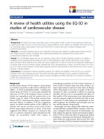
disc, while the global environment can be considered to be the entire body operating in space (see
figure 1).
Figure 1. Diagram of the systems approach that may be applied to explain the multifactorial aetiology
underlying osteoporotic vertebral fracture. Bone, macroscopic, local and global factors may interact to
influence the risk of sustaining a vertebral fracture.
Bone
properties
Macroscopic
properties
Local
environment
Global
environment
Cellular
activity
Trabecular
architecture
Subregional
BMD
Vertebral
geometry
Neuromuscular
control
Muscle force &
strength
Intervertebral
disc integrity
Body
position
External
loads
Vertebral body
integrity
Int. J. Med. Sci. 2004 1: 170-180
172
2. Bone Structural Features
2.1. Trabecular micro-architecture
The design of the trabecular network allows for the efficient distribution of compressive and shear
forces. This efficiency exists via the orientation and spacing of the three dimensional arrangement of
osseous plates and struts that are continuous with the inner surface of the cortex. Vertically-orientated
trabeculae attenuate axial forces, while horizontal plates respond to the tensile (shear) stresses
transferred from the intervertebral disc [88]. An excess of bone resorption over formation leads to
trabecular deterioration and loss. Deterioration in the trabecular architecture transforms the densely
connected, plate-like trabeculae into discontinuous rod-like structures [1, 82]. Such transformations, if
severe enough, are a hallmark of osteoporosis.
Trabecular bone strength is in part attributable to its large surface to volume ratio. This ratio is
approximately four times greater than that observed in cortical bone [72]. With age and menopause, the
trabecular bone mass in the vertebral body (centrum) declines at a faster rate than cortical bone mass.
Cortical bone therefore dominates in the elderly spine. Generally, women loose 20% of peak cortical
bone mass and 40-50% of trabecular mass by 90 years of age [58]. This results in a decline in the load-
bearing capacity of the vertebral body. For example, load bearing capacity in a lumbar vertebra can
decrease from 1000kg in a young individual to 60-90kg with age [62]. In osteoporosis, the normal age-
related changes in cortical thickness and trabecular density become more pronounced resulting in a
significant decline of compressive strength in the vertebral body, increasing the likelihood of fracture.
A loss of trabecular bone in particular, will contribute significantly to a decline in mechanical strength
of the vertebral body [94]. Ex vivo investigations demonstrate that mechanical characteristics of
vertebral bone to strain is dominated by trabecular bone properties [43, 81].
2.2. Subregional Bone Mineral Density (srBMD)
The characteristic reduction in bone mass per unit of volume, i.e. volumetric BMD in osteoporosis
interferes with the mechanical support properties of the bone. Various measures of BMD correlate
strongly with compressive failure loads of vertebrae and can therefore be used as convenient and
generally reliable indicators of compressive strength of vertebrae in vivo when measured with tools
such as dual energy x-ray absorptiometry (DXA) and quantitative computed tomography (QCT) [31,
65]. Approximately 75-90% of the variance in bone strength is accounted for by apparent BMD [53,
87]. Low BMD is one of the most important risk factors for osteoporotic fracture [13]. However,
examination of BMD alone is insufficient to explain the basis for vertebral fracture. Studies have
shown differences in prevalence rates for fracture among individuals with comparable BMD [1, 12, 15,
18, 28, 46, 61, 73, 79, 89, 90]. These observations suggest that a single measure of BMD may not be
representative of vertebral capacity and/or that other factors may operate independent of BMD
measures. For example, risk variation between individuals may lie in the more subtle subregional
density differences within the vertebral body. Subregional differences are hidden when whole vertebral
body BMD is measured [46].
During aging, there is a non-uniform loss of bone within the vertebral body, resulting in trabecular
bone density becoming non-homogenous throughout the centrum [2, 3, 56]. BMD therefore becomes
disproportionate between different regions of the vertebral body. In an examination of cancellous bone
morphometry of lumbar vertebrae, Simpson and colleagues [82] discovered that BMD, as measured by
the ratio of bone volume to total volume in the vertebra, showed significant subregional differences.
Posterior margins of the vertebrae showed greater BMD than anterior regions, while central zones had
the lowest BMD, lowest trabecular number and greatest trabecular separation. Generally vertebrae are
less dense anteriorly and superiorly and more dense posteriorly and inferiorly with heterogeneity
between symmetric sites within the vertebral body [2, 3, 25, 56, 67, 78]. The lower density and
therefore lower compressive strength of the anterior zone of the vertebral body may account for the
greater incidence of anterior crush fracture in individuals with osteoporosis. Therefore, a better
understanding of the mechanics of vertebral fracture may be ascertained by investigating differences in
subregional BMD between individuals with and without vertebral fracture. Overall vertebral BMD may
remain the same in the presence of differential responses in trabecular architecture to mechanical
loading [82]. The mechanisms underlying variation in subregional BMD remain unknown. However, it
is likely that axial force transmission through the vertebral body may play a role. Adjacent musculature
and the intervertebral disc modulate axial force.
3. Macroscopic factors
3.1. Vertebral Geometry
The shape of a vertebra is well designed to accept axial loads. The orientation and framework of
the vertebral body assures that in normal circumstances, compressive load is transferred through the
Int. J. Med. Sci. 2004 1: 170-180
173
vertebral centrum, which by design, is the primary load bearing structure. This is highlighted by the
observation that vertebral centrum size increases cranio-caudally as body weight percentage increases
[9, 23, 87]. The vertebral body has a ratio of trabecular : cortical bone volume of 95:5, which explains
its load bearing capacity when considering the function of the trabecular network [2]. Its design
therefore provides the requirements for optimal load transfer by ensuring maximal strength with
minimal weight [38].
Fracture risk is commonly based on measurements of vertebral BMD. However, other factors such
as bone size and thickness of the cortical rim and endplates need to also be considered [16].
Consideration of these simple engineering principles may assist in fracture risk prediction [31]. Each of
these factors is influenced independently with age, immobilisation, metabolic bone disease, disc
degeneration and osteophyte formation. It is therefore difficult to determine the exact role of each factor
and their individual contribution to the aetiology of vertebral fracture. The size of a vertebral body,
often measured using computed tomography (CT) to calculate cross sectional area (CSA), has been
found to be a predictor of risk of fracture [28, 65]. CSA provides an index of the ability to resist fracture
given that bone strength is positively correlated with bone size [20].
Mechanical stress in a vertebral body during axial loading is inversely proportional to the CSA of
the bone [16, 27]. Mazess et al. [54] found that individuals with osteoporosis had a significantly smaller
vertebral CSA than individuals without osteoporosis. In a study comparing osteoporotic women with
and without vertebral fracture, it was found that those with fracture had a reduced vertebral CSA in
T12-L4 when matched for age, height, weight and BMD [28]. Another study comparing women with
and without vertebral fracture found that vertebral volume was significantly lower in the fracture group
when matched with controls for age, height, weight and BMD [15]. The smaller vertebral size in the
fracture group would increase mechanical stress in the vertebral body, thereby limiting the loading
capacity of the spine. Superimposing smaller vertebral size with the pathology of osteoporosis could
increase fracture risk substantially.
4. Local Environment
4.1. Muscle Force
Muscles that attach to the vertebral column have a role in producing movement and providing
protection to the spine by stabilising its structures. Without muscular support, the spine has a
compression threshold of just 2kg before buckling [6, 66]. The paraspinal extensor muscles play an
important role in maintaining static equilibrium of the trunk by resisting the flexion moment imposed
by gravity and any mass carried anterior to the spine (see figure 2). The role of the erector spinae as a
tonic muscle group has been confirmed by a histological study that found a predominance of type I
muscle fibres (slow twitch, associated with endurance) at both thoracic and lumbar levels [50].
However, forces produced by muscle contraction may also be injurious to spinal structures. The
muscles responsible for balancing spinal gravitational moments attach closely to the axis of rotation of
the segment, therefore being at a mechanical disadvantage when considering the lever arm length.
Relative to other muscles in the body, the spinal extensors have a very short lever arm. Therefore the
force generated by these muscles must be sufficiently large to stabilise the trunk and overcome this
mechanical disadvantage. The result is a higher compressive reaction force delivered to the
intervertebral discs and vertebral centrum [14, 45, 68, 92]. Lever arm length can be affected by
vertebral geometry and vertebral body position, as a function of spinal posture. Women have a smaller
vertebral body size than men [6]. This helps to explain the observation that women have a 9% smaller
lever arm from the erector spinae muscle to the centre of the vertebral body [27]. Gilsanz et al. [28]
discovered that women with osteoporotic fractures had smaller lever arms (by 5.8%) than osteoporotic
women with no fractures. The combination of reduced vertebral CSA and shorter lever arm resulted in
increased mechanical loading by 8% during erect stance and by 15% during trunk flexion in the fracture
cohort.
In a flexed spinal posture, the lever arm lengths of the lumbar erector spinae may decrease by up to
13.3% [9, 92]. Therefore, in order to maintain the same torque, muscle force must increase since torque
is the product of force and lever length. Under normal conditions, the musculoskeletal system is able
make this adjustment safely. However, given the pathology of osteoporosis, vertebral bone may be
incapable of sustaining the resultant increase in compressive load. Vertebral fracture has been
associated with an increase in thoracic kyphosis and a decrease in lumbar lordosis [84]. This change in
spinal alignment will shorten the lever arm lengths since the spine will be positioned in greater flexion.
Additionally, flexion will shift the body’s line of gravity further anteriorly from the vertebral bodies.
This will increase flexion moments, requiring large extensor counter-moments and therefore an increase
in vertebral compression load. Compression force may also be increased due to the change in fibre
orientation of the erector spinae when the spine flexes. Lumbar flexion is associated with a change in
the line of action of longissimus and iliocostalis, thus reducing their capacity to resist shear force and
Int. J. Med. Sci. 2004 1: 170-180
174
increasing the compression vector [57]. Spinal posture and geometry may therefore significantly
influence the risk of osteoporotic vertebral fracture.
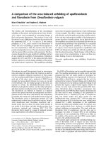
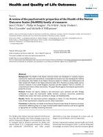
Figure 2. Calculation of segmental vertebral load using a flexion moment at T5. The length of the lever
arm (r) to the force vector (F) is partly related to the curvature of the spine at that level. The T12 lever
arm is less than the T5 lever arm.
4.2. Muscle Strength
Spinal posture may be influenced by the strength of paraspinal muscles. Back extensor strength is
negatively correlated with thoracic kyphosis in some studies [39, 84, 85], but not others [17]. The
strength of muscles around the thoracic spine is believed to protect against thoracic fracture in patients
with osteoporosis [85]. Several studies have shown back extensor strength decreases with age [17, 47,