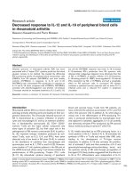
Báo cáo y học: "Decreased response to IL-12 and IL-18 of peripheral blood cells in rheumatoid arthritis" doc
Bạn đang xem bản rút gọn của tài liệu. Xem và tải ngay bản đầy đủ của tài liệu tại đây (84.08 KB, 7 trang )
Introduction
Rheumatoid arthritis (RA) is a chronic disorder of unknown
aetiology primarily affecting joints and leading to their pro-
gressive destruction. The chronically inflamed synovium of
RA is characterized by a massive infiltration of lympho-
cytes and macrophages [1] and by an extensive prolifera-
tion of fibroblast-like synoviocytes [2]. CD4
+
CD45
+
memory T cells are the major cellular component and
show signs of activation [3], but their exact role in the
pathogenesis of RA remains controversial [3,4]. In particu-
lar, the important biologic mediators produced by acti-
vated T cells, such as IL-2, IL-4, and IFN-γ, have been
detected only at low levels in RA joints [5–8], in contrast
to the abundance of cytokines from macrophages and
synoviocytes, such as IL-1, TNF-α, and IL-6 [3]. However,
some studies of T-cell cytokine patterns in the RA joint at
the mRNA level and others using T-cell clones indicate the
predominance of IFN-γ-producing T helper (Th)1 cells
[9–11]. By intracellular cytokine staining of peripheral
blood and synovial tissue T cells from RA patients, we
have confirmed the selective accumulation of Th1 and Th0
cells in the synovium [12]. In addition, IL-12, which plays a
critical role in the differentiation of IFN-γ-producing Th1
cells, is produced predominantly by macrophages local-
ized adjacent to lymphatic aggregates. IL-12 can potently
and selectively stimulate IFN-γ production by RA synovial
tissue, mainly by acting on synovial T cells [13].
IL-18, initially described as an IFN-γ-inducing factor, is a
novel cytokine of the IL-1 family [14]. IL-18 stimulates the
synthesis of IFN-γ in T cells and natural killer (NK) cells,
leading to the development of Th1-type immune
responses. In addition, IL-18 also activates the prolifera-
tion of activated T cells and their production of IL-2 and
granulocyte/macrophage-colony-stimulating factor, and
the cytotoxic activity of NK cells through up-regulation of
Fas ligand [15]. Early studies suggested that the effects of
IL-18 on Th1 differentiation were independent of IL-12.
ELISA = enzyme-linked immunosorbent assay; H-PBMCs = PBMCs from healthy controls; IFN = interferon; IL = interleukin; IL-18BP = IL-18-
binding protein; MTX = methotrexate; NK = natural killer (cells); PBMCs = peripheral blood mononuclear cells; PHA = phytohemagglutinin; PMA =
phorbol 12-myristate 13-acetate; RA = rheumatoid arthritis; RA-PBMCs = PBMCs from RA patients; Th = T helper (cells); VCAM-1 = vascular cell
adhesion molecule-1.
Available online />Research article
Decreased response to IL-12 and IL-18 of peripheral blood cells
in rheumatoid arthritis
Masanori Kawashima and Pierre Miossec
Department of Immunology and Rheumatology and INSERM U-403, Pavillon F, Hospital Edouard Herriot, 69437 Lyon Cedex 03, France
Correspondence: Pierre Miossec ()
Received: 8 May 2003 Revisions requested: 11 Jun 2003 Revisions received: 29 Sep 2003 Accepted: 13 Oct 2003 Published: 4 Nov 2003
Arthritis Res Ther 2004, 6:R39-R45 (DOI 10.1186/ar1020)
© 2004 Kawashima and Miossec, licensee BioMed Central Ltd (Print ISSN 1478-6354; Online ISSN 1478-6362). This is an Open Access article:
verbatim copying and redistribution of this article are permitted in all media for any purpose, provided this notice is preserved along with the
article's original URL.
Abstract
Inflamed synovium of rheumatoid arthritis (RA) has been
associated with a T helper (Th)1 cytokine profile but the blood
situation remains to be clarified. We studied the differential
IFN-γ producing activity of peripheral blood mononuclear cells
(PBMCs) from RA patients (RA-PBMCs) and from healthy
controls (H-PBMCs) in response to IL-12 and IL-18.
RA-PBMCs had a decreased IFN-γ production in response to
IL-12 and IL-18 when compared with H-PBMCs. RA-PBMCs
activated with phytohemagglutinin and phorbol 12-myristate
13-acetate showed an increased sensitivity to IL-12 and IL-18,
but still the RA-PBMC response was lower. IL-18 increased
IL-12-stimulated IFN-γ production from RA synovium cells
obtained after collagenase digestion more effectively than that
of RA- or H-PBMCs. A specific inhibitor of IL-18 bioactivity,
IL-18-binding protein (IL-18BP), down-regulated IL-12-induced
IFN-γ production by RA- or H-PBMCs and had a remarkable
effect on RA synovium cells. In conclusion, RA disease
combines a polarized immune response with an active Th1 in
inflamed joints and a reduced Th1 pattern in peripheral
circulation.
Keywords: interferon-γ, interleukin-12, interleukin-18, interleukin-18-binding protein, rheumatoid arthritis
Open Access
R39
R40
Arthritis Research & Therapy Vol 6 No 1 Kawashima and Miossec
However, later studies showed that exogenous IL-18 in
the absence of IL-12 failed to drive the differentiation of
naive T cells to Th1 cells [16] and that IL-18 was a potent
inducer of IFN-γ from established Th1 cells only in combi-
nation with IL-12 [15,17–19].
Accordingly, IL-18 may be involved in various immune-
mediated inflammatory conditions. Significant levels of its
expression have been demonstrated in the synovium of
patients with RA [20,21]. It was suggested that IL-18 in
synergy with IL-12 and IL-15 could be involved in both
Th1 immune responses and macrophage production of
inflammatory cytokines such as TNF-α [20]. To control
some of the potentially deleterious properties of IL-18,
IL-18-binding protein (IL-18BP) has been identified as a
specific inhibitor of its bioactivity. IL-18BP, though it lacks
significant homology with IL-18 receptor components, can
bind to IL-18 protein with high affinity, thereby acting as a
soluble decoy receptor [22,23].
In the present study, we looked at the differential effects of
IL-18 alone or in combination with IL-12 on RA cells from
blood versus synovium. Our results show that the PBMCs
of RA patients produced less IFN-γ in response to IL-12
and IL-18 than those of healthy volunteers. In addition, dif-
ferences were observed between blood and synovium RA
cells. Finally, differential regulatory effects of IL-18BP on
these cells were also observed.
Materials and methods
Cytokines and reagents
Recombinant human IL-12 was purchased from R&D
Systems (Abingdon, UK). Recombinant human IL-18 was
from MBL (Nagoya, Japan). IL-18BP, kindly provided by Dr
John Sims, Immunex/Amgen (Seattle, WA, USA), was pro-
duced in COS cells as a fusion protein combining
IL-18BP and the CH2 and CH3 domains of human IgG1
and purified by protein A affinity. RPMI (Roswell Park
Memorial Institute) 1640 culture media was purchased
from Invitrogen SARL (Cergy Pontoise, France) and sup-
plemented with 100 units/ml penicillin, 100 µg/ml strepto-
mycin, and 10% fetal calf serum (Invitrogen).
Phytohemagglutinin (PHA) and phorbol 12-myristate
13-acetate (PMA) were purchased from Sigma-Aldrich
SARL (St Quentin Fallavier, France).
Preparation of synovium and cell cultures
Peripheral blood samples were obtained from 14 patients
(2 men and 12 women) with RA who fulfilled the 1987
revised criteria of the American College of Rheumatology
[24], and 12 healthy volunteers (2 men and 10 women). The
mean ages ±
SEM of RA patients and healthy controls were
52.9 ± 4.8 and 49.3 ± 1.9 years, respectively. The mean
disease duration was 12.4 years (range 1–53 years). The
majority of patients were being treated with nonsteroidal
anti-inflammatory drugs, prednisone (n = 7; range
2–20 mg/day), methotrexate (n = 11) alone or combined
with cyclosporin (n = 1) or anti-TNF treatment (n = 1), and
cyclophosphamide (n = 2). PBMCs were isolated from
heparinized blood by Ficoll density-gradient centrifugation,
washed twice with phosphate-buffered serum, and re-sus-
pended in RPMI 1640 medium. Synovium samples were
obtained from patients with RA who were undergoing
wrist, elbow, or hip synovectomy, or joint replacement.
For the isolation of synovium cells, samples were minced
with scissors and digested for 1 hour with collagenase
(Sigma, St Louis, MO, USA) and DNase (Invitrogen) in
RPMI 1640 medium at 37°C. After removing tissue debris
through a cell strainer, the resulting cell suspensions were
washed twice with medium. The resultant single-cell sus-
pensions and PBMC suspensions were dispensed into
the wells of 96-well plates (Nunc, Roskilde, Denmark) at a
density of 1 × 10
6
cells/ml in 200 µl of RPMI medium. Cul-
tures were at 37°C in 5% CO
2
/95% humidified air.
Determination of IFN-
γγ
levels by ELISA
IFN-γ levels were measured by quantitative sandwich
ELISA, using a commercially available ELISA kit (DuoSet
ELISA Development System human IFN-γ, R&D Systems).
The detection limit of the assay was 20 pg/ml. IFN-γ values
below this limit were regarded as 0.
Statistical analysis
Results were expressed as mean ± SEM of the indicated
number of experiments. The statistical significance of dif-
ferences between two groups was determined by the
Mann–Whitney U test. The Wilcoxon signed-rank test was
used to analyse matched pairs.
Results
Effect of IL-12 and/or IL-18 on IFN-
γγ
production by
normal PBMCs and its modulation by IL-18BP
Peripheral blood mononuclear cells from healthy controls
(H-PBMCs) were cultured for 7 days for IFN-γ production
with or without IL-12 and IL-18 alone or in combination. As
shown in Fig. 1a, IL-18 alone up to 50 ng/ml had only a
negligible effect on IFN-γ production. In contrast, IL-12
alone (1 ng/ml) induced a high IFN-γ production from
peripheral blood mononuclear cells (PBMCs). However,
there was no clear difference between 1 ng/ml vs 5 ng/ml
of IL-12 on IFN-γ production (data not shown). IL-12-stim-
ulated IFN-γ production was increased by IL-18 dose
dependently in a synergistic fashion (Fig. 1b).
To study the regulation of IFN-γ production by IL-18BP,
IL-18 and IL-18BP were pre-incubated together for
30 minutes at 37°C before addition to the culture.
IL-18BP could successfully neutralize IFN-γ production
from PBMCs stimulated by IL-12 and IL-18. IFN-γ produc-
tion induced by IL-12 (1 ng/ml) and IL-18 (5 ng/ml) was
reduced by IL-18BP in a dose-dependent manner
R41
(Fig. 1c). PBMCs were cultured for 3, 5, and 7 days with
or without IL-12 (1 ng/ml), or IL-12 (1 ng/ml) + IL-18
(5 ng/ml). IFN-γ production by spontaneous PBMC culture
could not be detected at day 3 or 5 (Fig. 1d). Accordingly,
concentrations of 1 ng/ml of IL-12, 5 ng/ml of IL-18, and
2 µg/ml of IL-18BP for 7 days of culture were selected for
the following experiments, when these results were
extended to more individuals.
Unstimulated H-PBMCs (n = 12) were cultured for 7 days
with or without IL-12 and IL-18 alone and in combination
with or without IL-18BP, and IFN-γ levels were compared
by ELISA. IL-12 induced significant levels of IFN-γ produc-
tion from H-PBMCs compared with medium alone
(P < 0.001). As shown in Fig. 2a (white bars), IL-12-stimu-
lated IFN-γ production from H-PBMCs was significantly
augmented by IL-18 (P < 0.01) but not by IL-18BP.
Effect of IL-12 and IL-18 on unstimulated PBMCs from
RA patients
PBMCs from patients with RA (RA-PBMCs; n = 14) were
tested in the same experiments (Fig. 2a, black bars). In
culture with medium alone (0.07 ± 0.05 vs
0.05 ± 0.02 ng/ml), H-PBMCs produced very low levels of
IFN-γ similar to those produced by RA-PBMCs. IL-12-
induced IFN-γ production was reduced in RA-PBMCs,
being less than half that of H-PBMCs (P < 0.01). Exoge-
nous IL-18 slightly augmented the IL-12-induced IFN-γ pro-
duction from RA-PBMCs, but the effect was smaller than
that seen in H-PBMCs (RA-PBMCs, +22.6% vs
H-PBMCs, +52.3%). In cultures with medium alone, RA-
PBMCs produced lower levels of IFN-γ than did H-PBMCs,
and this was not corrected by stimulation with IL-12 or
IL-18. Although the difference in the percentage increase
was not significant, RA-PBMCs produced less IFN-γ than
H-PBMCs in response to IL-12 alone or combined with
IL-18 when results were expressed as concentrations.
Effect of IL-12 and IL-18 on PHA/PMA-stimulated PBMCs
Because IFN-γ production is associated with T-cell activa-
tion, and to further sensitize the cells to the action of IL-12
and IL-18, H- or RA-PBMCs were cultured with the same
conditions as above, but in the presence of 200 ng/ml of
PHA and 2 ng/ml of PMA. At these suboptimal concentra-
Available online />Figure 1
Synergistic effect of IL-12 and IL-18 on IFN-γ production by peripheral blood mononuclear cells (PBMCs) from healthy donors and its modulation
by IL-18-binding protein (IL-18BP). PBMCs (1 × 10
6
/ml in RPMI 1640 medium with 10% fetal calf serum) were cultured in triplicate for IFN-γ
production with or without IL-12, IL-18, the combination of IL-12 and IL-18, or the combination of IL-12, IL-18, and IL-18BP. (a) PBMCs were
cultured for 7 days with 1 ng/ml of IL-12, and various concentrations of IL-18. (b) PBMCs were cultured for 7 days with the combination of IL-12
and IL-18. (c) PBMCs were cultured for 7 days with 1 ng/ml of IL-12 and 5ng/ml of IL-18, with or without various concentrations of IL-18BP. (d)
PBMCs were cultured for 3 days (white bars), 5 days (gray bars), or 7 days (black bars) with or without IL-12 (1 ng/ml) or IL-12 (1ng/ml) + IL-18
(5 ng/ml). Bars show mean values ± SEM of triplicate cultures.
IL-18 (ng/ml) – 0.5 5 50
IL-12 (ng/ml) – 1–––
–
0
0.3
0.6
0.9
1.2
IFN-γ (ng/ml)
(a)
IL-18 (ng/ml) 5 5555
IL-12 (ng/ml) 1 1 1 1 1
IL-18BP (µg/ml)
– 0.08 0.4 2 10
0
0.2
0.4
0.6
0.8
1.0
1.2
1.4
(c)
IL-18 (ng/ml) – 50–0.5 5
IL-12 (ng/ml) – 1 1 1 1
0
1
2
8
9
IF (ng/ml)
(b)
(d)
0
0.3
0.6
0.9
1.2
IL-18 (ng/ml)
5
IL-12 (ng/ml) 1 1
–
–
–
1.5
10
N-γ
IFN-γ (ng/ml)
IFN-γ (ng/ml)
tions, additional PHA and PMA alone had only a small
effect on IFN-γ production by PBMCs (Fig. 2b). Despite
this stimulation, IL-18 by itself did not induce either H- or
RA-PBMCs to produce IFN-γ. In contrast, activated H- or
RA-PBMCs (Fig. 2b) produced greater amounts of IFN-γ
than resting ones (Fig. 2a) in response to IL-12. The syner-
gistic effect of IL-12 and IL-18 on IFN-γ production by H-
or RA-PBMCs was also augmented by PHA and PMA
treatment. These findings indicate that activated cells are
more sensitive to IL-12 and IL-18 than resting ones.
However, even with the stimulation of PHA and PMA, the
IFN-γ production by RA-PBMCs in response to IL-12 and
IL-18 was lower than that of H-PBMCs (Fig. 2b).
Regulation of IFN-
γγ
production from H- or RA-PBMCs
by IL-18BP
While exogenous IL-18 showed a synergistic effect with
IL-12 on IFN-γ production, levels of IL-12-stimulated IFN-γ
production from resting PBMCs were not influenced by
IL-18BP treatment (Fig. 2a). On the other hand, IL-12-stimu-
lated IFN-γ production was decreased by IL-18BP in
PBMCs activated by PHA/PMA (reduction with IL-18BP
was 47.2% in H-PBMCs [P <0.01] and 53.0% in
RA-PBMCs [P <0.01]). These observations indicate that
the stimulation of PHA/PMA induces PBMCs to produce
significant amounts of IL-18. Exogenous IL-18 increased
IL-12-induced IFN-γ production by 47% for activated
H-PBMCs versus 25% for activated RA-PBMCs. This differ-
ence indicated that RA-PBMCs had a defective response to
IL-18 even when these cells were activated by PHA/PMA.
Aging of RA PBMCs and sensitivity to Th1-inducing
cytokine
Since immune defects have been associated with age, we
classified the patients into two groups according to their
age and compared the IFN-γ production by their PBMCs in
response to IL-12 and IL-18. The mean age, disease dura-
tion, and C-reactive-protein levels at the date of the blood
sampling are shown in Table 1. The values of C-reactive
protein were almost identical in the two groups. The older
RA group showed a greater decrease in response to IL-12
and IL-18 than the younger RA group, indicating that this
defect was disease related but increased with age,
although without reaching significance.
IFN-
γγ
production by RA synovium cells in response to
IL-12 and IL-18
T cells from RA synovium are characterized by a reduced
production of IFN-γ, contrasting with the large production
of IFN-γ by T-cell clones isolated from RA synovium. To
further explore this contrast, total RA synovial cells (n = 7),
obtained after collagenase digestion, were exposed to
IL-12 and IL-18 using the same conditions as were used
with the PBMCs (Fig. 3a). IL-12 alone but not IL-18
induced total RA synovium cells to produce significant
amounts of IFN-γ. However, IL-18 increased IL-12-stimu-
lated IFN-γ production from total RA synovium cells more
effectively (+78.4%, P < 0.05) than those of H- or
RA-PBMCs. In cells activated with PHA and PMA, IL-18
increased by 71.4% the IL-12-stimulated IFN-γ production
from total RA synovium cells, and IL-18BP decreased it by
38.5% (Fig. 3b). Thus, total RA synovium cells showed a
greater response to IL-18 than blood cells. Stimulation
with PHA and PMA further augmented the magnitude of
IFN-γ production, but the percentage increase of IFN-γ
production by exogenous IL-18 was not changed by
PHA/PMA. Accordingly, total RA synovium cells were con-
sidered to respond better to exogenous IL-18 than the
blood cells did. IL-18BP decreased IL-12-stimulated IFN-γ
production from total RA synovium cells cultured with or
without PHA/PMA (–38.5% and –46.2%, respectively).
RA synovium is known to produce significant amounts of
IL-18 spontaneously [21]. Addition of IL-18BP could
Arthritis Research & Therapy Vol 6 No 1 Kawashima and Miossec
R42
Figure 2
Differences in response to IL-12 or IL-18 between peripheral blood
mononuclear cells (PBMCs) from healthy donors (H-PBMCs; n =15)
and from patients with RA (RA-PBMCs; n =14). (a) H- or RA-PBMCs
(1×10
6
/ml in RPMI 1640 medium with 10% fetal calf serum) were
cultured in triplicate for 7 days with or without 1ng/ml of IL-12, 5 ng/ml
of IL-18, 2 µg/ml of IL-18 binding protein (IL-18BP), or their
combination. (b) Cells were activated with 200 ng/ml of
phytohemagglutinin (PHA) and 2 ng/ml of phorbol myristate acetate
(PMA). IFN-γ concentrations in culture supernatants were measured by
ELISA. Black bars represent RA-PBMCs and white bars represent
H-PBMCs. *P <0.05, **P< 0.01. NS, statistically not significant.
medium
IL-12
IL-18
IL-18BP
IL-12+IL-18
IL-12+IL-18BP
0123
(a)
medium
IL-12
IL-18
IL-18BP
IL-12+IL-18
IL-12+IL-18BP
051015
IFN-γ (ng/ml)
(b)
With PHA/PMA
4
NS
NS
**
NS
NS
**
*
**
IFN-γ (ng/ml)
decrease the IL-12-induced IFN-γ production by these
cells by neutralizing the endogenously produced IL-18,
indicating the importance of endogenous IL-18 in IFN-γ
production by RA synovium.
Discussion
Several lines of evidence have indicated that IFN-γ-produc-
ing Th1 cells predominate at the site of chronic inflamma-
tion in RA. IL-12 is considered to play a critical role in
inducing Th1-cell-mediated organ-specific autoimmune dis-
eases, as shown in several animal models [25–28]. In addi-
tion, we have already demonstrated the presence of IL-12
as a contributory factor in inducing the IFN-γ-dominant
T-cell cytokine response in joints of patients with long-
standing RA [13]. IL-18 is a proinflammatory cytokine that
plays an important role in the Th1-type immune response
through the induction of IFN-γ synthesis in T cells and NK
cells, T-cell proliferation, and cytokine production [15]. Sig-
nificant levels of expression of IL-18 have been previously
demonstrated in the synovium of RA patients [20,21], and
the major effect of IL-18 is to increase the IFN-γ-dominant
T-cell response induced by IL-12 [21].
T cells and NK cells are the major source of IFN-γ in
PBMC cultures, and IL-12 and IL-18 augment IFN-γ pro-
duction by these cells. Defects in IFN-γ production may
result from changes in cell number and/or in cell response.
Although differing conclusions have been reached regard-
ing the possible changes in numbers of CD4
+
T cells, NK
cells, and NK T cells between RA and healthy PBMCs
[29], the precise subpopulation with reduced ability to
produce IFN-γ remains to be clarified [29–31]. In order to
assess overall differences in systemic response to the
Th1-inducing cytokines, IL-12 and IL-18, we compared the
IFN-γ production by PBMCs from RA patients to that by
cells from healthy controls. The IFN-γ production of RA-
PBMCs in response to IL-12 and IL-18 was lower than
that of H-PBMCs, even with the activation of PHA and
PMA. This was still observed when the younger RA
patients were compared with age-matched controls. Fur-
thermore, PBMCs from the older RA patients demon-
strated a greater decrease in response to IL-12 and IL-18
than that of the younger patients. Indeed, a reduction of
the systemic Th1 functions with aging in normal individu-
als has been reported [32]. The same tendency was
observed for the response to Th1-inducing cytokines in
RA. These results indicate that RA-PBMCs are defective
in their response to Th1-inducing cytokines, with an addi-
tional effect related to age. When looking at a possible
effect of treatment on these defects, we found no differ-
ence between patients treated or not treated with pred-
nisone (data not shown). The finding was similar for
patients receiving methotrexate. Preliminary results appear
to indicate that PBMCs from RA patients taking
Available online />R43
Table 1
IFN-γ production by PBMCs from patients with rheumatoid
arthritis in response to IL-12 and IL-18
Variable Younger patients Older patients
No. of patients 7 7
Mean age (years) 40.1 ± 5.0 65.7 ± 4.6
Disease duration (years) 5.5 ± 2.3 17.7 ± 6.4
C-reactive protein (mg/l) 13.4 ± 2.0 13.9 ± 5.0
IFN-γ production (ng/ml)
In medium alone 0.05 ± 0.03 0.05 ± 0.03
With IL-12 0.56 ± 0.14 0.50 ± 0.17
With IL-12 + IL-18 0.83 ± 0.17 0.48 ± 0.14
With IL-12 + IL-18BP 0.53 ± 0.19 0.36 ± 0.15
Values are means ± SEM. IL-18BP, IL-18-binding protein; PBMCs,
peripheral blood mononuclear cells.
Figure 3
Effect of IL-12 and IL-18 on rheumatoid arthritis synovium cells.
(a) Rheumatoid arthritis synovium cells (1 ×10
6
/ml in RPMI 1640
medium with 10% fetal calf serum) were cultured in triplicate for
7 days with or without 1ng/ml of IL-12, 5 ng/ml of IL-18, 2 µg/ml of
IL-18 binding protein (IL-18BP), or a combination of these. (b) Cells
were activated with 200 ng/ml of phytohemagglutinin (PHA) and
2 ng/ml of phorbol 12-myristate 13-acetate (PMA). IFN-γ concentrations
in culture supernatants were measured by ELISA. *P <0.05.
medium
IL-12
IL-18
IL-18BP
IL-12+IL-18
IL-12+IL-18BP
(a)
medium
IL-12
IL-18
IL-18BP
IL-12+IL-18
IL-12+IL-18BP
(b)
012
0246
With PHA/PMA
*
*
*
*
IFN-γ (ng/ml)
IFN-γ (ng/ml)
methotrexate produced greater amounts of IFN-γ after suc-
cessful anti-TNF treatment. This result, if it proves to be
correct, would further indicate that the defective IFN-γ pro-
duction by RA-PBMCs might be related to disease activity.
Next, we compared the response to TH1-inducing
cytokines of blood versus synovium cells from RA patients.
Judging from the effect on IFN-γ production, total RA syn-
ovium cells showed a greater response to exogenous
IL-18 in the presence of IL-12 than resting PBMCs. The
pattern of response to IL-12 and IL-18 of total RA syn-
ovium cells was similar to that of activated H-PBMCs, sug-
gesting that these cells had been activated and were more
sensitive to TH1-inducing cytokines.
IL-18BP has been identified as a specific inhibitor of IL-18
[22,23]. There is no significant similarity between IL-18BP
and IL-18 receptor components. Since it lacks a transmem-
brane domain, IL-18BP appears to exist only as a soluble,
circulating protein. Its major function is to regulate the
inflammatory activity of IL-18 by acting as its soluble decoy
receptor [22,23]. Our results showed that exogenous IL-18
alone did not induce total RA synovium cells to produce
detectable levels of IFN-γ, but the neutralization of endoge-
nous IL-18 by IL-18BP reduced by about 50% the IL-12-
induced IFN-γ production by these cells, suggesting a role
for endogenously produced IL-18 in IFN-γ production. The
effect of IL-18BP was much higher in RA synovium cells
than in PBMCs, probably because RA synovium produced
more endogenous IL-18 and responsed more strongly to
IL-18. Although an IL-18-neutralizing activity was found in
RA synovial fluid samples, IL-18 bioactivity was still
detectable [21]. These findings indicate that endogenous
IL-18 is an important contributory factor to IL-12-induced
IFN-γ production in RA synovium.
In a previous study, we used flow cytometry to examine
the ability of CD4
+
T cells of blood and synovium samples
from RA patients to produce IFN-γ and/or IL-4. Total RA
synovium cells showed a higher Th1 and a lower Th2 fre-
quency than peripheral blood cells [12]. In addition,
Haddad and colleagues reported that activated whole
blood cells from RA patients produced higher levels of
IL-4 and lower IFN-γ than did cells from healthy controls
[33]. Accordingly, the IL-4 : IFN-γ ratio, which reflects the
Th2 : Th1 cytokine balance in blood, was higher in RA
patients. These findings suggest that in RA the Th1 : Th2
ratio in the blood and that in the synovium are different.
Accumulation of Th2 vs Th1 cells in blood may result from
changes in T-cell migration. Indeed, IFN-γ producing
T cells were significantly increased in the peripheral blood
of RA patients shortly after anti-TNF-α treatment, resulting
in a shift of the Th1 : Th2 ratio in favor of Th1 in peripheral
blood [34]. Adhesion molecules, such as P- and E-selectin
or VCAM-1 (vascular cell adhesion molecule-1), are con-
sidered to be important for the selective homing of Th1
cells and not Th2 cells [35]. Expression of E-selectin and
VCAM-1 was significantly reduced by anti-TNF therapy
[36]. Thus, anti-TNF treatment may suppress the selective
migration of Th1 cells into RA synovium through the rapid
down-regulation of adhesion molecules. It remains to be
clarified whether this is associated with an increased
migration of Th2 cells with anti-inflammatory properties.
Recently, cases of severe tuberculosis have been
reported in patients with Crohn’s disease and RA receiv-
ing anti-TNF treatment [37]. Tuberculosis was the most
common serious opportunistic infection reported in those
patients. Similar observations were made in an HIV popu-
lation where a secondary cell-mediated immune defect is
present, as demonstrated by a defect in IFN-γ production.
It is noteworthy that tuberculosis developed in RA patients
shortly after the beginning of anti-TNF treatment. Our
results showed a decreased response of RA peripheral
blood to IL-12 and IL-18, leading to a reduced production
of IFN-γ compared with that of healthy controls. These
findings could explain to some extent the occurrence of
tuberculosis during anti-TNF treatment.
Conclusion
Our data demonstrate that compared with H-PBMCs,
RA-PBMCs have a lower response to IL-12 and IL-18,
which are both important in Th1 development via T-bet
expression. These results are in contrast with the finding
that synovium cells in RA show an increased response to
IL-12 and IL-18. Thus, RA patients have a polarized
immune response, Th1 being overexpressed in inflamed
joints and Th1 defects in peripheral circulation.
Competing interests
None declared.
References
1. Janossy G, Panayi G, Duke O, Bofill M, Poulter LW, Goldstein G:
Rheumatoid arthritis: a disease of T-lymphocyte/macrophage
immunoregulation. Lancet 1981, 2:839-842.
2. Qu Z, Garcia CH, O’Rourke LM, Planck SR, Kohli M, Rosenbaum
JT: Local proliferation of fibroblast-like synoviocytes con-
tributes to synovial hyperplasia. Results of proliferating cell
nuclear antigen/cyclin, c-myc, and nucleolar organizer region
staining. Arthritis Rheum 1994, 37:212-220.
3. Firestein GS, Zvaifler NJ: How important are T cells in chronic
rheumatoid synovitis? Arthritis Rheum 1990, 33:768-773.
4. Panayi GS, Lanchbury JS, Kingsley GH: The importance of the T
cell in initiating and maintaining the chronic synovitis of
rheumatoid arthritis. Arthritis Rheum 1992, 35:729-735.
5. Firestein GS, Xu WD, Townsend K, Broide D, Alvaro-Gracia J,
Glasebrook A, Zvaifler NJ: Cytokines in chronic inflammatory
arthritis. I. Failure to detect T cell lymphokines (interleukin 2
and interleukin 3) and presence of macrophage colony-stimu-
lating factor (CSF-1) and a novel mast cell growth factor in
rheumatoid synovitis. J Exp Med 1988, 168:1573-1586.
6. Miossec P, van den Berg W: Th1/Th2 cytokine balance in
arthritis. Arthritis Rheum 1997, 40:2105-2115.
7. Miossec P, Naviliat M, Dupuy d’Angeac A, Sany J, Banchereau J:
Low levels of interleukin-4 and high levels of transforming
growth factor beta in rheumatoid synovitis. Arthritis Rheum
1990, 33:1180-1187.
Arthritis Research & Therapy Vol 6 No 1 Kawashima and Miossec
R44
8. Chen E, Keystone EC, Fish EN: Restricted cytokine expression
in rheumatoid arthritis. Arthritis Rheum 1993, 36:901-910.
9. Miltenburg AM, van Laar JM, de Kuiper R, Daha MR, Breedveld
FC: T cells cloned from human rheumatoid synovial mem-
brane functionally represent the Th1 subset. Scand J Immunol
1992, 35:603-610.
10. Quayle AJ, Chomarat P, Miossec P, Kjeldsen-Kragh J, Forre O,
Natvig JB: Rheumatoid inflammatory T-cell clones express
mostly Th1 but also Th2 and mixed (Th0-like) cytokine pat-
terns. Scand J Immunol 1993, 38:75-82.
11. Simon AK, Seipelt E, Sieper J: Divergent T-cell cytokine pat-
terns in inflammatory arthritis. Proc Natl Acad Sci USA 1994,
91:8562-8566.
12. Morita Y, Yamamura M, Kawashima M, Harada S, Tsuji K, Shibuya
K, Maruyama K, Makino H: Flow cytometric single-cell analysis
of cytokine production by CD4+ T cells in synovial tissue and
peripheral blood from patients with rheumatoid arthritis.
Arthritis Rheum 1998, 41:1669-1676.
13. Morita Y, Yamamura M, Nishida K, Harada S, Okamoto H, Inoue
H, Ohmoto Y, Modlin RL, Makino H: Expression of interleukin-
12 in synovial tissue from patients with rheumatoid arthritis.
Arthritis Rheum 1998, 41:306-314.
14. Nakamura K, Okamura H, Wada M, Nagata K, Tamura T: Endo-
toxin-induced serum factor that stimulates gamma interferon
production. Infect Immun 1989, 57:590-595.
15. Dinarello CA: Interleukin-18. Methods 1999, 19:121-132.
16. Robinson D, Shibuya K, Mui A, Zonin F, Murphy E, Sana T, Hartley
SB, Menon S, Kastelein R, Bazan F, O’Garra A: IGIF does not
drive Th1 development but synergizes with IL-12 for inter-
feron-gamma production and activates IRAK and NFkappaB.
Immunity 1997, 7:571-581.
17. Kohno K, Kataoka J, Ohtsuki T, Suemoto Y, Okamoto I, Usui M,
Ikeda M, Kurimoto M: IFN-gamma-inducing factor (IGIF) is a
costimulatory factor on the activation of Th1 but not Th2 cells
and exerts its effect independently of IL-12. J Immunol 1997,
158:1541-1550.
18. Micallef MJ, Ohtsuki T, Kohno K, Tanabe F, Ushio S, Namba M,
Tanimoto T, Torigoe K, Fujii M, Ikeda M, Fukuda S, Kurimoto M:
Interferon-gamma-inducing factor enhances T helper 1
cytokine production by stimulated human T cells: synergism
with interleukin-12 for interferon-gamma production. Eur J
Immunol 1996, 26:1647-1651.
19. Ahn HJ, Maruo S, Tomura M, Mu J, Hamaoka T, Nakanishi K, Clark
S, Kurimoto M, Okamura H, Fujiwara H: A mechanism under-
lying synergy between IL-12 and IFN-gamma-inducing factor
in enhanced production of IFN-gamma. J Immunol 1997, 159:
2125-2131.
20. Gracie JA, Forsey RJ, Chan WL, Gilmour A, Leung BP, Greer MR,
Kennedy K, Carter R, Wei XQ, Xu D, Field M, Foulis A, Liew FY,
McInnes IB: A proinflammatory role for IL-18 in rheumatoid
arthritis. J Clin Invest 1999, 104:1393-1401.
21. Yamamura M, Kawashima M, Taniai M, Yamauchi H, Tanimoto T,
Kurimoto M, Morita Y, Ohmoto Y, Makino H: Interferon-gamma-
inducing activity of interleukin-18 in the joint with rheumatoid
arthritis. Arthritis Rheum 2001, 44:275-285.
22. Aizawa Y, Akita K, Taniai M, Torigoe K, Mori T, Nishida Y, Ushio S,
Nukada Y, Tanimoto T, Ikegami H, Ikeda M, Kurimoto M: Cloning
and expression of interleukin-18 binding protein. FEBS Lett
1999, 445:338-342.
23. Novick D, Kim SH, Fantuzzi G, Reznikov LL, Dinarello CA, Rubin-
stein M: Interleukin-18 binding protein: a novel modulator of
the Th1 cytokine response. Immunity 1999, 10:127-136.
24. Arnett FC, Edworthy SM, Bloch DA, McShane DJ, Fries JF,
Cooper NS, Healey LA, Kaplan SR, Liang MH, Luthra HS,
Medsger Jr T, Mitchell DM, Neustadt DH, Pinals RS, Schaller JG,
Sharp JT, Wilden RL, Hunder GG: The American Rheumatism
Association 1987 revised criteria for the classification of
rheumatoid arthritis. Arthritis Rheum 1988, 31:315-324.
25. Germann T, Szeliga J, Hess H, Storkel S, Podlaski FJ, Gately MK,
Schmitt E, Rude E: Administration of interleukin 12 in combi-
nation with type II collagen induces severe arthritis in DBA/1
mice. Proc Natl Acad Sci USA 1995, 92:4823-4827.
26. Trembleau S, Penna G, Bosi E, Mortara A, Gately MK, Adorini L:
Interleukin 12 administration induces T helper type 1 cells
and accelerates autoimmune diabetes in NOD mice. J Exp
Med 1995, 181:817-821.
27. Leonard JP, Waldburger KE, Goldman SJ: Prevention of experi-
mental autoimmune encephalomyelitis by antibodies against
interleukin 12. J Exp Med 1995, 181:381-386.
28. Neurath MF, Fuss I, Kelsall BL, Stuber E, Strober W: Antibodies
to interleukin 12 abrogate established experimental colitis in
mice. J Exp Med 1995, 182:1281-1290.
29. Yanagihara Y, Shiozawa K, Takai M, Kyogoku M, Shiozawa S:
Natural killer (NK) T cells are significantly decreased in the
peripheral blood of patients with rheumatoid arthritis (RA).
Clin Exp Immunol 1999, 118:131-136.
30. Maldonado A, Mueller YM, Thomas P, Bojczuk P, O’Connors C,
Katsikis PD: Decreased effector memory CD45RA+ CD62L-
CD8+ T cells and increased central memory CD45RA-
CD62L+ CD8+ T cells in peripheral blood of rheumatoid
arthritis patients. Arthritis Res Ther 2003, 5:R91-96.
31. Ezawa K, Yamamura M, Matsui H, Ota Z, Makino H: Comparative
analysis of CD45RA- and CD45RO-positive CD4+T cells in
peripheral blood, synovial fluid, and synovial tissue in patients
with rheumatoid arthritis and osteoarthritis. Acta Med
Okayama 1997, 51:25-31.
32. Rink L, Cakman I, Kirchner H: Altered cytokine production in the
elderly. Mech Ageing Dev 1998, 102:199-209.
33. Haddad A, Bienvenu J, Miossec P: Increased production of a
Th2 cytokine profile by activated whole blood cells from
rheumatoid arthritis patients. J Clin Immunol 1998, 18:399-
403.
34. Maurice MM, van der Graaff WL, Leow A, Breedveld FC, van Lier
RA, Verweij CL: Treatment with monoclonal anti-tumor necro-
sis factor alpha antibody results in an accumulation of Th1
CD4+ T cells in the peripheral blood of patients with rheuma-
toid arthritis. Arthritis Rheum 1999, 42:2166-2173.
35. Austrup F, Vestweber D, Borges E, Lohning M, Brauer R, Herz U,
Renz H, Hallmann R, Scheffold A, Radbruch A, Hamann A: P- and
E-selectin mediate recruitment of T-helper-1 but not T-helper-
2 cells into inflammed tissues. Nature 1997, 385:81-83.
36. Tak PP, Taylor PC, Breedveld FC, Smeets TJ, Daha MR, Kluin PM,
Meinders AE, Maini RN: Decrease in cellularity and expression
of adhesion molecules by anti-tumor necrosis factor alpha
monoclonal antibody treatment in patients with rheumatoid
arthritis. Arthritis Rheum 1996, 39:1077-1081.
37. Keane J, Gershon S, Wise RP, Mirabile-Levens E, Kasznica J,
Schwieterman WD, Siegel JN, Braun MM: Tuberculosis associ-
ated with infliximab, a tumor necrosis factor alpha-neutraliz-
ing agent. N Engl J Med 2001, 345:1098-1104.
Correspondence
Prof Pierre Miossec, 5 place d’Arsonval, Department of Immunology
and Rheumatology and INSERM U-403, Pavillon F, Hospital Edouard
Herriot, 69437 Lyon Cedex 03, France. Tel: +33 472 117487; fax:
+33 472 117429; e-mail
Available online />R45