Báo cáo y học: "α Does protein kinase R mediate TNF-α- and ceramide-induced increases in expression and activation of matrix metalloproteinases in articular cartilage by a novel mechanism" pps
Bạn đang xem bản rút gọn của tài liệu. Xem và tải ngay bản đầy đủ của tài liệu tại đây (591.54 KB, 10 trang )
R46
Introduction
Rheumatoid arthritis and osteoarthritis each affects a sig-
nificant proportion of the population and the resulting loss
of articular cartilage and inflammation causes severe pain
and disability. There are no effective treatments for repair
of the damaged articular cartilage in these diseases and
while their likely aetiologies are very different, common
pathways of degradation are important in both. Cartilage
degradation occurs as a result of an imbalance of extracel-
lular matrix proteinases and their inhibitors, in particular
the matrix metalloproteinases (MMPs) and the tissue
inhibitors of MMPs (TIMPs). Specifically, MMP-2 and -9
have been reported to be elevated in osteoarthritis carti-
lage [1,2] and within the synovial fluid of patients with
rheumatoid arthritis [3], suggesting critical roles for these
degradative enzymes in arthritic disease. In addition to its
ability to degrade the cartilage matrix directly, MMP-2
plays a significant role in the activation of collagenases
that are also strongly implicated in arthritic disease. MMPs
and TIMPs, in turn, are regulated via induction of the early
response genes c-fos and c-jun and by proinflammatory
cytokines that are known to be involved in arthritic dis-
eases [4,5], such as interleukin-1 and tumour necrosis
factor α (TNF-α). TNF-α is capable of inducing cartilage
catabolism in vitro [5] via increased MMP expression and
activation [4] and is elevated in the synovial fluids from
2-AP = 2-aminopurine; DMEM = Dulbecco’s Modified Eagle’s Medium; DMMB = dimethylmethylene blue; eIF2α = eukaryotic initiation factor 2α;
FCS = fetal calf serum; MMP = matrix metalloproteinase; MT1-MMP = membrane type 1 MMP; NFκB = nuclear factor κB; PACT = PKR-activating
protein; PKR = protein kinase R; SDS = sodium dodecyl sulfate; sGAG = sulfated glycosaminoglycan; TIMPs = tissue inhibitors of MMPs; TNF-α =
tumour necrosis factor α; TNF-R55 = tumour necrosis factor receptor-55.
Arthritis Research & Therapy Vol 6 No 1 Gilbert et al.
Research article
Does protein kinase R mediate TNF-
αα
- and ceramide-induced
increases in expression and activation of matrix
metalloproteinases in articular cartilage by a novel mechanism?
Sophie J Gilbert, Victor C Duance and Deborah J Mason
Connective Tissue Biology Laboratories, School of Biosciences, Cardiff University, Cardiff, Wales, UK
Correspondence: Sophie J Gilbert (e-mail: )
Received: 24 Jul 2003 Revisions requested: 15 Sep 2003 Revisions received: 14 Oct 2003 Accepted: 21 Oct 2003 Published: 12 Nov 2003
Arthritis Res Ther 2004, 6:R46-R55 (DOI 10.1186/ar1024)
© 2004 Gilbert et al., licensee BioMed Central Ltd (Print ISSN 1478-6354; Online ISSN 1478-6362). This is an Open Access article: verbatim
copying and redistribution of this article are permitted in all media for any purpose, provided this notice is preserved along with the article's original
URL.
Abstract
We investigated the role of the proinflammatory cytokine TNF-α,
the second messenger C
2
-ceramide, and protein kinase R
(PKR) in bovine articular cartilage degradation. Bovine articular
cartilage explants were stimulated with C
2
-ceramide or TNF-α
for 24 hours. To inhibit the activation of PKR, 2-aminopurine was
added to duplicate cultures. Matrix metalloproteinase (MMP)
expression and activation in the medium were analysed by
gelatin zymography, proteoglycan release by the
dimethylmethylene blue assay, and cell viability by the
Cytotox 96
®
assay. C
2
-ceramide treatment of cartilage explants
resulted in a significant release of both pro- and active MMP-2
into the medium. Small increases were also seen with TNF-α
treatment. Incubation of explants with 2-aminopurine before
TNF-α or C
2
-ceramide treatment resulted in a marked reduction
in expression and activation of both MMP-2 and MMP-9. TNF-α
and C
2
-ceramide significantly increased proteoglycan release
into the medium, which was also inhibited by cotreatment with
2-aminopurine. A loss of cell viability was observed when
explants were treated with TNF-α and C
2
-ceramide, which was
found to be regulated by PKR. We have shown that
C
2
-ceramide and TNF-α treatment of articular cartilage result in
the increased synthesis and activation of MMPs, increased
release of proteoglycan, and increased cell death. These effects
are abrogated by treatment with the PKR inhibitor
2-aminopurine. Collectively, these results suggest a novel role
for PKR in the synthesis and activation of MMPs and support
our hypothesis that PKR and its activator, PACT, are implicated
in the cartilage degradation that occurs in arthritic disease.
Keywords: articular cartilage, ceramide, matrix metalloproteinase, PKR, TNF-α
Open Access
Available online />R47
patients with arthritic disease [6,7]. It signals via a number
of pathways including activation of sphingomyelinases,
which degrade the membrane phospolipid sphingomyelin
into phosphocholine and ceramide [8]. In turn, ceramide
exerts its effects in a variety of ways depending on cell type
(for a review see [9]), but several studies have implicated
this second messenger in the regulation of MMPs [10–13].
In fibroblasts, the cell-permeable ceramide analogue
C
2
-ceramide has been shown to stimulate mRNA expres-
sion for MMP-1 and MMP-3 through activation of signal
pathways that ultimately lead to the induction of c-jun and
c-fos and AP-1-dependent transcription of MMP genes
[10]. In addition, triggering of the ceramide pathway in
human keratinocytes results in overexpression of MMP-9
[11]. Recently it was shown that ceramide stimulates pro-
teoglycan degradation and mRNA expression of MMP-1,
-3, and -13 in rabbit articular cartilage [12,13]. This finding
is important because it establishes a direct link between
cartilage degradation and the ceramide pathway.
Previously, we have shown that the protein kinase R (PKR)-
activating protein (PACT) [14] is up-regulated in regions
of cartilage that subsequently develop osteoarthritis-like
changes in vivo [15] and that PACT and PKR are involved
in the TNF-α signalling pathway in articular chondrocytes
[16]. The PKR pathway is also known to be activated by
the sphingolipid ceramide [17]. This has led us to the
hypothesis that TNF-α induces MMP expression in chon-
drocytes via ceramide-mediated activation of PKR. In the
current study we have therefore investigated the role of
TNF-α, the cell-permeable ceramide analogue
C
2
-ceramide, and PKR in a well-characterised in vitro
model of articular cartilage degradation [18]. We have
used this model to activate degradative pathways in other-
wise healthy cartilage to reveal potential signalling mecha-
nisms that may be important in arthritic disease.
Materials and methods
Materials
All chemicals were obtained from Sigma (Poole, UK)
unless otherwise stated and were of analytical grade.
Recombinant human cytokines were purchased from
Peprotech EC Ltd (London, UK). Culture medium con-
sisted of Dulbecco’s Modified Eagle’s Medium with Gluta-
max-I (DMEM; Gibco BRL, Paisley, UK) containing 10 IU
penicillin and 10 µg/ml streptomycin.
In vitro cartilage samples
Cartilage was taken from the metacarpophalangeal joint of
7-day-old bovine calves within 12 hours of slaughter using
a scalpel and full-depth cartilage explants (20–70 mg) cul-
tured overnight at 37°C in a humidified atmosphere of 5%
CO
2
, 95% air, in 1 ml of DMEM supplemented with 10%
fetal calf serum (FCS). DMEM containing FCS was
removed and explants were incubated overnight in serum-
free DMEM with insulin–transferrin–sodium selenite liquid
supplement; they were then cultured in the presence of
TNF-α (20–100 ng/ml) or cell-permeable C
2
-ceramide
(10–100 µ
M) (Tocris, Bristol, UK) for 24 hours. These con-
centrations of TNF-α and C
2
-ceramide are known to stim-
ulate catabolism in cartilage [12,13,18]. To inhibit the
activation of PKR, 2-aminopurine (2-AP) (1–10 m
M) was
added to duplicate cultures 1 hour before the addition of
treatments. This range of concentrations has been shown
to inhibit activation of PKR in a number of studies [19–21]
and did not affect chondrocyte viability in explants over
24 hours of culture (data not shown). Untreated explants
or explants treated with 2-AP only served as controls. At
the end of each experiment, explants were snap frozen in
liquid nitrogen and media collected for analysis as
described below.
Gelatin substrate zymography
MMP-2 and MMP-9 activities were detected in media
samples by gelatin substrate zymography as described
previously [22]. Briefly, gelatin (porcine skin, BDH, Poole.
Dorset, UK) was incorporated into 7.5% (w/v) sodium
dodecyl sulfate (SDS)–polyacrylamide gels at a final sub-
strate concentration of 1.0 mg/ml. Media samples (20 µl,
diluted 1 : 1 with sample buffer (0.12
M
Tris/HCl (pH 6.8),
containing 4% (w/v) SDS, 20% (v/v) glycerol, and 0.01%
(w/v) bromophenol blue), a recombinant MMP-9 standard
(1 µl) (Oncogene, Nottingham, UK), bovine-fibroblast-con-
ditioned medium (5 µl) known to contain MMP-2 activity,
and protein molecular weight markers (5 µl; molecular
weight 14–200 kDa, BioRad, Hertfordshire, UK) were
loaded onto the gels and resolved by electrophoresis.
Then the gels were washed, with agitation, in 2.5% (v/v)
Triton X-100 for 1 hour with at least three changes of solu-
tion and subsequently incubated for 16–20 hours at 37°C
in 50 m
M Tris/HCl (pH 7.8), containing 50 mM CaCl
2
and
0.5
M NaCl. Gels were stained with Coomassie
®
Brilliant
Blue R 250 (2 g/l) in distilled water, methanol, and acetic
acid (4.5 : 4.5 : 1) for at least 1 hour and were destained in
methanol, acetic acid, water (1 : 0.75 : 8.25) until the zones
of proteolysis had cleared. The addition of molecular-
weight markers and conditioned media to the gels facili-
tated identification of the enzymes and allowed
comparisons between gels. The relative quantities of pro-
teolytic enzymes were analysed by scanning densitometry
(Umax Colour Scanner, GMbH, Willich, Germany) and
NIH image software (National Institutes of Health,
Bethesda, MD, USA) [23] and expressed as absorbance
units per milligram wet weight of explant.
Proteoglycan release
Proteoglycan release into the medium of cartilage explant
cultures was measured by the dimethylmethylene blue
(DMMB) assay for sulfated glycosaminoglycan (sGAG),
using chondroitin sulfate C from shark cartilage as a stan-
dard, as described previously [18]. After treatment,
explants were digested in 300 µg/ml of papain in 20 m
M
Arthritis Research & Therapy Vol 6 No 1 Gilbert et al.
R48
sodium phosphate (pH 6.8), 1 m
M EDTA, 2 mM dithiothreitol
at 60°C for 1 hour and sGAG was determined as above.
Differences in the levels of sGAG associated with culture
treatment were expressed as micrograms of GAG per mil-
ligram wet weight of cartilage.
Cytotoxicity assay
Cell death was measured using the CytoTox 96
®
assay
(Promega, Southampton, UK). This assay quantitatively
measures lactate dehydrogenase present in the culture
medium that has been released upon natural lysis of cells
during the culture period [24]. Differences in the release
of lactate dehydrogenase associated with culture treat-
ment were expressed as absorbance units per milligram
wet weight of cartilage.
Statistical analysis
Data are presented as mean ± SEM (n ≥ 4 cartilage
explants) of a representative experiment and analysed,
where appropriate, by Student’s unpaired two-sample
t-test or Mann–Whitney test (MMP-9 data). Differences
were considered significant at P values less than 0.05.
Where significance was not obtained, but the P value was
considered informative, this is also shown. All experiments
were performed three times with similar results.
Results
C
2
-ceramide and TNF-
αα
increase both synthesis and
activation of MMP-2 in cartilage explants
Articular cartilage explants were cultured in the presence
of C
2
-ceramide or TNF-α with and without the addition of
the PKR inhibitor 2-AP for 24 hours and media were
analysed for MMP-2 and MMP-9 activity by gelatin sub-
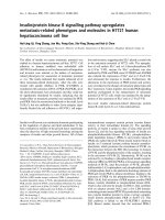
strate zymography. The presence of MMP-2 (Fig. 1a) and
of MMP-9 (Fig. 1b) was confirmed by comparisons with
standards known to contain both enzymes. An additional
proteolytic band was detected that appeared to be
increased by TNF-α treatment and migrated at a molecular
weight previously reported to be a rat/murine-specific gly-
cosylated form of MMP-2 [25].
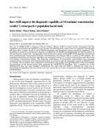
The relative levels of MMP-2 were determined by scanning
densitometry of the zymograms (Fig. 2a). C
2
-ceramide
(50 µ
M) treatment of cartilage explants resulted in a signifi-
cant release of both pro- (15.8 ± 1.8; P = 0.004) and
active (3.4 ± 1.0; P = 0.036) MMP-2 into the culture
medium over 24 hours in comparison with basal levels
produced by unstimulated control explants (8.2 ± 1.0 and
1.4 ± 0.3 respectively). TNF-α (100 ng/ml) treatment
resulted in a small, but not significant, increase in levels of
proMMP-2 and active MMP-2 in the media.
C
2
-ceramide-induced regulation of MMP-2 is abrogated
by inhibition of PKR
To determine a role for PKR in ceramide-induced regula-
tion of MMP-2, we treated explant cultures with
C
2
-ceramide in the presence of the PKR inhibitor 2-AP.
With the addition of 10 m
M 2-AP, the C
2
-ceramide-
induced increase in proMMP-2 (15.8 ± 1.8) was signifi-
cantly reduced (10.6 ± 0.9; P = 0.029) to near basal levels
(8.2 ± 1.0) and activation of MMP-2 was completely abol-
ished (Fig. 2a). A reduction in the activation of MMP-2 was
also observed when 2-AP was added to explants treated
with TNF-α, although this was not statistically significant
(P = 0.109). Addition of 2-AP (1, 5, or 10 m
M) to control
cultures alone had no significant effect on MMP-2 (data
not shown).
De novo
synthesis and activation of MMP-9 is regulated
by PKR
The relative levels of MMP-9 were determined by scanning
densitometry (Fig. 2b). A two-fold increase in the produc-
tion (P = 0.196) and activation (P = 0.123) of MMP-9 was
observed in cartilage explants stimulated with TNF-α
(100 ng/ml) in comparison with levels produced by unstim-
ulated control cultures. C
2
-ceramide (50 µM) had no effect
on the levels of MMP-9 produced by cartilage explants.
However, in contrast, the addition of 2-AP (10 m
M) to
explant cultures completely abolished the production and
activation of MMP-9 irrespective of ceramide or TNF treat-
ment. Addition of 2-AP (1, 5, or 10 m
M) to control cultures
Figure 1
Detection of (a) MMP-2 and (b) MMP-9 activity in media collected
from bovine articular cartilage explants treated with TNF-α or
C
2
-ceramide for 24 hours. Medium was collected 24 hours after
treatment of explants with 100 ng/ml TNF-α or 50 µM
C
2
-ceramide in
the presence or absence of the PKR inhibitor 2-AP (10 mM) and
analysed by gelatin substrate zymography. A standard known to
contain either MMP-2 or MMP-9 was included (Std). An unidentified
enzyme is indicated by ‘?’. 2-AP, 2-aminopurine; MMP, matrix
metalloproteinase; TNF, tumour necrosis factor.
(a)
proMMP-2
active
MMP-2
Std
TNF-α
(100 ng/ml)
C
2
-ceramide (50 µM)
2-AP (10 mM)
+
–
–
–
–
–
+
+
–
+
+
––
+
–
?
(b)
Std
TNF-α (100 ng/ml)
C
2
-ceramide (50 µM)
2-AP (10 mM)
+
–
–
–
–
–
+
+
–
+
+
––
+
–
proMMP-9
active
MMP-9
alone had no significant effect on MMP-9 (data not
shown).
Treatment with TNF-
αα
or C
2
-ceramide increases the
release of proteoglycan from bovine articular cartilage
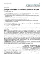
An increase in sGAG release from cartilage explants was
observed following 24 hours of treatment with 50 ng/ml
TNF-α (1.78 ± 0.5; P = 0.06), which was highly significant
at 100 ng/ml TNF-α (2.44 ± 0.5; P < 0.001) in compari-
son with release from control cultures (0.73 ± 0.07)
(Fig. 3a).
The release of sGAG into the medium increased 1.5-fold
after treatment with C
2
-ceramide at 10 µM (P = 0.092) and
50 µ
M (P = 0.05) and two-fold with C
2
-ceramide at 100 µM
(P = 0.029) (Fig. 3b).
2-aminopurine inhibits proteoglycan release from
bovine articular cartilage
A role for PKR in C
2
-ceramide- and TNF-α-induced pro-
teoglycan release was examined using the PKR inhibitor
2-AP. Control cultures were treated with 2-AP alone to
determine the effects of the inhibitor on basal sGAG
release (Fig. 4a). No effect was seen at 1 and 5 m
M 2-AP.
However, a two-fold inhibition of GAG release occurred
when control cultures were treated with 10 m
M 2-AP
(P = 0.01).
A concentration of 1 m
M 2-AP was therefore subsequently
used to determine whether inhibition of PKR can abrogate
the effects of ceramide or TNF-α on GAG release. The
addition of 2-AP (1 m
M) to explants 1 hour before treat-
ment with TNF-α (100 ng/ml) significantly inhibited sGAG
release into the medium (2-AP 1.03 ± 0.5 vs TNF-α
2.44 ± 0.5; P = 0.027) (Fig. 4b). A significant inhibition of
sGAG release was also observed when explants were
incubated with 2-AP (1 m
M) before the addition of 100 µM
C
2
-ceramide (2-AP 0.65 ± 0.14 vs ceramide 0.82 ± 0.1;
P = 0.005) (Fig. 4c). These concentrations of TNF-α and
C
2
-ceramide were chosen because they stimulated the
largest proteoglycan release (Fig. 3).
TNF-
αα
and C
2
-ceramide-induced increases in cartilage
proteoglycan are PKR dependent
The amount of sGAG remaining within the cartilage
explant after 24 hours of treatment with TNF-α and
C
2
-ceramide in the presence and absence of 2-AP was
determined by papain digestion and DMMB assay (Fig. 5).
Interestingly, over this period, both TNF-α and
C
2
-ceramide treatment resulted in a small (1.3-fold,
P = 0.02, and 1.8-fold, P = 0.003, respectively) but signifi-
cant increase in the sGAG content of the cartilage
explants. Treatment with 2-AP alone (1 m
M) had no effect
on the amount of sGAG left within the explant and so this
concentration was used to determine whether inhibition of
Available online />R49
Figure 2
Quantitative analysis of (a) MMP-2 and (b) MMP-9 synthesis by bovine articular cartilage. Medium was collected 24 hours after treatment of
explants with 100 ng/ml TNF-α or 50 µM
C
2
-ceramide in the presence or absence of 2-AP (10 m
M) and analysed by gelatin substrate zymography.
The area (absorbance units) of substrate gel cleared by proMMP-9 and active MMP-9 was measured by scanning densitometry. Each area
obtained was related to the gel clearance obtained with the fibroblast-conditioned-medium standard in order to facilitate comparisons between
gels. Data shown are arbitrary units per milligram of wet weight of tissue and are expressed as means ±
SEM. *Significantly different from control
explants at P <0.05; **P <0.01. 2-AP, 2-aminopurine; MMP, matrix metalloproteinase; TNF, tumour necrosis factor.
TNF-α (100 ng/ml)
C
2
-ceramide (50 µ
µ
M)
2-AP (10 mM)
–
–
–
+
–
–
–
+
–
+
–
+
–
+
+
0
5
10
15
20
*
Arbitrary units
per mg tissue
**
*
proMMP-2
active MMP-2
(a)
0
0.2
0.4
0.6
0.8
Arbitrary units
per mg tissue
–
–
–
+
–
–
–
+
–
+
–
+
–
+
+
proMMP-9
active MMP-9
(b)
TNF-α (100 ng/ml)
C
2
-ceramide (50 µ
µ
M)
2-AP (10 mM)
PKR can abrogate the effect of TNF-α or C
2
-ceramide.
The addition of 2-AP (1 m
M) to explants 1 hour before the
addition of TNF-α or C
2
-ceramide blocked their effect on
sGAG synthesis within the cartilage.
TNF-
αα
and C
2
-ceramide induce chondrocyte cell death
via a mechanism involving PKR
The viability of articular cartilage explants after treatments
was assessed by quantitatively measuring lactate dehy-
drogenase released into the medium, reflecting cell lysis
during the culture period. Treatment of cartilage explants
with 100 ng/ml TNF-α resulted in a 4.5-fold increase
(P = 0.058) in cell death in comparison with control
explants (Fig. 6a). The addition of 2-AP (10 m
M) 1 hour
before the treatment of cartilage with TNF-α (100ng/ml)
significantly inhibited TNF-α-induced cell death (5.6-fold;
P = 0.002) and appeared to promote cell survival in com-
parison with levels of cell death in untreated controls
(P = 0.032).
C
2
-ceramide induced a 1.7-fold loss of cell viability at
50 µ
M (P = 0.141) (Fig. 6b). This loss became significant at
100 µ
M of C
2
-ceramide (P = 0.014) (data not shown). The
addition of 2-AP (1 m
M) before the addition of
C
2
-ceramide abrogated the effect on cell death induced
by C
2
-ceramide (2.3-fold; P = 0.05) and again appeared to
promote cell survival.
Discussion
PKR is a serine/threonine protein kinase that can phos-
phorylate a limited number of cellular proteins, including
the eukaryotic initiation factor 2α (eIF2α), resulting in a
block on translation. Present at low levels in all cells, PKR
is activated by double-stranded RNA, other polyanionic
molecules such as heparin, and the protein activator
PACT, and has been shown to be important in transcrip-
tional pathways activated by specific cytokines [21,26],
growth factors [27], and extracellular stresses [28]. PKR is
involved in a number of cellular responses, including signal
transduction, differentiation, and apoptosis [29–31], that
may be involved in cartilage degradation. In the current
study, we used the nucleoside analogue 2-AP as an
Arthritis Research & Therapy Vol 6 No 1 Gilbert et al.
R50
Figure 3
Treatment with TNF-α or ceramide induces proteoglycan release from
articular cartilage. Cartilage explants were cultured for 24 hours in the
presence of (a) TNF-α (0–100 ng/ml) or (b) C
2
-ceramide (0–100 µM
)
and media were analysed for release of sulfated GAGs by
dimethylmethylene blue assay. Differences in the release of sGAG
associated with culture treatment are expressed as micrograms GAG
per milligram wet weight of cartilage. *Significantly different from
untreated, control explants at P <0.05; **P <0.001. sGAG, sulfated
glycosaminoglycan; TNF, tumour necrosis factor.
0
1
2
3
4
0
2 0 5 0 100
TNF-α (ng/ml)
sGAG (µg/mg)
**
(b)
(a)
0
0.5
1
1.5
2
0
10 50 100
C
2
-ceramide (µM)
sGAG (µg/mg)
*
*
Figure 4
The PKR inhibitor 2-AP blocks both basal and TNF-α and
C
2
-ceramide-induced proteoglycan release from articular cartilage.
Bovine articular cartilage explants were cultured for 24 hours in the
presence of (a) various concentrations of 2-AP (0–10 mM) alone, (b)
TNF-α (100 ng/ml) with or without 2-AP (1 mM) or (c) C
2
-ceramide
(100 µM
) with or without 2-AP (1 mM). Medium was analysed for
release of sGAGs by dimethylmethylene blue assay, expressed as
micrograms of glycosaminoglycan released per milligram wet weight of
cartilage. *Significantly different from control explants at P <0.05;
**P <0.01; ***P <0.001. 2-AP, 2-aminopurine; PKR, protein kinase R;
sGAG, sulfated glycosaminoglycan; TNF, tumour necrosis factor.
0
0.2
0.4
0.6
0.8
1
01510
**
2-aminopurine (mM)
sGAG (µg/mg)
(a)
(c)
0
0.5
1
1.5
2
sGAG (µg/mg)
+
+
–
–+
–
C
2
-ceramide (100 µM)
2-AP (1 mM)
***
0
0. 5
1
1. 5
2
2. 5
3
3. 5
TNF-α (100 ng/ml)
2-AP (1 mM)
–
(b)
–
–
++
+
sGAG (µg/mg)
****
inhibitor of PKR to investigate the role of PKR in the
TNF-α- and ceramide-signalling pathways in cartilage
degradation. The use of 2-AP as an inhibitor of PKR has
been widely reported and there remains little doubt as to
the importance of this compound in identifying PKR-
dependent pathways in many cell types [17,19–21,32].
Since PKR has been shown to mediate TNF-α signalling
in other cell types, and we had detected an up-regulation
of PACT at the onset of osteoarthritis [15], we investi-
gated whether PKR mediates TNF-α-induced degradative
pathways in chondrocytes. Our data demonstrate that
TNF-α treatment of bovine cartilage explants resulted in a
Available online />R51
Figure 5
The PKR inhibitor 2-AP blocks TNF-α and C
2
-ceramide-induced proteoglycan synthesis. Cartilage explants were cultured for 24 hours in the
presence of 2-AP (1 mM) alone, TNF-α (100 ng/ml) with or without 2-AP (1 mM) or C
2
-ceramide (100 µM) with or without 2-AP (1 mM). Explants
were digested by papain (300 µg/ml) as described in Materials and methods and the amount of sGAG present determined as described above and
expressed as micrograms glycosaminoglycan per milligram wet weight of cartilage. *Significantly different from control explants at P <0.05;
**P <0.01. 2-AP, 2-aminopurine; PKR, protein kinase R; sGAG, sulfated glycosaminoglycan; TNF, tumour necrosis factor.
0
10
20
30
40
50
60
70
**
*
**
TNF-α (100 ng/ml)
2-AP (1 mM)
C
2
-ceramide (100 µM)
–
–
–
–
–
+
–
+
–
–
+
+
+
–
+
+
–
–
sGAG (µg/mg)
Figure 6
TNF-α and C
2
-ceramide induce cell death in articular cartilage via a mechanism involving PKR. Bovine articular cartilage explants were cultured for
24 hours in the presence of (a) TNF-α (100 ng/ml) or (b) C
2
-ceramide (50 µM). 2-AP (1 or 10 mM) was added 1 hour before the addition of treatments.
Viability of explant tissue was determined using the CytoTox 96
®
assay, which quantitatively measures lactate dehydrogenase released into culture
media upon cell death during the culture period. Data shown are absorbance units per milligrams of starting tissue and are expressed as means ± SEM.
*Significantly different from control explants at P <0.05; **P <0.01. 2-AP, 2-aminopurine; PKR, protein kinase R; TNF, tumour necrosis factor.
0
0.0005
0.001
0.0015
0.002
Cell death (absorbance
units/mg tissue)
TNF-α (100 ng/ml)
2-AP (10 mM)
–
–
+
–
+
+
**
*
0
0.0005
0.001
0.0015
0.002
C
2
-ceramide (50 µM)
2-AP (1 mM)
Cell death (absorbance
units/mg tissue)
–
–
+
–
+
+
*
(a)
(b)
small increase in the expression and activation of MMP-2
and -9. The low level of expression of MMP-9 within our
control explants may represent activation of stress
response pathways in chondrocytes that have been dis-
rupted at the explant edge, since MMP-9 is not constitu-
tively expressed in chondrocytes and its presence usually
represents a degradative or diseased state [1]. The com-
plete loss of expression of both pro- and active MMP-9 in
the presence of 2-AP is an interesting finding and sug-
gests that PKR may be a critical regulator of
TNF/ceramide induced MMP-9 expression, although this
will require further investigation. Inactivation of PKR sig-
nalling, therefore, due to TNF/ceramide is likely to abro-
gate MMP-9 expression below control levels. This
regulation of MMP-9 may be through the nuclear factor κB
(NFκB) response element in the promoter region of its
gene, since NFκB is known to be a transcriptional activa-
tor of MMP expression in chondrocytes [33,34] and PKR
is known to mediate TNF-α activation of NFκB in a number
of cell types [21,26].
TNF-α signalling has been linked to the ceramide pathway
in other cell types [9] and ceramide has been shown to
increase MMP expression and activation in rabbit cartilage
[12,13]. Importantly, binding of TNF-α to its cell-surface
receptor (TNF-R55) activates neutral sphingomyelinase,
which in turn releases ceramide as a second messenger
[10]. TNF-R55 is known to be increased in arthritic disease
[5]. We therefore tested whether the catabolic effects of
C
2
-ceramide are also mediated through PKR in cartilage.
Our studies show that C
2
-ceramide increased pro- and
active MMP-2 and -9 in bovine cartilage explants and that
this effect was significantly diminished (MMP-2) or com-
pletely abolished (MMP-9) by treatment with the PKR
inhibitor 2-AP. The mechanism of MMP-2 activation
observed in our study remains to be elucidated, but involve-
ment of membrane type 1 MMP (MT1-MMP) seems likely,
given that previous studies in hepatic myofibroblasts show
that ceramide induces apoptosis and MMP-2 activation
through increased MT1-MMP expression [35]. In addition,
studies within our own laboratory suggest that C
2
-ceramide
treatment of chondrocytes increases levels of MT1-MMP
(data not shown). The increased expression and activation
of MMP-2 and -9, induced by C
2
-ceramide treatment of
chondrocytes, is a novel finding and the significant inhibitory
effects of 2-AP provide compelling evidence that PKR is a
critical mediator of this response in chondrocytes. Studies
are being carried out to determine whether the effects of
inhibiting PKR on MMPs are mediated by decreased levels
of MT1-MMP or increased levels of TIMPs.
Previous work has shown that treatment of cartilage
explants with TNF-α [36] or ceramide [12,13] results in
proteoglycan release. Our data confirmed this in bovine
cartilage explants, in which either TNF-α or ceramide treat-
ment for 24 hours significantly enhanced proteoglycan
release. Somewhat surprisingly, exposure to TNF/
ceramide over 24 hours resulted in an increase in the
amount of sGAG left within the cartilage, a finding that is
suggestive of an anabolic response. Previous studies have
shown that exposure to TNF-α or C
2
-ceramide over periods
of 3 days or longer results in a net loss of proteoglycan from
the cartilage, which is reflected in the increased levels found
within the culture medium [13,18]. The short-term period of
culture used in this study may reflect events that occur in
the early stages of osteoarthritis where, as a response to
damaged matrix, chondrocytes show enhanced production
of collagen and proteoglycans [37].
Both TNF-α- and C
2
-ceramide-induced proteoglycan
release from, and synthesis within, the explants was signifi-
cantly reduced by treatment with the PKR inhibitor 2-AP at
a concentration (1 m
M) that does not affect constitutive pro-
teoglycan release/synthesis. This suggests a novel role for
PKR in proteoglycan metabolism in chondrocytes. Treat-
ment of cartilage explants with a higher concentration of
2-AP (10 m
M), in the absence of other treatments, blocked
basal sGAG release without affecting cell viability. Since
10 m
M 2-AP did not affect basal levels of MMP production,
sGAG release must be due to the activity of alternative
enzymes such as the aggrecanases ADAMTS4 and 5. Here
we have shown a novel mechanism for proteoglycan catab-
olism involving PKR that may be important in cartilage
degradation. Others have shown that ceramide stimulates
aggrecanase-mediated degradation of proteoglycans in
articular cartilage, but the mechanism of action remains
unknown [13]. Our future studies will therefore investigate
whether this occurs via the PKR signalling pathway.
In the current study, we aimed to determine whether
TNF-α and C
2
-ceramide can induce cell death in our in
vitro model of cartilage degradation and whether any such
effect is mediated by PKR in chondrocytes. Changes in
chondrocyte proliferation and viability are thought to be
important in arthritic disease (for a review see [38]) and in
animal models of osteoarthritis [39], although the role of
apoptosis in arthritis remains controversial. In other cell
types, PKR activation has been reported to mediate TNF-α-
and ceramide-induced apoptosis [17,40–42]. We show
that both TNF-α and C
2
-ceramide increase chondrocyte
death and that this death can be significantly reduced by
the addition of 2-AP, confirming a role for PKR in this
event. Previous studies have shown that TNF-α treatment
of primary chondrocytes and chondrocyte cell lines results
in increased apoptosis and caspase activity [43,44]. Since
we have previously shown that TNF-α increases PACT
protein expression and phosphorylation of PKR and eIF2α
in chondrocytes [16] and that this is known to trigger the
apoptotic pathway in other cell types [40,41,45], it is
tempting to speculate that the cell death observed in this
current study is due to apoptosis. This will be confirmed in
future studies.
Arthritis Research & Therapy Vol 6 No 1 Gilbert et al.
R52
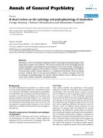
Conclusion
Our data have shown that PKR potentially is an impor-
tant mediator of degradative and death pathways in
chondrocytes. We hypothesise that the TNF-α signalling
pathway depicted in Fig. 7 that has been elucidated for
other cell types also operates in chondrocytes. Since
both the expression/activation of degradative enzymes
and the number of chondrocytes committed to cell death
are important determinants of cartilage integrity, our
results suggest a pivotal role for the PKR pathway in
arthritic disease onset and progression that requires
further investigation.
Competing interests
None declared.
Acknowledgements
The authors would like to thank the Arthritis Research Campaign for
funding this work (Grant number M0650) and Dr Emma Blain for her
expertise.
References
1. Dean DD, Martel-Pelletier J, Pelletier JP, Howell DS, Woessner JF
Jr: Evidence for metalloproteinase and metalloproteinase
inhibitor imbalance in human osteoarthritic cartilage. J Clin
Invest 1989, 84:678-685.
2. Aigner T, Zien A, Gehrsitz A, Gebhard PM, McKenna L: Anabolic
and catabolic gene expression pattern analysis in normal
Available online />R53
Figure 7
The potential role of PKR in cartilage degradation. Results from the current study have led us to hypothesise that TNF-α-induced degradative
pathways in cartilage may be mediated via activation of PKR. TNF-α binding to its receptor (TNF-R55) may activate PKR either directly or through
ceramide (CER) to increase transcription and activation of MMPs via induction of NFκB and early response genes (c-fos and c-jun). Activation of
PKR and subsequent phosphorylation of eIF2α would lead to an inhibition of protein synthesis and increased apoptosis, which may also affect
cartilage integrity. Increased expression and activation of MMPs, in the absence of a corresponding increase in their inhibitors, would shift the
balance of homeostasis towards matrix catabolism. AP-1, activator protein-1; eIF2α, eukaryotic initiation factor 2α; IκBα, inhibitor kappa B alpha;
MMP, matrix metalloproteinase; NFκB, nuclear factor κB; P, phosphorylated; PACT, PKR-activating protein; PKR, protein kinase R; TIMP, tissue
inhibitors of MMP; TNF, tumour necrosis factor; TNF-R55, tumour necrosis factor receptor-55.
versus osteoarthritic cartilage using complementary DNA-
array technology. Arthritis Rheum 2001, 44:2777-2789.
3. Yoshihara Y, Nakamura H, Obata K, Yamada H, Hayakawa T,
Fujikawa K, Okada Y: Matrix metalloproteinases and tissue
inhibitors of metalloproteinases in synovial fluids from
patients with rheumatoid arthritis or osteoarthritis. Ann
Rheum Dis 2000, 59:455-461.
4. Goldring MB: Osteoarthritis and cartilage: The role of
cytokines. Curr Rheumatol Rep 2000, 2:459-465.
5. Westacott CI, Barakat AF, Wood L, Perry MJ, Neison P, Bisbinds
I, Armstrong L, Millar AB, Elson CJ: Tumour necrosis factor-
alpha can contribute to focal loss of cartilage in osteoarthritis.
Osteoarthritis Cartilage 2000, 8:213-221.
6. Westacott CI, Whicher JT, Barnes IC, Thompson D, Swan AJ,
Dieppe PA: Synovial fluid concentration of five different
cytokines in rheumatic diseases. Ann Rheum Dis 1990,
49:676-681.
7. Feldmann M, Maini RN: The role of cytokines in the pathogene-
sis of rheumatoid arthritis. Rheumatology (Oxford) 1999, Suppl
2:3-7.
8. Serhan CN, Haeggstrom JZ, Leslie CC: Lipid mediator networks
in cell signaling: update and impact of cytokines. FASEB J
1996, 10:1147-1158.
9. Ruvolo PP: Intracellular signal transduction pathways acti-
vated by ceramide and its metabolites. Pharmacol Res 2003,
47:383-392.
10. Reunanen N, Westermarck J, Hakkinen L, Holmstrom TH, Elo I,
Eriksson JE, Kahari VM: Enhancement of fibroblast collagenase
(matrix metalloproteinase-1) gene expression by ceramide is
mediated by extracellular signal-regulated and stress-acti-
vated protein kinase pathways. J Biol Chem 1998, 273:5137-
5145.
11. Buisson-Legendre N, Bernard P, Bobichon H, Emonard H,
Schneider C, Maquart FX, Haye B, Hornebeck W: Involvement
of the 92-kDa gelatinase (matrix metalloproteinase-9) in the
ceramide-mediated inhibition of human keratinocyte growth.
Biochem Biophys Res Commun 1999, 260:634-640.
12. Sabatini M, Rolland G, Leonce S, Thomas M, Lesur C, Perez V, de
Nanteuil G, Bonnet J: Effects of ceramide on apoptosis, pro-
teoglycan degradation, and matrix metalloproteinase expres-
sion in rabbit articular cartilage. Biochem Biophys Res
Commun 2000, 267:438-444.
13. Sabatini M, Thomas M, Deschamps C, Lesur C, Rolland G, de
Nanteuil G, Bonnet J: Effects of ceramide on aggrecanase
activity in rabbit articular cartilage. Biochem Biophys Res
Commun 2001, 283:1105-1110.
14. Patel RC, Sen GC: PACT, a protein activator of the interferon-
induced protein kinase, PKR. EMBO J 1998, 17:4379-4390.
15. Gilbert SJ, Duance VC, Mason DJ: Identification of changes in
chondrocyte gene expression in an in vivo animal model of
osteoarthritis [abstract]. Osteoarthritis Cartilage 2000, Suppl
B:S33.
16. Gilbert SJ, Duance VC, Mason DJ: Tumour necrosis factor
alpha up-regulates protein kinase R (PKR)-activating protein
(PACT) and increases phosphorylation of PKR and eukaryotic
initiation factor 2-alpha in articular chondrocytes. Biochem
Soc Trans 2002, 30:886-889.
17. Ruvolo PP, Gao FG, Blalock WL, Deng X, Stratford May W:
Ceramide regulates protein synthesis by a novel mechanism
involving the cellular PKR activator RAX. J Biol Chem 2001,
276:11754-11758.
18. Little CB, Flannery CR, Hughes CE, Mort JS, Roughly PJ, Dent C ,
Caterson B: Aggrecanase versus matrix metalloproteinases in
the catabolism of the interglobular domain of aggrecan in
vitro. Biochem J 1999, 344:61-68.
19. Pataer A, Vorburger SA, Barber GN, Chada S, Mhashilkar AM,
Zou-Yang H, Stewart AL, Balachandran S, Roth JA, Hunt KK,
Swisher SG: Adenoviral transfer of the melanoma differentia-
tion-associated gene 7 (mda7) induces apoptosis of lung
cancer cells via up-regulation of the double-stranded RNA-
dependent protein kinase (PKR). Cancer Res 2002, 62:2239-
2243.
20. Osman F, Jarrous N, Ben-Asouli Y, Kaempfer R: A cis-acting
element in the 3
′′
-untranslated region of human TNF-alpha
mRNA renders splicing dependent on the activation of protein
kinase PKR. Genes Dev 1999, 1324:3280-3293.
21. Cheshire JL, Williams BRG, Baldwin AS Jr: Involvement of
double-stranded RNA-activated protein kinase in the syner-
gistic activation of nuclear factor-kB by tumour necrosis
factor-
αα
and
γγ
-interferon in preneuronal cells. J Biol Chem
1999, 274:4801-4806.
22. Blain EJ, Gilbert SJ, Wardale RJ, Capper SJ, Mason DJ, Duance
VC: Up-regulation of matrix metalloproteinase expression
and activation following cyclical compressive loading of artic-
ular cartilage in vitro. Arch Biochem Biophys 2001, 396:49-55.
23. Vaughan-Thomas A, Gilbert SJ, Duance VC: Elevated levels of
proteolytic enzymes in the aging human vitreous. Invest Oph-
thalmol Vis Sci 2000, 41:3299-3304.
24. Milner JM, Rowan AD, Elliott SF, Cawston TE: Inhibition of furin-
like enzymes blocks interleukin-1alpha/oncostatin M-stimu-
lated cartilage degradation. Arthritis Rheum 2003, 48:
1057-1066.
25. Lalu MM, Csonka C, Giricz Z, Csont T, Schulz R, Ferdinandy P:
Preconditioning decreases ischemia/reperfusion-induced
release and activation of matrix metalloproteinase-2. Biochem
Biophys Res Commun 2002, 296:937-941.
26. Zamanian-Daryoush M, Mogensen TH, DΙDonato JA, Williams
BRG: NF-
κκ
B activation by double-stranded-RNA-activated
protein kinase (PKR) is mediated through NF-
κκ
B-inducing
kinase and I
κκ
B kinase. Mol Cell Biol 2000, 20:1278-1290.
27. Mundschau LJ, Faller DV: Platelet-derived growth factor signal
transduction through the interferon-inducible kinase PKR. J
Biol Chem 1995, 270:3100-3106.
28. Ito T, Jagus R, May WS: Interleukin 3 stimulates protein syn-
thesis by regulating double-stranded RNA-dependant protein
kinase. Proc Natl Acad Sci USA 1994, 91:7455-7459.
29. Carpick BW, Graziano V, Schneider D, Maitra RK, Lee X, Williams
BRG: Characterization of the solution complex between the
interferon-induced, double-stranded RNA-activated protein
kinase and HIV-I trans-activating region RNA. J Biol Chem
1997, 272:9510-9516.
30. Chu W-M, Ostertag D, Li Z-W, Chang L, Chen Y, Hu Y, Williams
B, Perrault J, Karin M: JNK2 and IKK
ββ
are required for activating
the innate response to viral infection. Immunity 1999, 11:721-
731.
31. Clemens MJ, Elia A: The double-stranded RNA–dependant
protein kinase PKR: structure and function. J Interferon
Cytokine Res 1997, 17:503-524.
32. Williams BR: Signal integration via PKR. Sci STKE 2001, 89:
RE2.
33. Borden P, Heller RA: Transcriptional control of matrix metallo-
proteinases and the tissue inhibitors of matrix metallopro-
teinases. Crit Rev Eukaryot Gene Expr 1997, 7:159-178.
34. Mengshol JA, Vincenti MP, Coon CI, Barchowsky A, Brinckerhoff
CE: Interleukin-1 induction of collagenase 3 (matrix metallo-
proteinase 13) gene expression in chondrocytes requires p38,
c-Jun N-terminal kinase, and nuclear factor kappaB: differen-
tial regulation of collagenase 1 and collagenase 3. Arthritis
Rheum 2000, 43:801-811.
35. Preaux AM, D’ortho MP, Bralet MP, Laperche Y, Mavier P: Apop-
tosis of human hepatic myofibroblasts promotes activation of
matrix metalloproteinase-2. Hepatology 2002, 363:615-622.
36. Curtis CL, Hughes CE, Flannery CR, Little CB, Harwood JL,
Caterson B: n-3 fatty acids specifically modulate catabolic
factors involved in articular cartilage degradation. J Biol Chem
2000, 275:721-724.
37. van der Kraan PM, van den Berg WB: Anabolic and destructive
mediators in osteoarthritis. Curr Opin Clin Nutr Metab Care
2000, 3:205-211.
38. Aigner T, Kim HA: Apoptosis and cellular vitality. Issues in
osteoarthritic cartilage degeneration. Arthritis Rheum 2002,
46:1986-1996.
39. Hashimoto S, Takahashi K, Amiel D, Coutts RD, Lotz M: Chon-
drocyte apoptosis and nitric oxide production during experi-
mentally induced osteoarthritis. Arthritis Rheum 1998, 41:
1266-1274.
40. Der SD, Yang YL, Weissmann C, Williams BR: A double-
stranded RNA-activated protein kinase-dependant pathway
mediating stress-induced apoptosis. Proc Natl Acad Sci USA
1997, 94:8841-8845.
41. Srivastava SP, Kumar KU, Kaufman RJ: Phosphorylation of
eukaryotic translation initiation factor 2 mediates apoptosis in
response to activation of the double-stranded RNA-depen-
dent protein kinase. J Biol Chem 1998, 273:2416-2423.
Arthritis Research & Therapy Vol 6 No 1 Gilbert et al.
R54
42. Yeung MC, Liu J, Lau AS: An essential role for the interferon-
inducible, double-stranded RNA-activated protein kinase PKR
in the tumor necrosis factor-induced apoptosis in U937 cells.
Proc Natl Acad Sci USA1996, 93:12451-12455.
43. Nuttal ME, Nadeau DP, Fisher PW, Wang F, Keller PM. DeWolf
WE, Goldring MB, Badger AM, Lee D, Levy MA, Gowen M, Lark
MW: Inhibition of caspase-3-like activity prevents apoptosis
while retaining functionality of human chondrocytes in vitro. J
Orthop Res 2000, 18:356-363.
44. Aizawa T, Kon T, Einhorn TA, Gerstenfeld LC: Induction of apop-
tosis in chondrocytes by tumour necrosis factor-alpha. J
Orthop Res 2001, 19:785-796.
45. Patel CV, Handy I, Goldsmith T, Patel RC: PACT, a stress-modu-
lated cellular activator of interferon-induced, double-stranded
RNA activated protein kinase, PKR. J Biol Chem 2000, 275:
37993-37998.
Correspondence
Dr Sophie J Gilbert, School of Biosciences, Cardiff University, Museum
Avenue, Cardiff CF10 3US, Wales, UK. Tel: +44 (0) 29 20875419;
fax: +44 (0) 29 20874594; e-mail:
Available online />R55