Báo cáo Y học: The sodium pump Its molecular properties and mechanics of ion transport potx
Bạn đang xem bản rút gọn của tài liệu. Xem và tải ngay bản đầy đủ của tài liệu tại đây (254.84 KB, 10 trang )
MINIREVIEW
The sodium pump
Its molecular properties and mechanics of ion transport
Georgios Scheiner-Bobis
From the Institut fu
¨
r Biochemie und Endokrinologie, Fachbereich Veterina
¨
rmedizin, Justus-Liebig-Universita
¨
t Giessen, Germany
The sodium pump (Na
+
/K
+
-ATPase; sodium- and potas-
sium-activated adenosine 5¢-triphosphatase; EC 3.6.1.37)
has been under investigation for more than four decades.
During this time, the knowledge about the structure and
properties of the enzyme has increased to such an extent that
specialized groups have formed within this field that focus on
specific aspects of the active ion transport catalyzed by this
enzyme. Taking this into account, this review, while some-
what speculative, is an attempt to summarize the informa-
tion regarding the enzymology of the sodium pump with the
hope of providing to interested readers from outside the field
a concentrated overview and to readers from related fields a
guide in their search for gathering specific information
concerning the structure, function, and enzymology of this
enzyme.
Keywords: ATPase; P-type; ouabain; palytoxin; ion
transport.
THE SODIUM PUMP: A BRIEF
RETROSPECTIVE
Today there is a vast amount of information concerning ion
transport through biological membranes and primary
structures, crystals, mutants, and chimeras of ion trans-
porters. It is difficult to imagine that the impressive progress
achieved thus far was originally generated by a few
researchers who had the ability to observe simple phenom-
ena connected with ion distribution, to question their origin,
and to assemble experimental evidence in ways that did not
allow any other conclusion but that there must a mechanism
that enables ions to be actively transported against their
electrochemical gradients. This mechanism, termed a
Ôsodium pumpÕ by Dean in 1941, originates from the
observation that sodium ions within muscle fibers can
exchange with radioactive sodium added to their environ-
ment. Nevertheless, although a large amount of data and
interpretation of it followed Dean’s proposal, it was not
until 1954 that Gardos discovered that ion pumping in red
blood cell ghosts was supported by ATP, which in turn
became hydrolyzed. (Due to space limitations, some of the
early, seminal work is not included in the reference list;
instead, an up-to-date selection of papers from a variety of
groups from which both the current progress in the field can
be assessed and in which earlier, landmark discoveries are
fully referenced is provided.)
These observations, together with the finding that 18
sodium ions were transported for each molecule of oxygen
consumed (4.5 Na
+
per electron or, in other words, 3 Na
+
per ATP) and the fact that ouabain had already been shown
to inhibit sodium fluxes on frog skin, contributed to the
overall acceptance of Skou’s conclusion from 1957, which
identified in crab nerve membrane preparations the sodium
pump as an ATPase that was activated by Na
+
and K
+
and inhibited by ouabain [1].
Undoubtedly, however, all of these findings helped to
lay the cornerstone in the research field of ion transport,
which currently includes a vast number of primarily and
secondarily active transporters or ion channels. Among
them, the Na
+
/K
+
-ATPase takes its place within the
family of the so-called P-type ATPases, enzymes that
become autophosphorylated by the gamma phosphate
group of the ATP molecule that they hydrolyze. The Na
+
/
K
+
-ATPase was the first discovered ion transporter, and
indeed the first-discovered P-type ATPase. It is still,
however, not well understood; after many years of
investigation, the sodium pump is still at the center
of researchers’ attention.
Na
+
/K
+
-ATPASE: SUBUNIT
COMPOSITION
Every living cell is negatively charged in comparison with its
environment. Thus, in principle, the cell/environment pair
constitutes a battery. Just as a battery can be used to
perform work, a cell uses this electrochemical gradient to
obtain nutrients, ionic or nonionic, from its environment
and to extrude metabolites and ions from its interior. In this
fashion, the composition of the intracellular milieu remains
constant while allowing for adaptation to a changing
environment to occur.
Correspondence to G. Scheiner-Bobis, Institut fu
¨
r Biochemie und
Endokrinologie, Fachbereich Veterina
¨
rmedizin,
Justus-Liebig-Universita
¨
t Giessen, Frankfurter Str. 100,
D-35392 Giessen, Germany.
Fax: + 49 641 9938189, Tel.: + 49 641 9938180,
E-mail:
Abbreviations:Na
+
/K
+
-ATPase, sodium- and potassium-activated
adenosine 5¢-triphosphatase; FSBA, 5¢-p-fluorosulfonylbenzoyl-
adenosine; ClR-ATP, c-[4-(N-2-chloroethyl-N-methylamino)]benzyl-
amide ATP; FITC, 5¢-isothiocyanate.
Enzyme: sodium- and potassium-activated adenosine
5¢-triphosphatase (EC 3.6.1.37).
(Received 15 October 2001, revised 11 December 2001,
accepted 28 January 2002)
Eur. J. Biochem. 269, 2424–2433 (2002) Ó FEBS 2002 doi:10.1046/j.1432-1033.2002.02909.x
The sodium pump, also known as the Na
+
/K
+
-ATPase,
is responsible for establishing and maintaining this electro-
chemical gradient in animal cells. This enzyme is a
component of the plasma membrane and transports Na
+
and K
+
using ATP hydrolysis. For every molecule of ATP
hydrolyzed, three Na
+
ions from the intracellular space and
two K
+
ions from the external medium are exchanged.
Thus, the sodium pump contributes substantially to the
maintenance of the membrane potential of the cell, provides
the basis for neuronal communication, and contributes to
the osmotic regulation of the cell volume. In addition, the
electrochemical Na
+
gradient is the driving force behind
secondary transport systems.
The Na
+
/K
+
-ATPase belongs to the P-type ATPases, a
family of enzymes that become phosphorylated during
transport by the c-phosphate group of ATP at an aspartic
acid localized within the highly conserved sequence
DKTGS/T [2]. This family, which contains more than 50
members, includes membrane-bound enzymes responsible
for the transport of heavy metal ions (P
1
-type ATPases),
other metal ions (P
2
-type ATPases), and the K
+
-selective
Kdp-ATPase of Escherichia coli (P
3
-type ATPase).
Within the group of the P
2
-type ATPases, the Na
+
/
K
+
-ATPase, together with the colonic or gastric H
+
/
K
+
-ATPases, constitute a subgroup of oligomeric enzymes
consisting of a and b subunits. A third peptide referred to as
the c subunit appears in some tissues to be involved in
regulating the activity of the sodium pump and its
interactions with Na
+
or K
+
ions.
A number of isoforms of the a and b subunits has been
isolated from various tissues of numerous species, and it has
been repeatedly demonstrated that the function of Na
+
/
K
+
-ATPase requires the presence of both subunits.
The a subunit, which is referred to as the catalytic
subunit, has a relative molecular mass of 100–113 kDa,
depending on the presence of different isoforms: a1, a2, a3,
or a4. It crosses the membrane 10 times, forming trans-
membrane domains M1 to M10; both N- and C-termini are
localized on the cytosolic side [3]. Various studies have
shown that both ATP binding and ion occlusion occurs in
this subunit.
The b subunit is highly glycosylated and has a relative
molecular mass of about 60 kDa. The mass of the protein
moiety of this subunit is 36–38 kDa, depending on the
isoforms b1, b2, or b3. The bsubunit crosses the membrane
only once, and the N-terminus is localized on the intracel-
lular side of the membrane. The respective roles of these
proteins is still not entirely clear. More recent results have
shown that the b subunit makes direct contact with the
a subunit [4], thereby stabilizing the a subunit and assisting
in its transport from the endoplasmic reticulum to the
plasma membrane [5]. In addition, numerous experiments
have shown that the b subunit is important for ATP
hydrolysis, ion transport, and the binding of inhibitors such
as ouabain.
The third subunit of Na
+
/K
+
-ATPase, the csubunit of
7–11 kDa, was first identified as a component involved in
the binding of [
3
H]ouabain. The c subunit specifically
associates with the sodium pump [6], possibly via interac-
tions with the C-terminal domain of the a subunit [7]. The
c subunit belongs to type I membrane proteins and is
related to phospholemman and to the human Mat8
protein, a type I membrane protein associated with
mammary tumors. The availability of the cDNA coding
for the peptide permitted analysis of the role of the
c subunit in the function of the enzyme. Consistent with
the fact that c expression is not seen in all tissues where
a or b expression is otherwise easily identified, the presence
of the c peptide is not essential for obtaining Na
+
/
K
+
-ATPase activity in heterologous expressions systems
of the enzyme [8]. Nevertheless, c subunit expression in
HEK cells apparently modifies the affinity of the enzyme
for ATP, and its expression in different segments of the
nephron is associated with modulation of the affinity of
Na
+
/K
+
-ATPase for Na
+
or K
+
ions [9,10]. These data,
together with the fact that several peptides similar to the
c subunit have already been determined to interact with
and influence the sodium pump [11] confirm that the ion
pumping activity can be finely modulated by type I
membrane peptides and also offers the possibility
of addressing physiologically relevant questions in connec-
tion with the regulation of the expression of this type
of protein.
THE CATALYTIC MECHANISM
OF THE Na
+
/K
+
-ATPASE
The Na
+
/K
+
-ATPase has two conformational states, E
1
and E
2
. These states are not only characterized by differ-
ences in their interactions with Na
+
,K
+
, ATP, or ouabain,
they also have been clearly defined by tryptic cleavage
experiments.
In the first step of the reaction sequence, Na
+
and ATP
bind with very high affinity (K
d
values of 0.19–0.26 m
M
and
0.1–0.2 l
M
, respectively) to the E
1
conformation of the
enzyme (Fig. 1, step 1), during which phosphorylation at an
aspartate residue occurs via the transfer of the c-phosphate
of ATP (Fig. 1, step 2) [12,13]. Magnesium is very
important for this reaction. Thereafter, three Na
+
ions
are occluded while the enzyme remains in a phosphorylated
condition. After the E
2
-P3Na
+
conformation is attained,
the enzyme loses its affinity for Na
+
(K
0.5
¼ 14 m
M
)and
the affinity for K
+
is increased (K
d
0.1 m
M
). Thus, three
Na
+
ions are released to the extracellular medium (Fig. 1,
step 3) and K
+
ions are taken up (Fig. 1, step 4). The
binding of K
+
to the enzyme induces a spontaneous
dephosphorylation of the E
2
-P conformation. The
dephosphorylation of E
2
-P leads to the occlusion of two
K
+
ions, leading to E
2
(2K
+
) (Fig. 1, step 5) [12,13].
Intracellular ATP increases the extent of the release of
K
+
from the E
2
(2K
+
) conformation (Fig. 1, step 6) and
thereby also the return of the E
2
(2K
+
) conformation to the
E
1
ATPNa conformation. The affinity of the E
2
(2K
+
)
conformation for ATP, with a K
0.5
value of 0.45 m
M
,is
very low [12,13].
Through the juxtapositioning of these three reaction
sequences, the full catalytic cycle of Na
+
/K
+
-ATPase is
obtained (Fig. 1).
All P-type ATPases function in a similar way: they all
hydrolyze ATP and occlude ions during the translocation
process within the membrane-inserted segment of the
protein. Through this process, the ionophore of every
ion-transporting ATPase is accessible from only one side of
themembraneatanygiventime.
The sequential model presented above, however, often
referred to as the Albers–Post scheme [13] does not take into
Ó FEBS 2002 Sodium pump structure and properties (Eur. J. Biochem. 269) 2425
consideration that the sodium pump might exist as a
diprotomer of cooperating (ab)
2
subunits and thus contain
two binding sites for ATP.
The concentration–effect curve for ATP hydrolysis is
biphasic, which can be explained by an extrapolation of the
single-site model shown in Fig. 1. Each ab protomer has a
single ATP binding site that changes from high affinity to
low affinity with changes in conformation. This model is
strongly supported by experiments showing that the
stoichiometry of binding for either ATP, phosphate, or
ouabain is 1 per a subunit, and that solubilized enzyme
retains its catalytic activity [14]. Results obtained with highly
purified enzyme from duck salt gland lend credence to this
hypothesis [15].
A second model, which was originally put forward by
Repke, postulates that the biphasic nature of the ATP
concentration curve is due to the presence of two catalytic a
subunits that work cooperatively [16]. Each catalytic
subunit goes through the same conformational changes
that are described in the single-site model but in such a way
that they are shifted 180° from each other. Thus, in this
model the high affinity and low affinity ATP binding sites
occur simultaneously, and there is also simultaneous
transportofNa
+
out of the cell and K
+
into the cell.
Several experimental results support this model.
In a third model proposed by Plesner, the cooperativity of
the a subunits described by Repke occurs only in the
presence of Na
+
and K
+
[17]. The partial reactions of the
Na
+
/K
+
-ATPase are catalyzed by the ab protomeric
enzyme, as is the case with Na
+
-ATPase or K
+
-stimulated
phosphatase.
The models of Repke and Plesner differ from the single-
site model in that they predict the presence of two binding
sites on each functional enzyme entity. The results of many
investigations support the existence of two binding sites on
one (ab)
2
diprotomer. Kinetic studies have shown that the
single-site model is not sufficient to explain the coupling of
ATP hydrolysis to ion transport [18]. Moreover, crystallo-
graphic studies have demonstrated that Na
+
/K
+
-ATPase
crystallizes in a way that allows ab protomers to be in close
contact with each other [19]. Finally, radiation inactivation
has shown in several cases that the target size is consistent
with that of an (ab)
2
diprotomeric structure. These data,
however, are not compelling proof of the simultaneous
existence of two ATP binding sites and therefore do not
definitively establish the (ab)
2
diprotomer as the basic
functional unit of Na
+
/K
+
-ATPase. Alternative proposals
suggest the existence of (ab)
4
tetrameric enzymes [20] or
enzymes with two ATP binding sites per a subunit [21].
THE K
+
-STIMULATED PHOSPHATASE
ACTIVITY
A special characteristic of the Na
+
/K
+
-ATPase is its ability
to hydrolyze phosphoesters and phosphoanhydrides in the
presence of K
+
ions [22]. This so-called K
+
-stimulated
phosphatase activity is ouabain-sensitive. The physiological
relevance of this reaction is unknown.
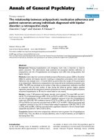
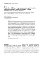
Fig. 1. Reaction cycle of Na
+
/K
+
-ATPase. Na
+
/K
+
-ATPase binds Na
+
and ATP in the E
1
conformational state (step 1) and is phosphorylated at
an aspartate residue by the c-phosphate of ATP. This leads to the occlusion of three Na
+
ions (step 2) and then to their release to the extracellular
side (step 3). This new conformational state (E
2
-P) binds K
+
with high affinity (step 4). Binding of K
+
leads to dephosphorylation of the enzyme
andtotheocclusionoftwoK
+
cations (step 5). K
+
is then released to the cytosol after ATP binds to the enzyme with low affinity (step 6). The
dashed box highlights the electrogenic steps of the catalytic cycle.
2426 G. Scheiner-Bobis (Eur. J. Biochem. 269) Ó FEBS 2002
THE ATP BINDING DOMAIN
The cytosolic protein structure between membrane domains
M4 and M5 (L4/5) is of great importance for the function of
the enzyme, because a series of amino acids within this
region have been identified to be either essential for or
highly involved in ATP hydrolysis and enzyme function.
(The prefix L stands for loop, a transmembrane domain-
connecting peptide. L2/3, L4/5, L6/7, and L8/9 are localized
on the cytosolic side, and L1/2, L3/4, L5/6, L7/8, and L 9/10
are accessible from the extracellular side.)
First, the ATP phosphorylation site is localized within
this loop as a part of the sequence DKTGT/S that is highly
conserved among all P-type ATPases. In addition, all ATP
analogs used thus far label peptide structures within this
loop, and the recently published Ca
2+
-ATPase crystal
structure was shown to contain TNP-AMP bound within
this L4/5 peptide. Therefore, it is justified to refer to this part
of the enzyme as the ATP binding domain.
By using the protein-reactive ATP analogs 2-azido-ATP
and 8-azido-ATP, it was possible to label and identify
Gly502 and Lys480, respectively, as possible recognition
sites for the adenosine moiety of ATP [23,24]. (Hereafter,
the amino-acid sequence numbers refer to that of the a1
isoform of the sheep.) The fact that Lys480 is also labeled by
both pyridoxal 5¢-diphospho-5¢-adenosine and pyridoxal
5¢-phosphate suggests that this amino acid might be
involved additionally in the recognition of phosphate
groups, as proposed by Hinz & Kirley [25]. Thus, in this
point of view, the labeling of Lys480 by 8-azido-ATP [23]
does not necessarily indicate that this amino acid directly
interacts with the adenine moiety of the ATP molecule, but
that it is merely within reach of the highly reactive azido
group of 8-azido-ATP. In the crystal structure of the
Ca
2+
-ATPase, Lys492, the equivalent of Lys480 of
the sodium pump a1 subunit, seems to interact with the
phosphate group of TNP-AMP [26]. Site-directed muta-
genesis experiments have confirmed the importance of
Lys480 for ATP hydrolysis and enzyme function [27].
Various other ATP analogs such as 5¢-p-fluoro-
sulfonylbenzoyl-adenosine (FSBA) or c-[4-(N-2-chloro-
ethyl-N-methylamino)]benzylamide ATP (ClR-ATP) were
successfully used for identifying amino acids within the
L4/5 peptide. Nevertheless, although these substances
resemble nucleotide triphosphates and their interaction with
the enzyme can be prevented by ATP, they are not
substrates of the sodium pump. Thus, it was still uncertain
whether Cys656 and Lys719, the FSBA labeling sites [28],
and Asp710, the ClR-ATP labeling site [29], were truly
constituents of the ATP binding site. In contrast to these
ATP-like substances, fluorescein 5¢-isothiocyanate (FITC),
a protein-reactive probe, was shown to modify Lys501 of
the sodium pump a1 subunit [30]. Although there is no
apparent similarity between FITC and ATP, the fact that
ATP prevents modification of Lys501 by FITC led to the
conclusion that Lys501 is localized within the adenosine-
recognizing moiety of the a1 subunit. This proposal has
been supported by findings concerning the conformation of
Mg
2+
-complexed ATP analyzed by
1
H-NMR and ultra-
violet spectrophotometric methods. According to these
reports, the a-phosphate group of the ATP molecule is
in close proximity to the C8 atom of the adenine
moiety. Therefore, if ATP is assumed to retain a similar
conformation when bound within the ATP binding site, one
can imagine that the C8-azido group of 8-azido-ATP labels
Lys480, which originally interacts with the a-phosphate
group of ATP. Taking into account that the distance
between Lys501 and Lys480, as determined by labeling
experiments with dihydro-4,4¢-diisothiocyanostilbene-2,2¢-
disulfonate, is approximately 1.4 nm [31], it is conceivable
that the azido group of 8-azido-ATP labels Lys480 while the
azido group of 2-azido-ATP labels Gly502.
The recently resolved crystal structure of Ca
2+
-ATPase
demonstrates that all ATP analogs used so far label
functional areas of the a subunit. The azido derivatives of
ATP, pyridoxal 5¢-diphospho-5¢-adenosine and pyridoxal
5¢-phosphate, or FITC label near the adenosine binding
pocket, as demonstrated for the binding of TNP-AMP
within the crystal structure of Ca
2+
-ATPase. This area is
referred to as the N (nucleotide binding) domain of the L4/5
peptide. Other ATP analogs such as FSBA or ClR-ATP
label the enzyme in the vicinity of the phosphorylation site,
within a substructure of the L4/5 peptide referred to as the P
(phosphorylation) domain. This area of the protein, consti-
tuting a Rossman fold, was first identified as being
conserved among various hydrolases by comparison of
the primary sequences of P-type ATPases with the primary
sequence of the
L
-2-haloacid dehalogenase from Pseudo-
monas sp. and was thought to directly participate in the
phosphorylation/dephosphorylation of Asp369 via the
terminal phosphate of ATP. More recent studies, however,
have suggested that this area of the protein is a Mg
2+
binding site [32].
The distance between the adenosine binding area of the
N domain and the phosphorylation site in the P domain is
rather large (2.5 nm) to be bridged by the ATP molecule.
Thus, some conformational transition must occur prior to
ATP hydrolysis, which results in the two domains
approaching each other. A third subdomain formed by
the L2/3 peptide might be involved in these conforma-
tional changes. This area of the protein is referred to as the
actuator domain (A domain). No functional analysis has
yet been published, however, that supports this proposal.
Nevertheless, the A domain undoubtedly contributes to
the conformational transitions associated with ATP
hydrolysis, ion transport, and dephosphorylation of the
phosphoenzyme formed by the transfer of the c phosphate
group of ATP. In experiments involving ascorbate/
H
2
O
2
-catalyzed peptide cleavage in the presence of
ATP-Fe
2+
, it was demonstrated that the peptide
TGESE(212–216) from the A domain moves towards
the phosphorylation site in the P domain, supporting
the dephosphorylation of the enzyme during the
E
2
-P fi E
2
(K
+
)-transition [33]. Because this peptide
(TGES/A) is highly conserved among all known P-type
ATPases, transport catalyzed by these other enzymes is
likely to take place by similar mechanisms.
MEMBRANE-SPANNING DOMAINS
AND THEIR INVOLVEMENT IN THE
CATION TRANSLOCATION PROCESS
Investigations using isolated Na
+
/K
+
-ATPase have shown
that after tryptic removal of the hydrophilic part of the
enzyme, the remaining C-terminal, membrane-spanning
segment (so-called Ô19-kDa membranesÕ) is still able to
Ó FEBS 2002 Sodium pump structure and properties (Eur. J. Biochem. 269) 2427
occlude Na
+
or the K
+
analog Rb
+
[34], indicating that
the ionophore, as expected, must consist of membrane-
spanning domains. Negatively charged amino acids within
this structure are viewed as possible interfaces between the
protein and ions being transported. However, analysis of
mutants has not always demonstrated that substitution of
acidic amino acids within the membrane-spanning domains
has a marked effect on enzyme activity. Substitution at
Glu327 (within the fourth membrane-spanning domain,
denoted M4), Asp926 (M8), Glu953, or Glu954 (both M9)
does not lead to significant changes in the affinity of the
mutant enzyme for Na
+
or K
+
or affect its electrical
properties [35–37]. Mutation of Glu953 or Glu954 also has
no effect on the interaction of the enzyme with palytoxin (G.
Scheiner-Bobis, unpublished observations).
Mutation of Glu779 from the sixth membrane-spanning
domain has a number of effects, depending on the nature of
the substitution. A Glu779Ala mutant has an ATPase
activity that is independent of K
+
(a Na
+
-ATPase) [38];
here, it may be that Na
+
mimics the binding of K
+
at
extracellular sites. Nevertheless, mutation of this Glu779 to
Gln, Asp, or Lys leads to only moderate changes in the K
0.5
for the cation activation of Na
+
/K
+
-ATPase. For this
reason, and because the Glu779fiLys mutants have a
slightly higher affinity for Na
+
, a direct role for Glu779 in
the cation binding process is fairly unlikely. Rather, it may
be assumed that Glu779 is a part of the overall structure
that participates in the formation of an ion coordination
complex involved in cation selectivity and activation of the
sodium pump.
Of all acidic amino acids examined thus far, only
nonconservative mutation of Asp804 and Asp808 leads to
a nonfunctional enzyme. It is possible that these mutations
have a deleterious effect on K
+
recognition at the
extracellular face of the enzyme [39]. The interaction with
the conservative mutation Asp808fiGlu. The conclusion
drawn from these studies is that Asp804 and Asp808 from
the sixth membrane-spanning domain of the a1 subunit
are involved in cation coordination [39]. The data
reported thus far, however, give the impression that the
mutations have an effect only on K
+
and not on Na
+
recognition.
The examination of various acidic residues from the
transmembrane domains of the sodium pump has not
brought us closer to the goal of identifying amino acids that
are essential for ion transport. In general, it would appear as
if it weren’t the individual negatively charged amino acids of
the membrane-spanning domains that were directly
involved in ion transfer, but larger peptide structures that
contain these amino acids. This conclusion, as unsatisfac-
tory as it may be, agrees well with investigations of a
considerable number of mutants of the Ca
2+
-ATPase that
clearly demonstrate that numerous amino acids within the
transmembrane domains M4, M5, M6, and M8 are
important for the function of the enzyme, independent of
whether they are charged or not [40].
If no single acidic residue from the transmembrane
domains is essential for ion transport, then which structures
are important?
It is known that cations are transferred along the
backbone of carbonyl groups by ion/dipole interactions
from studies of the ionophores valinomycin and gramicidin
[40a]. This general preference for ion/dipole instead of ion/
ion interactions has also been noted for soluble enzymes
that bind monovalent cations. Should ion translocation by
the sodium pump also occur by ion/dipole interactions, one
would assume that cations interact with carbonyl or
hydroxyl groups and not just with carboxyl groups.
In analogy to the Ca
2+
-ATPase, these amino acids
would be in the membrane-spanning domains M4, M5,
M6, and M8 of the a subunit of the sodium pump. In fact,
the crystal structure of Ca
2+
-ATPase, which was recently
reported with a resolution of 2.6 A
ˆ
, shows two binding
sites for Ca
2+
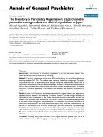
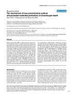
within the transmembrane region (Fig. 2).
One calcium ion is bound within a pocket formed by
Asn768 and Glu771 (M5), Thr799 and Asp800 (M6), and
Glu908 (M8) [26]. These results agree well with previous
conclusions drawn from mutation experiments [40].
A second Ca
2+
binds via interaction with the carbonyl
groups of Val304, Ala305, and Ile307 (M4) and through
the side-chain oxygen atoms of Asn796 and Asp800 (M6)
and Glu309 (M4) [26].
A similar situation could be assumed for the coordination
of cations within the membrane-spanning domains of the
Na
+
/K
+
-ATPase, because several structures are, as dem-
onstrated in extensive and thorough theoretical work, very
similar to those of the Ca
2+
-ATPase. This constellation
would also explain why single mutations within this region
do not lead to a complete loss of transport, because the
cations are coordinated simultaneously by several amino
acids. If this is the case, then only the mutation of several
amino acids concomitantly would lead to a marked change
in ion transport properties.
Fig. 2. Cation coordination sites of Ca
2+
-ATPase. The view is a cross-
section of the protein from the lumen of the sarcoplasmic reticulum.
Areas of the protein not involved in Ca
2+
coordination have been
eliminated. Two Ca
2+
ions shown in green are coordinated by Val304,
Ala305, Ile307 and Glu309 (M4), Asn768 and Glu771 (M5), Thr799
and Asp800 (M6), and Glu908 (M8). The side chain carboxyl group of
Asp800 participates in the coordination of both Ca
2+
ions. The cor-
responding amino acids of the sodium pump a1 subunit of the sheep
are given in parentheses. Atoms of interest: oxygen, red; nitrogen, blue;
calcium, green.
2428 G. Scheiner-Bobis (Eur. J. Biochem. 269) Ó FEBS 2002
COUPLING OF ATP HYDROLYSIS
TO ION TRANSPORT
Despite the appreciable amount of knowledge about the
ATP-recognition area of the protein or its ion coordination
sites, the molecular mechanisms that couple ATP hydrolysis
to the opening of the ionophore for the translocation of ions
against their electrochemical gradient are not well under-
stood. Comparison with some other known ion transporters
might be helpful in understanding the translocation process,
or at least in gaining some room for speculation.
The Kdp-ATPase of bacteria is a particularly interesting
K
+
-transporting ATPase made up of three protein
components: KdpA, KdpB, and KdpC. KdpA is inserted
into the membrane and is similar in sequence to the
hydrophobic portion of other P-type ATPases. KdpB is
hydrophilic and analogous to the hydrophilic, ATP-binding
L4/5 domains of other P-type ATPases. Finally, the KdpC
protein is equivalent to the b subunit of K
+
-transporting
P-type ATPases [41]. Furthermore, the KdpA component
has similarities to K
+
channels [42]. Taking into account
these observations, one could speculate that during evolu-
tion an ion channel has, together with the help of an ATP
hydrolase, been selected to move ions against their electro-
chemical gradients. In the further development of P-type
ATPases, ATP hydrolases and ion channels became phys-
ically fused.
InthecaseofNa
+
/K
+
-ATPase, by taking into consid-
eration Armstrong’s proposal regarding the selectivity of
ion channel ionophores for Na
+
or K
+
[43], such trans-
formations in the ion binding structure could explain how
one single structure could coordinate Na
+
in one instance
and K
+
in another. In the E
1
conformation, Na
+
is bound
by ion/dipole interactions to carbonyl groups of the M4,
M5, M6, and M8 domains. This applies for a sodium ion in
an aqueous milieu. Because K
+
is larger (r ¼ 1.33 A
˚
)than
Na
+
(r ¼ 0.95 A
˚
), K
+
would not fit into the Na
+
binding
site. Phosphorylation of Na
+
/K
+
-ATPase causes a
conformational change that brings about an alteration in
the Na
+
binding site, allowing Na
+
to exit toward the
extracellular side. One can assume that this conformational
change occurs concomitantly with an expansion of the
cation binding site (E
2
conformation of Na
+
/K
+
-ATPase),
so that now the larger K
+
can be accommodated. The ion/
dipole interactions in this case are also those of K
+
in an
aqueous environment. This newly expanded binding site
does not bind Na
+
well because Na
+
cannot be adequately
coordinated by the carbonyl groups. In this state, an
exchange of the water molecules surrounding Na
+
for
carbonyl groups would be thermodynamically unfavorable.
For the rigid pore opening of K
+
channels, Armstrong [43]
calculated that an energy expenditure of approximately
10 kcalÆmol
)1
would be required to remove two water
molecules 0.38 A
˚
(difference in ionic radii between Na
+
and
K
+
)fromNa
+
. This results in a preference for selecting K
+
over Na
+
of 10
6
: 1. The mechanism of ion selectivity
proposed by Armstrong guarantees that despite an
enormous excess of Na
+
in the extracellular medium, the
binding of K
+
is preferred. Thus, the Eisenman hypothesis,
which dictates that smaller ions pass more easily through a
pore than larger ones, does not apply for all ion channels
or pores. It is conceivable that after the release of Na
+
,
the selectivity for K
+
at the extracellular side of
Na
+
/K
+
-ATPase is maintained by such a rigid pore
opening, which may be formed by the L7/8 peptide of the
a subunit as well as the b subunit.
THEROLEOFTHEa / b SUBUNIT
INTERACTIONS FOR ION TRANSPORT
The a and b subunits of the sodium pump must interact with
each other in order to accomplish ion transport. In several
reports from the laboratory of Fambrough and colleagues,
it was shown that 26 amino acids from within the L7/8
peptide loop of the a subunit interact with extracellular
parts of the bsubunit [4]. Such interactions appear not only
to stabilize the a/b heterodimer but also to have functional
relevance, as ATP hydrolysis, ouabain binding, and paly-
toxin-induced K
+
efflux occur only in the presence of both
subunits and are markedly influenced by mutations in this
region of the enzyme.
Moreover, the bsubunit appears to influence the confor-
mation and ion sensitivity of the sodium pump. If the b
subunit of the sodium pump is replaced by that of the H
+
/
K
+
-ATPase, Na
+
-independent specific ouabain binding
can still be measured in the presence of Mg
2+
and ATP [44].
Apparently, the b subunit of the H
+
/K
+
-ATPase confers a
conformational change on the a subunit that enhances the
binding of ouabain.
Besides verifying that the interaction between a and
b subunits involves the L7/8 region, our own investigations
using an NGH26 chimera have additionally shown that the
binding of specific inhibitors is mediated through this
interaction. Thus, an NGH26/HKb heterodimer recognizes
not only palytoxin and ouabain but also the gastric
H
+
/K
+
-ATPase-specific inhibitor SCH 28080 [45].
Taken together, these results point to the function of the
b subunit as being more than just a vehicle for the transport
of the a subunit from the ER to the plasma membrane [46].
This hypothesis is supported by the fact that there are three
or possibly even four isoforms of the b subunit. Besides the
b1 isoform, which is the most widely distributed isoform,
there is the b2 isoform that is found in excitable tissues
(muscle and nervous tissue), the b3intestes,adrenal,and
brain, and the bm in skeletal and heart muscle. In view of
the variety of isoforms that have been identified, it is not
unreasonable to speculate that this multiplicity has a
physiological relevance.
Interestingly, the b2 isoform was known for some time in
glial cells as Ôadhesion molecule on gliaÕ [47]. This lends
further support to the idea that the b2 isoform has a
function besides that of stabilizing the a subunit. For
example, in tissue sections from cerebellum, Fab fragments
of monoclonal antibodies against adhesion molecule on glia
inhibit the migration of granulocytes. In the cochlea, the
expression of b2 is specifically associated with the striata
vulgaris, a tissue that forms the barrier between endolymph
and extracellular fluid. The endolymph contains a high
concentration of K
+
andalmostnoNa
+
. It is also strongly
electropositive, and K
+
must be transported against this
potential (+80 mV). Thus, it appears that b2 expression is
associated with structures that have a high K
+
-transporting
capability. Finally, a dual function for the b2 isoform is also
suggested by the fact that it is expressed in tissues that
contain no b1 isoform, including pineal gland, photorecep-
tor cells, and astrocytes, and also in tissues in the CNS
Ó FEBS 2002 Sodium pump structure and properties (Eur. J. Biochem. 269) 2429
(glia, choroid plexus, arachnoid membrane) that have
specialized ion-translocating characteristics. Nevertheless,
although these observations suggest that the b2 subunit
influences ion transport via the sodium pump, data that
confirm this function are still lacking.
An extracellularly localized peptide composed of 34
amino acids of the b1 subunit (Val93-Asp126) interacts with
the 26 amino-acid peptide of the a1 subunit already
mentioned [48]. The corresponding fragment of the b2
subunit (Val96-Arg129) has only 29% identity with the
Val93-Asp126 fragment of the b1, and 47% homology.
Whether these differences in the primary structure of these
two regions are responsible for any differences in enzyme
characteristics has yet to be investigated.
Nevertheless, the overall impression is that the 26 amino-
acid peptide and possibly the entire L7/8 region are
somehow involved in ion conduction by the pump. Our
own results show that mutations of Asp884 and Asp885
from within the L7/8 peptide to Arg considerably affect the
interactions of the enzyme with Na
+
, while, if anything, the
affinity for K
+
increases [49]. Notably, an SYG motif is
present within the 26-amino-acid peptide that somewhat
resembles the GYG motif of the P-loop of K
+
channels.
There, this tyrosine is essential for ion translocation.
Although it is not clear yet whether the corresponding
tyrosine of the asubunit is also involved in K
+
conduction,
it is certainly interesting to note that all but one of the
K
+
-transporting P-type ATPases, which always have a and
b subunits, have this tyrosine residue conserved (in Hydra, it
is a phenylalanine). A further point worth mentioning is that
naturally occurring mutation of the highly conserved GYG
sequence of the pore opening of K
+
channels to SYG
(which is the sequence in the Na
+
/K
+
-ATPase) leads to a
reduction in K
+
selectivity and an increase in Na
+
permeability [50]. Although there are currently no data
directly indicating a role for the SYG(894–896) sequence of
the Na
+
/K
+
-ATPase in ion transport, Cu
2+
-catalyzed
cleavage of the L7/8 loop (possibly near His875) results in
the loss of Rb
+
occlusion [51] usually obtained with the
19-kDa-membrane preparations of the a subunit. This,
together with the likelihood that the b subunit may play a
roleincationocclusion[52],makestheL7/8areaandthe
26 amino-acid peptide within this region attractive for
further investigation.
Besides this peptide, aromatic amino acids from the
transmembrane domain of the b subunit might be import-
ant for a/b subunit interactions and might influence the
properties of the enzyme. In the membrane-spanning
domains of the b1, b2, and b3 subunits of the sodium
pump, there is a relatively high number of amino acids with
aromatic side chains (phenylalanine, tyrosine, tryptophan)
whose position is conserved in almost all isoforms. In a
more recent study it was confirmed that Tyr40 and Tyr44 of
the membrane-spanning domain of the b1 subunit influence
the transport kinetics of the Na
+
/K
+
-ATPase and its
affinity towards K
+
[53]. However, the mechanism by
which the tyrosine residues might influence interactions of
theenzymewithK
+
are not yet understood.
SPECIFIC INHIBITORS
Possibly due to its key function in cellular physiology and
indeed the entire organism, the sodium pump has been a
target of a vast number of toxins produced by both plants
and animals. Thus, its ion pumping activity is specifically
inhibited by a series of naturally occurring steroids, termed
cardiac steroids or cardiac glycosides, such as ouabain and
digitalis. Other substances, like palytoxin from marine
corals of the genus Palythoa or sanguinarine from the plant
Sanguinaria canadensis, are also specific inhibitors of the
sodium pump. Unlike the cardioactive steroids, which
inhibit ion flow through the pump, palytoxin and possibly
also sanguinarine convert the enzyme into an open channel
that allows ions to flow down their concentration gradient.
In all cases, however, the toxin/receptor interactions result
in loss of the membrane potential, a fatal situation for the
cell or organism.
Cardioactive steroids bind reversibly to the extracellular
side of the Na
+
/K
+
-ATPase and inhibit ATP hydrolysis
and thus ion transport. The Na
+
/K
+
-ATPase is the only
enzyme known to interact with this class of substances.
Cardioactive steroids, especially water-soluble ouabain
(g-strophanthine), have often been used to identify
Na
+
/K
+
-ATPase and to study ion transport mechanisms
involved in this system. Under optimal conditions, 1 mole of
Na
+
/K
+
-ATPase binds 1 mol of ouabain. Optimal binding
occurs when the incubation medium contains one of the
following groups of ligands: (a) Mg
2+
,Na
+
,andATPor
(b) Mg
2+
and P
i
. Because both conditions can induce the
E
2
-P conformation of the enzyme, this is the conformation
to which the cardioactive steroids bind, resulting in the
formation of a stable phosphoenzyme/cardioactive steroid
complex, termed [E
2
–P*Æouabain]. The presence of the ions
to be transported influences the dissociation constant of the
enzyme–ouabain complex of the Na
+
/K
+
-ATPase: K
+
lowers the affinity of the enzyme for cardioactive steroids at
their high affinity, extracellular binding site. The presence of
extracellular Na
+
competitively inhibits this effect of K
+
,
and high concentrations of Na
+
enhance cardioactive
steroid binding. This probably occurs via interaction with
sites from which Na
+
is released to the extracellular
medium. On the other hand, with purified enzyme in the
presence of Mg
2+
and P
i
, low concentrations of Na
+
have
the effect of lowering the affinity of Na
+
/K
+
-ATPase for
cardioactive steroids when K
+
is present.
Inhibition of the sodium pump by cardiac steroids is
clinically relevant. Application of these substances, especi-
ally of digitalis and its congeners, helps to increase muscular
contractility of the failing heart, possibly by indirectly
inducing an elevation in the Ca
2+
concentration in the
myocardium. The wide use of digitalis for many centuries in
medicine, the great therapeutic impact of these substances,
and the need for a regulatory substance that increases heart
tonus without influencing its beating frequency led more
than 50 years ago to the proposal that endogenous factors
must exist that either have a similar structure or act in a
similar way to the cardiac steroids currently in use for clinical
purposes. The discovery of various isoforms of the sodium
pump that are specifically expressed in discrete tissues
indirectly supports this concept of an endogenous digitalis-
like factor, especially because in some cases distinctive
differences were found in the interaction of the various pump
isoforms with cardiac steroids and transported cations.
Recently, various research groups have succeeded in both
isolating endogenous circulating factors that interact with
the sodium pump and inhibit
86
Rb
+
uptake (Rb
+
is a
2430 G. Scheiner-Bobis (Eur. J. Biochem. 269) Ó FEBS 2002
surrogate for K
+
) and also in identifying several of them as
ouabain or its congeners [54]. In addition, evidence was
provided in several investigations that the concentration of
so-called endogenous ouabain increases in plasma upon
excessive work and is present at higher levels in the serum of
hypertensive patients [54].
All these data indicate that ouabain might be directly or
indirectly involved in the regulation of vascular tone and
possibly also in the pathogenesis of hypertension. Never-
theless, the mechanisms that might be relevant have not yet
been elucidated, and ouabain or cardiac glycosides do not
appear in the list of vasoactive endogenous substances that
includes such agents as endothelin and nitric oxide. Recent
experiments demonstrating mitogen-activated protein kin-
ase activation in rat cardiomyocytes by low concentrations
of ouabain [55,56], however, indicate that investigating
signal cascades induced by the glycoside might be helpful in
understanding its potential physiological relevance and its
possible involvement in vascular tone regulation or in the
pathogenesis of hypertension.
The Na
+
/K
+
-ATPase is a target of other substances
besides the cardiac glycosides. Palytoxin, produced by
corals of the genus Palythoa, is the most potent toxin of
animal origin. The LD
50
for rodents is 10–250 ngÆkg
)1
[57].
Previous investigations demonstrated that palytoxin opens
ion channels in vertebrate cells with a conductance of
approximately 10 pS. These channels remain open for some
time and allow K
+
ions to flow out of the cytosol. This is
probably the reason for the high toxicity of palytoxin, as the
outflow of K
+
and the resulting collapse of the membrane
potential lead to a general loss of basic cell functions.
Furthermore, depolarization is a key event that affects
numerous secondary systems. Thus, the concentration of
Ca
2+
becomes elevated in several organs through the
opening of Ca
2+
channels and leads to the production of
inositol trisphosphate [57], the activation of phospholi-
pase A
2
and metabolism of arachidonic acid, and numerous
other physiological responses that all stem from the
increased Na
+
influx and the ensuing increase in the
concentration of cytosolic Ca
2+
that accompany the initial
K
+
outflow [57].
The actual binding site for palytoxin has been the subject
of controversy for some time, despite the fact that the Na
+
/
K
+
-ATPase was known to be inhibited by the toxin. This
issue was resolved by expressing Na
+
/K
+
-ATPase hetero-
logously in yeast [58]. Untransformed yeast cells are
insensitive to palytoxin, whereas cells transformed with
both subunits of the Na
+
/K
+
-ATPase show a marked
efflux of K
+
in response to the toxin. This fact, and the
observation that this palytoxin-induced K
+
efflux is inhib-
ited by ouabain and other cardiotonic steroids, confirmed
that the sodium pump is the target of palytoxin. In vitro
expression experiments have lent further support to this
theory by showing that the palytoxin-induced channel is
directly associated with the presence of the Na
+
/K
+
-
ATPase [59]. Through its binding to the Na
+
/K
+
-ATPase,
the toxin appears to convert the enzyme into a permanently
open conformation that allows K
+
to flow down its
concentration gradient out of the cell. This channel is
possibly the permanently open state of the natural iono-
phore of the sodium pump.
Palytoxin binds predominantly to the E
1
-P conformation
of the pump. This observation results from experiments
demonstrating that ATP and Na
+
, which first induce the
E
1
-P conformation, enhance the binding of
125
I-labeled
palytoxin. Mg
2+
and P
i
, which support the direct formation
of the E
2
-P conformation, decrease binding [57]. ATP
hydrolysis or enzyme autophosphorylation, however, are
not necessary for the formation of the palytoxin-induced
channel because palytoxin produces K
+
efflux in yeast cells
expressing an Asp369Ala mutant of the a1 subunit that is
enzymatically inactive.
Palytoxin is apparently not the only molecule that
converts the sodium pump into an ion channel. Sanguin-
arine, one of a number of alkaloids developed by the
plant Sanguinaria canadensis in the course of evolution to
protect itself from herbivores, was described about
25 years ago as an inhibitor of the sodium pump.
Nevertheless, the interactions between sanguinarine and
the pump were not pursued because at that time
experiments that would yield conclusive results were not
possible. Using the yeast expression system for the sodium
pump, we recently showed that sanguinarine induces the
formation of a ouabain- or proscillaridin A-sensitive
channel in the sodium pump that allows K
+
ions to
flow out of the cell cytosol [60]. Sanguinarine also appears
to bind primarily to the E
1
-P conformation of the enzyme
and to inhibit the binding of [
3
H]ouabain, although,
as with palytoxin, phosphorylation is not absolutely
required.
The experiments with palytoxin and sanguinarine show
that under the appropriate conditions an ion channel can be
created within an ion pump. This ion channel, which is
possibly the ionophore of the pump arrested into a
permanently open state, is regulated under normal, physio-
logical conditions so that at any given time it is open to only
one side of the membrane. Interestingly, the electrogenic
step in the catalytic cycle of the sodium pump is associated
with the E
1
-P conformation of the enzyme.
Viewed from this standpoint, the reaction cycle of the
sodium pump (Fig. 1) takes on a new aspect: in the first part
of the reaction up to the occlusion of Na
+
, the pump can be
seen as a ligand-inactivated ion channel where P
i
is the
ligand that blocks the backflow of Na
+
out of the occlusion
pocket. In the last part of the reaction sequence, the release
of K
+
into the intracellular medium, the enzyme can be
viewed as a ligand-activated ion channel where ATP is the
ligand whose binding opens the occlusion pocket and allows
the release of K
+
to the cytosol.
PROSPECTS FOR FUTURE RESEARCH
Although much has been learned about the mechanics of
the transport of ions against their electrochemical gradients
by ATPases or the role of these enzymes as targets of either
endogenous or foreign toxins, the picture is still not
complete. The resolution of the crystal structure of Ca
2+
-
ATPase has appeared at a time when it was being suggested
that additional efforts might only result in semantic
refinements rather than the gain of new information. This
structure has provided new hope that the mechanisms of
this enzyme can be unveiled by addressing new questions in
new projects, and with the expectation of gaining new
perspectives. Thus, although they are long-known enzymes,
ATPases remain a fresh target for researchers and may soon
be discovered anew.
Ó FEBS 2002 Sodium pump structure and properties (Eur. J. Biochem. 269) 2431
ACKNOWLEDGEMENTS
The author has been supported through DFG, grants Sche 307/5-1 and
307/5-2. He wishes to thank Drs W. Schoner and R. A. Farley for many
constructive discussions.
REFERENCES
1. Skou, J.C. (1957) The influence of some cations on adenosine-
triphosphatase from peripheral nerves. Biochim. Biophys. Acta
23, 394–401.
2. Lutsenko, S. & Kaplan, J.H. (1995) Organization of P-type
ATPases: significance of structural diversity. Biochemistry 34,
15607–15613.
3. Antolovic,R.,Bruller,H.J.,Bunk,S.,Linder,D.&Schoner,W.
(1991) Epitope mapping by amino-acid-sequence-specific anti-
bodies reveals that both ends of the a subunit of Na
+
/K
+
-
ATPase are located on the cytoplasmic side of the membrane.
Eur. J. Biochem. 199, 195–202.
4. Lemas, M.V., Hamrick, M., Takeyasu, K. & Fambrough, D.M.
(1994) 26 Amino acids of an extracellular domain of the Na,
K-ATPase a-subunit are sufficient for assembly with the
Na,K-ATPase b-subunit. J. Biol. Chem. 269, 8255–8259.
5. Geering, K., Meyer, D.I., Paccolat, M.P., Kraehenbuhl, J.P. &
Rossier, B.C. (1985) Membrane insertion of a-andb-subunits of
Na
+
,K
+
-ATPase. J. Biol. Chem. 260, 5154–5160.
6. Beguin,P.,Wang,X.,Firsov,D.,Puoti,A.,Claeys,D.,Horis-
berger, J.D. & Geering, K. (1997) The c subunit is a specific
component of the Na,K-ATPase and modulates its transport
function. EMBO J. 16, 4250–4260.
7. Donnet, C., Arystarkhova, E. & Sweadner, K.J. (2001) Thermal
denaturation of the Na,K-ATPase provides evidence for a–a
oligomeric interaction and c subunit association with the C-ter-
minal domain. J. Biol. Chem. 276, 7357–7365.
8. Scheiner-Bobis, G. & Farley, F.A. (1994) Subunit requirements
for the expression of functional sodium pumps in yeast cells.
Biochim. Biophys. Acta 1193, 226–234.
9. Therien, A.G., Karlish, S.J. & Blostein, R. (1999) Expression and
functional role of the c subunit of the Na,K-ATPase in mam-
malian cells. J. Biol. Chem. 274, 12252–12256.
10. Arystarkhova, E., Wetzel, R.K., Asinovski, N.K. & Sweadner,
K.J. (1999) The c subunit modulates Na
+
and K
+
affinity of the
renal Na,K-ATPase. J. Biol. Chem. 274, 33183–33185.
11. Mahmmoud,Y.A.,Vorum,H.&Cornelius,F.(2000)Identifi-
cation of a phospholemman-like protein from shark rectal
glands. Evidence for indirect regulation of Na,K-ATPase by
protein kinase C via a novel member of the FXYDY family.
J. Biol. Chem. 275, 35969–35977.
12. Skou, J.C. (1988) Overview: the Na,K-pump. Methods Enzymol.
156, 1–25.
13. Glynn, I.M. (1993) Annual review prize lecture. ÔAll hands to the
sodium pumpÕ. J. Physiol. 462, 1–30.
14. Vilsen, B., Andersen, J.P., Petersen, J. & Jorgensen, P.L. (1987)
Occlusion of
22
Na
+
and
86
Rb
+
in membrane-bound and soluble
protomeric ab-subunits of Na,K-ATPase. J. Biol. Chem. 262,
10511–10517.
15. Martin, D.W., Marecek, J., Scarlata, S. & Sachs, J.R. (2000) ab
protomers of Na
+
,K
+
-ATPase from microsomes of duck salt
gland are mostly monomeric: formation of higher oligomers does
not modify molecular activity. Proc. Natl Acad. Sci. USA 97,
3195–3200.
16. Repke, K.R. & Schoen, R. (1973) Flip-flop model of (NaK)-
ATPase function. ActaBiol.Med.Ger.31, K19–K30.
17. Plesner, I.W., Plesner, L., Norby, J.G. & Klodos, I. (1981) The
steady-state kinetic mechanism of ATP hydrolysis catalyzed by
membrane-bound (Na
+
+K
+
)-ATPase from ox brain. III. A
minimal model. Biochim. Biophys. Acta 643, 483–494.
18. Askari, A. & Huang, W. (1982) Na
+
,K
+
-ATPase: evidence for
the binding of ATP to the phosphoenzyme. Biochem. Biophys.
Res. Commun. 104, 1447–1453.
19. Skriver,E.,Maunsbach,A.B.,Hebert,H.,Scheiner-Bobis,G.&
Schoner, W. (1989) Two-dimensional crystalline arrays of Na,
K-ATPase with new subunit interactions induced by cobalt-tet-
rammine-ATP. J. Ultrastruct. Mol. Struct. Res. 102, 189–195.
20. Taniguchi, K., Kaya, S., Abe, K. & Mardh, S. (2001) The oli-
gomeric nature of Na/K-transport ATPase. J. Biochem. 129,
335–342.
21. Ward, D.G. & Cavieres, J.D. (1993) Solubilized ab Na,K-
ATPase remains protomeric during turnover yet shows apparent
negative cooperativity towards ATP. Proc. Natl Acad. Sci. USA
90, 5332–5336.
22. Bader, H. & Sen, A.K. (1966) (K
+
)-Dependent acyl phosphatase
aspartofthe(Na
+
+K
+
)-dependent ATPase of cell mem-
branes. Biochim. Biophys. Acta 118, 116–123.
23. Tran, C.M., Scheiner-Bobis, G., Schoner, W. & Farley, R.A.
(1994) Identification of an amino acid in the ATP binding site of
Na
+
/K
+
-ATPase after photochemical labeling with 8-azido-
ATP. Biochemistry 33, 4140–4147.
24. Tran, C.M., Huston, E.E. & Farley, R.A. (1994) Photochemical
labeling and inhibition of Na,K-ATPase by 2-azido-ATP.
Identification of an amino acid located within the ATP binding
site. J. Biol. Chem. 269, 6558–6565.
25. Hinz, H.R. & Kirley, T.L. (1990) Lysine 480 is an essential
residue in the putative ATP site of lamb kidney (Na,K)-ATPase.
Identification of the pyridoxal 5¢-diphospho-5¢-adenosine and
pyridoxal phosphate reactive residue. J. Biol. Chem. 265, 10260–
10265.
26. Toyoshima, C., Nakasako, M., Nomura, H. & Ogawa, H. (2000)
Crystal structure of the calcium pump of sarcoplasmic reticulum
at 2.6 A
˚
resolution. Nature 405, 647–655.
27. Scheiner-Bobis, G. & Schreiber, S. (1999) Glutamic acid 472 and
lysine 480 of the sodium pump a1 subunit are essential for
activity. Their conservation in pyrophosphatases suggests their
involvement in recognition of ATP phosphates. Biochemistry 38,
9198–9208.
28. Ohta, T., Nagano, K. & Yoshida, M. (1986) The active site
structure of Na
+
/K
+
-transporting ATPase: location of the
5¢-(p-fluorosulfonyl)benzoyladenosine binding site and soluble
peptides released by trypsin. Proc.NatlAcad.Sci.USA83,
2071–2075.
29. Ovchinnikov, Y.A., Dzhandzugazyan, K.N., Lutsenko, S.V.,
Mustayef, A.A. & Modyanov, N.N. (1987) Affinity modification
of E1-form of Na
+
,K
+
-ATPase revealed Asp-710 in the cata-
lytic site. FEBS Lett. 217, 111–116.
30. Farley, R.A., Tran, C.M., Carilli, C.T., Hawke, D. & Shively,
J.E. (1984) The amino acid sequence of a fluorescein-labeled
peptidefromtheactivesiteof(Na,K)-ATPase.J. Biol. Chem.
259, 9532–9535.
31. Gatto, C., Lutsenko, S. & Kaplan, J.H. (1997) Chemical
modification with dihydro-4,4¢-diisothiocyanostilbene-2,2¢-
disulfonate reveals the distance between K480 and K501 in the
ATP-binding domain of the Na,K-ATPase. Arch. Biochem.
Biophys. 340, 90–100.
32. Jorgensen, P.L. & Pedersen, P.A. (2001) Structure–function
relationships of Na
+
,K
+
,ATP,orMg
2+
binding and energy
transduction in Na,K-ATPase. Biochim. Biophys. Acta 1505,
57–74.
33. Patchornik, G., Goldshleger, R. & Karlish, S.J. (2000) The
complex ATP-Fe
2+
serves as a specific affinity cleavage reagent
in ATP-Mg
2+
sites of Na,K-ATPase: altered ligation of Fe
2+
(Mg
2+
) ions accompanies the E1 fi E2 conformational change.
Proc. Natl Acad. Sci. USA 97, 11954–11959.
34. Shainskaya, A. & Karlish, S.J. (1994) Evidence that the cation
occlusion domain of Na/K-ATPase consists of a complex of
2432 G. Scheiner-Bobis (Eur. J. Biochem. 269) Ó FEBS 2002
membrane-spanning segments. Analysis of limit membrane-
embedded tryptic fragments. J. Biol. Chem. 269, 10780–10789.
35. Jewell-Motz, E.A. & Lingrel, J.B. (1993) Site-directed mutagen-
esis of the Na,K-ATPase: consequences of substitutions of
negatively-charged amino acids localized in the transmembrane
domains. Biochemistry 32, 13523–13530.
36. Vilsen, B. (1993) Glutamate 329 located in the fourth trans-
membrane segment of the a-subunit of the rat kidney Na
+
,
K
+
-ATPase is not an essential residue for active transport of
sodium and potassium ions. Biochemistry 32, 13340–13349.
37. Van Huysse, J.W., Jewell, E.A. & Lingrel, J.B. (1993) Site-
directed mutagenesis of a predicted cation binding site of Na,
K-ATPase. Biochemistry 32, 819–826.
38. Vilsen, B. (1995) Mutant Glu781 fi Ala of the rat kidney
Na
+
,K
+
-ATPase displays low cation affinity and catalyses ATP
hydrolysis at a high rate in the absence of potassium ions. Bio-
chemistry 34, 1455–1463.
39. Kuntzweiler,T.A.,Arguello,J.M.&Lingrel,J.B.(1996)Asp804
and Asp808 in the transmembrane domain of the Na,K-ATPase
alpha subunit are cation coordinating residues. J. Biol. Chem.
271, 29682–29687.
40. Rice, W.J. & MacLennan, D.H. (1996) Scanning mutagenesis
reveals a similar pattern of mutation sensitivity in transmem-
brane sequences M4, M5, and M6, but not in M8, of the Ca
2+
-
ATPase of sarcoplasmic reticulum (SERCA1a). J. Biol. Chem.
271, 31412–31419.
40a. Eisenmann, G. & Dani, J.A. (1987) An introduction to molecular
architecture and permeability of ion channels. Annu. Rev. Bio-
phys. Biomol. Struct. 16, 247–263.
41.Altendorf,K.,Siebers,A.&Epstein,W.(1992)TheKDP
ATPase of Escherichia coli. Ann. NY Acad. Sci. 671, 228–243.
42. Durell, S.R., Bakker, E.P. & Guy, H.R. (2000) Does the KdpA
subunit from the high affinity K
+
-translocating P-type KDP-
ATPase have a structure similar to that of K
+
channels? Biophys.
J. 78, 188–199.
43. Armstrong, C. (1998) The vision of the pore. Science 280, 56–57.
44. Eakle, K.A., Lyu, R M. & Farley, R.A. (1995) The influence of
b subunit structure on the interaction of Na
+
/K
+
-ATPase
complexes with Na
+
. A chimeric b subunit reduces the Na
+
dependence of phosphoenzyme formation from ATP. J. Biol.
Chem. 270, 13937–13947.
45. Farley,R.A.,Schreiber,S.,Wang,S G.&Scheiner-Bobis,G.
(2001) A hybrid between Na
+
,K
+
-ATPase and H
+
,K
+
-
ATPase is sensitive to palytoxin, ouabain, and SCH 28080. J.
Biol. Chem. 276, 2608–2615.
46. Geering, K. (1991) Posttranslational modifications and intra-
cellular transport of sodium pumps: importance of subunit
assembly. Soc. General Physiol. Series 46, 31–43.
47. Schmalzing, G., Kroner, S., Schachner, M. & Gloor, S. (1992)
The adhesion molecule on glia (AMOG/b2) and a1 subunits
assemble to functional sodium pumps in Xenopus oocytes.
J. Biol. Chem. 267, 20212–20216.
48. Colonna, T.E., Huynh, L. & Fambrough, D.M. (1997) Subunit
interactions in the Na,K-ATPase explored with the yeast two-
hybrid system. J. Biol. Chem. 272, 12366–12372.
49. Schneider, H. & Scheiner-Bobis, G. (1997) Involvement of the
M7/M8 extracellular loop of the sodium pump a subunit in ion
transport. Structural and functional homology to P-loops of ion
channels. J. Biol. Chem. 272, 16158–16165.
50. Silverman,S.K.,Kofuji,P.,Dougherty,D.A.,Davidson,N.&
Lester, H.A. (1996) A regenerative link in the ionic fluxes
through the weaver potassium channel underlies the pathophy-
siology of the mutation. Proc. Natl Acad. Sci. USA 93, 15429–
15434.
51. Shimon, M.B., Goldshleger, R. & Karlish, S.J. (1998) Specific
Cu
2+
-catalyzed oxidative cleavage of Na,K-ATPase at the
extracellular surface. J. Biol. Chem. 273, 34190–34195.
52. Lutsenko, S. & Kaplan, J.H. (1993) An essential role for the
extracellular domain of the Na,K-ATPase b-subunit in cation
occlusion. Biochemistry 32, 6737–6743.
53. Hasler, U., Crambert, G., Horisberger, J.D. & Geering, K.
(2001) Structural and functional features of the transmembrane
domain of the Na,K-ATPase b subunit revealed by tryptophan
scanning. J. Biol. Chem. 276, 16356–16364.
54. Schoner, W. (2002) Endogenous cardiac glycosides, a new class
of steroid hormones. Eur. J. Biochem. 269, 2440–2448.
55. Haas, M., Askari, A. & Xie, Z. (2000) Involvement of Src and
epidermal growth factor receptor in the signal-transducing
function of Na
+
/K
+
-ATPase. J. Biol. Chem. 275, 27832–27837.
56. Xie, Z. & Askari, A. (2002) Na
+
/K
+
-ATPase as a signal
transducer. Eur. J. Biochem. 269, 2434–2439.
57. Habermann, E. (1989) Palytoxin acts through Na
+
/K
+
-ATPase.
Toxicon 27, 1171–1187.
58. Scheiner-Bobis, G., Meyer zu Heringdorf, D., Christ, M. &
Habermann, E. (1994) Palytoxin induces K
+
efflux from yeast
cells expressing the mammalian sodium pump. Mol. Pharmacol.
45, 1132–1136.
59. Hirsh, J.K. & Wu, C.H. (1997) Palytoxin-induced single-channel
currents from the sodium pump synthesized by in vitro expres-
sion. Toxicon 35, 169–176.
60. Scheiner-Bobis, G. (2000) Sanguinarine induces K
+
outflow
from yeast cells expressing mammalian sodium pumps. Naunyn-
Schmiedeberg’s Arch. Pharmacol. 363, 203–208.
Ó FEBS 2002 Sodium pump structure and properties (Eur. J. Biochem. 269) 2433